. 1999 May 11;96(10):5826-31.
doi: 10.1073/pnas.96.10.5826.

Trading force for speed: why superfast crossbridge kinetics leads to superlow forces

Affiliations

Trading force for speed: why superfast crossbridge kinetics leads to superlow forces

L C Rome et al. Proc Natl Acad Sci U S A. .

Abstract

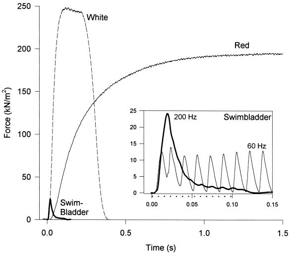
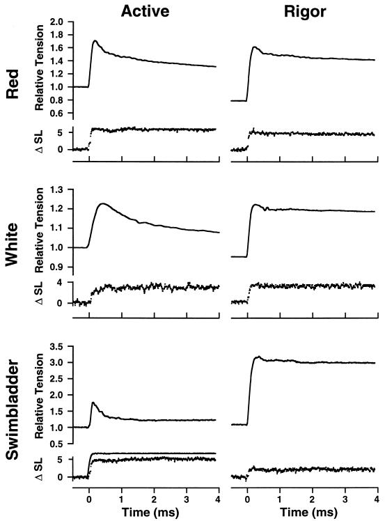
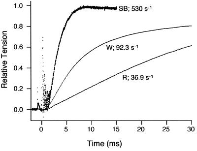
Superfast muscles power high-frequency motions such as sound production and visual tracking. As a class, these muscles also generate low forces. Using the toadfish swimbladder muscle, the fastest known vertebrate muscle, we examined the crossbridge kinetic rates responsible for high contraction rates and how these might affect force generation. Swimbladder fibers have evolved a 10-fold faster crossbridge detachment rate than fast-twitch locomotory fibers, but surprisingly the crossbridge attachment rate has remained unchanged. These kinetics result in very few crossbridges being attached during contraction of superfast fibers (only approximately 1/6 of that in locomotory fibers) and thus low force. This imbalance between attachment and detachment rates is likely to be a general mechanism that imposes a tradeoff of force for speed in all superfast fibers.


Figures

Figure 1
Figure 1
Force generation during repetitive stimulation in intact bundles of three fiber types of toadfish. Both the red muscle (stimulation frequency = 20 Hz) and the white muscle fibers (stimulation frequency = 50 Hz) produced classic tetanic responses, a smooth and steady force record. By contrast, at a stimulation frequency of 60 Hz, the swimbladder (Inset) produced nearly separate twitches, and at stimulation frequencies of 125 Hz and higher, the swimbladder fibers produced a smooth force record (same trace in main figure and Inset), but force fell rapidly after a peak despite continuous stimulation [the period of stimulation at 200 Hz is depicted by dotted line beneath the time axis (Inset)]. For purposes of illustration, amplitudes of individual traces were scaled to the mean isometric force for that fiber type. Maximum forces (mean ±SE, n = 4) are 192 ± 13 kN/m2, 244 ± 19 kN/m2 and 24 ± 1.4 kN/m2 for the red, white, and swimbladder muscle, respectively. All experiments were conducted at 15°C.
Figure 2
Figure 2
Active and rigor stiffness in skinned fibers of three muscle types from toadfish. Rapid length steps (complete in 200 μs) were applied by a servomotor while sarcomere length and force were monitored. Force is normalized to isometric tension in the active fiber. Note that because of the low active force in swimbladder, but normal Young’s modulus value in rigor, even a very small stretch applied to swimbladder fibers in rigor caused tension to rise to a level ≈3-fold higher than the active force. ΔSL is given in nm/half sarcomere. A sample motor trace (low noise) is shown (Bottom Left). For the red and white fibers, the force increase was slightly greater in the rigor than in the active fiber. In the case of the swimbladder, a 2-fold larger stretch was applied in the active fiber, but the force change was still just a small fraction of that in rigor. All experiments were conducted at 15°C.
Figure 3
Figure 3
Force records after photolysis of caged Ca2+ are shown for the red, white, and swimbladder fiber types. The rate constants for tension rise (kdevelop) are derived from the traces as described in Methods. All experiments were conducted at 15°C.

References

    1. Mendelson M. J Cell Biol. 1969;42:548–563. - PMC - PubMed
    1. Rosenbluth R. J Cell Biol. 1969;42:534–547. - PMC - PubMed
    1. Appelt D, Shen V, Franzini-Armstrong C. J Muscle Res Cell Motil. 1991;12:543–552. - PubMed
    1. Josephson R K, Young D. J Exp Biol. 1981;91:219–237.
    1. Schaeffer P J, Conley K E, Lindstedt S L. J Exp Biol. 1996;199:351–358. - PubMed

Publication types