. 1999 May 11;96(10):5862-7.
doi: 10.1073/pnas.96.10.5862.

Genetic engineering of the unsaturation of fatty acids in membrane lipids alters the tolerance of Synechocystis to salt stress

Affiliations

Genetic engineering of the unsaturation of fatty acids in membrane lipids alters the tolerance of Synechocystis to salt stress

S I Allakhverdiev et al. Proc Natl Acad Sci U S A. .

Abstract

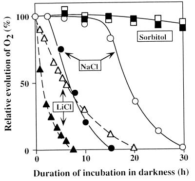
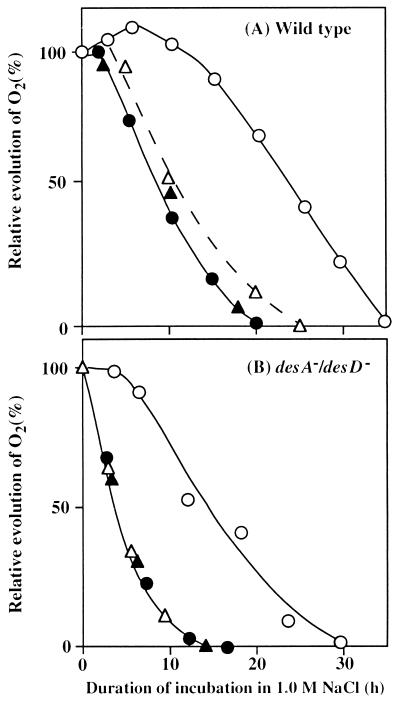
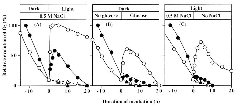
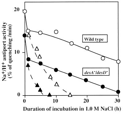
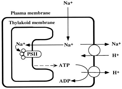
The role of unsaturated fatty acids in membrane lipids in the tolerance of the photosynthetic machinery to salt stress was studied by comparing the desA-/desD- mutant of Synechocystis sp. PCC 6803, which contained monounsaturated fatty acids, with the wild-type strain, which contained a full complement of polyunsaturated fatty acids. In darkness, the loss of oxygen-evolving photosystem II activity in the presence of 0.5 M NaCl or 0.5 M LiCl was much more rapid in desA-/desD- cells than in wild-type cells. Oxygen-evolving activity that had been lost during incubation with 0.5 M NaCl in darkness returned when cells were transferred to conditions that allowed photosynthesis or respiration. Recovery was much greater in wild-type than in desA-/desD- cells, and it was prevented by lincomycin. Thus, the unsaturation of fatty acids is important in the tolerance of the photosynthetic machinery to salt stress. It appears also that the activity and synthesis of the Na+/H+ antiporter system might be suppressed under high-salt conditions and that this effect can be reversed, in part, by the unsaturation of fatty acids in membrane lipids.


Figures

Figure 1
Figure 1
Changes in the photosynthetic oxygen-evolving activity of wild-type and desA/desD cells during incubation in darkness in the presence of 0.5 M NaCl, 0.5 M LiCl, or 1.0 M sorbitol. At designated times, a portion of the cell suspension was withdrawn and oxygen-evolving activity was examined at 35°C after addition of 1.0 mM BQ. The activities of wild-type and desA/desD cells that corresponded to 100% were 610 ± 24 and 556 ± 18 μmol of O2 per mg of Chl per h, respectively. Open symbols, wild-type cells; filled symbols, desA/desD cells. Each point represents the average of results from four independent experiments.
Figure 2
Figure 2
Effects of light and lincomycin on the NaCl-induced inactivation of the oxygen-evolving machinery. Wild-type (A) and desA/desD (B) cells were incubated with 1.0 M NaCl in darkness or in light at 50 μE⋅m−2⋅s−1 in the absence or presence of 25 μg/ml lincomycin. At designated times, a portion of the cell suspension was withdrawn and the oxygen-evolving activity was measured at 35°C after addition of 1.0 mM BQ. The activities of wild-type and desA/desD cells that corresponded to 100% were 590 ± 22 and 532 ± 16 μmol of O2 per mg of Chl per h, respectively. ○, Light in the absence of lincomycin; ▵, light in the presence of lincomycin; ●, darkness in the absence of lincomycin; and ▴, darkness in the presence of lincomycin. Each point represents the average of results from four independent experiments.
Figure 3
Figure 3
Effects of light, glucose, and removal of NaCl on restoration of oxygen-evolving activity in wild-type and desA/desD cells after NaCl-induced inactivation. (A) Wild-type and desA/desD cells were incubated for 25 h and 12 h, respectively, in darkness in the presence of 0.5 M NaCl. Then cells were exposed to light of 50 μE⋅m−2⋅s−1 in the presence of 25 μg/ml lincomycin (broken lines) or its absence (solid lines). (B) Wild-type and desA/desD cells were incubated in darkness in the presence of 0.5 M NaCl as in A. Then glucose was added to a final concentration of 5 mM. (C) Wild-type and desA/desD cells were incubated with 0.5 M NaCl in light at 50 μE⋅m−2⋅s−1 for 45 h and 25 h, respectively. Then cells were collected by centrifugation, resuspended in fresh BG-11 medium with no added NaCl, and incubated in light. ○, Wild-type cells; ●, desA/desD cells in the absence of lincomycin; ▵, wild-type cells; and ▴, desA/desD cells in the presence of lincomycin. Each point represents the average of results from four independent experiments.
Figure 4
Figure 4
Changes in the Na+/H+ activity of wild-type and desA/desD cells during incubation in the presence of 1.0 M NaCl in darkness and in light at 50 μE⋅m−2⋅s−1. At designated times, 20 μl of the suspension was withdrawn and diluted 100-fold with Na+-free medium that contained 5 μM acridine orange. The Na+/H+ antiport activity was determined as described in the text. ○, Wild-type cells in light; ▵, wild-type cells in darkness; ●, desA/desD cells in light; and ▴, desA/desD cells in darkness. Each point represents the average of results from four independent experiments.
Figure 5
Figure 5
Effects of light and removal of NaCl on restoration of the Na+/H+ antiport activity in wild-type and desA/desD cells after NaCl-induced inactivation. Wild-type and desA/desD cells were incubated for 20 h and 12 h, respectively, in darkness in the presence of 1.0 M NaCl. Then cells were exposed to light at 50 μE⋅m−2⋅s−1. After incubation in light for 30 h, cells were collected by centrifugation, resuspended in fresh BG-11 medium with no added NaCl, and incubated in light. Experimental conditions were the same as those described in the legend to Fig. 4. ○, Wild-type cells; and ●, desA/desD cells. Each point represents the average of results from three independent experiments.
Figure 6
Figure 6
A schematic explanation of the effects of Na+ ions and the unsaturation of fatty acids in membrane lipids on the oxygen-evolving activity of the PS II complex in the cyanobacterial cells.

References

    1. Nishida I, Murata N. Annu Rev Plant Physiol Plant Mol Biol. 1996;47:541–568. - PubMed
    1. Sato N, Murata N. Biochim Biophys Acta. 1980;619:353–366. - PubMed
    1. Berry J, Bjorkman O. Annu Rev Plant Physiol. 1980;31:491–543.
    1. Quinn P J, Williams W P. Biochim Biophys Acta. 1983;737:223–266.
    1. Murata N, Wada H. Biochem J. 1995;308:1–8. - PMC - PubMed

Publication types

MeSH terms

LinkOut - more resources