

Case Reports
Tissue response of a small saccular aneurysm after incomplete occlusion with a Guglielmi detachable coil
Affiliations
- PMID: 10319956
- PMCID: PMC7056010
Item in Clipboard
Case Reports
Tissue response of a small saccular aneurysm after incomplete occlusion with a Guglielmi detachable coil
AJNR Am J Neuroradiol.
1999 Apr.
Abstract
A 49-year-old woman had a small saccular aneurysm that was incompletely occluded with a Guglielmi detachable coil (GDC). She died from rupture of another aneurysm 42 days after the treatment. Autopsy for the embolized aneurysm revealed no neoendothelium at the aneurysmal neck, but an organized thrombus was observed limited to the periphery of the aneurysmal lumen. Although isolation of the aneurysm was not apparent, loose embolization with this method may help to reinforce the aneurysmal wall.
Figures

A 49-year-old woman with a small saccular aneurysm. A, Anteroposterior carotid angiogram shows the PTA (arrow) and a small saccular aneurysm at the basilar-superior cerebellar bifurcation (arrowhead). B, Angiogram, after embolization with a GDC, shows partial opacification of the fundus of the aneurysm.
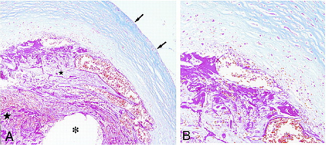
Microscopic examination of the small saccular aneurysm after GDC occlusion. A, Low-power photomicrograph shows a cross-section of the aneurysm at the fundus, with a fibrotic component (small star) adjacent to the aneurysmal wall (arrows) and an unformed thrombus at the center of the aneurysm (large star). The location of the coil is indicated (asterisk). (Azan stain; magnification ×100) B, Higher power magnification of the region adjacent to the wall shows fibrous connective tissue with rich capillaries. (Azan stain; magnification ×200)
Comment in
-
Tissue response to Guglielmi detachable coils: present implications and future developments.AJNR Am J Neuroradiol. 1999 Apr;20(4):533-4. AJNR Am J Neuroradiol. 1999. PMID: 10319951 Free PMC article. No abstract available.
References
-
- Graves VB, Strother CM, Duff TA, Perl J. Early treatment of ruptured aneurysms with Guglielmi detachable coils: effect on subsequent bleeding. Neurosurgery 1995;37:640-648 - PubMed
-
- Guglielmi G, Viñuela F, Dion J, Duckwiler G. Electrothrombosis of saccular aneurysms via endovascular approach. Part 2: Preliminary clinical experience. J Neurosurg 1991;75:8-14 - PubMed
-
- Malisch TW, Guglielmi G, Viñuela F, et al. Intracranial aneurysms treated with the Guglielmi detachable coil: midterm clinical results in a consecutive series of 100 patients. J Neurosurg 1997;87:176-183 - PubMed
-
- Molyneux AJ, Ellison DW, Morris J, Byrne JV. Histological findings in giant aneurysms treated with Guglielmi detachable coils. J Neurosurg 1995;83:129-132 - PubMed
Publication types
MeSH terms
LinkOut - more resources
Full Text Sources
Other Literature Sources
Medical