

Case Reports
Long-term histopathologic findings in two cerebral aneurysms embolized with Guglielmi detachable coils
Affiliations
- PMID: 10319957
- PMCID: PMC7056029
Item in Clipboard
Case Reports
Long-term histopathologic findings in two cerebral aneurysms embolized with Guglielmi detachable coils
AJNR Am J Neuroradiol.
1999 Apr.
Abstract
We present gross pathologic autopsy findings of a patient who was treated for two aneurysms with Guglielmi detachable coils (GDCs), and who died 33 months after the procedure. Histologic findings are also presented. In both aneurysms, the coils were firmly attached to the aneurysmal wall, making it impossible to remove them from the sac. The ostium of one aneurysm was covered by collagenous tissue and a single layer of endothelium.
Figures
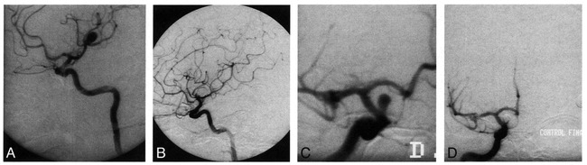
Left ICA angiogram shows MCA aneurysm before (A) and after (B) embolization with GDC. Right ICA angiogram shows carotid ophthlamic aneurysm before (C) and after (D) embolization with GDC
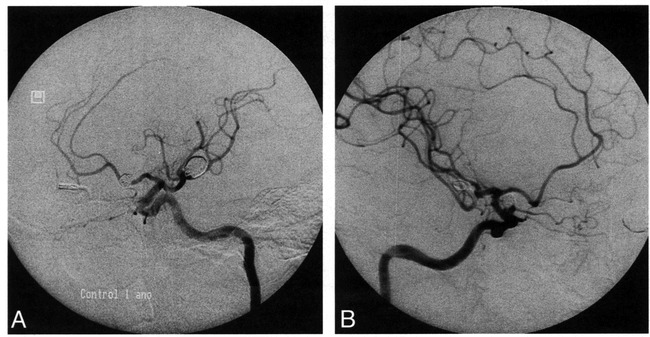
Left ICA angiogram done 1year after GDC treatment of left MCA. Aneurysm remains completely occluded (A). Right ICA angiogram done 1 year after GDC treatment of right carotid ophthalmic aneurysm (B). There is some coil compaction and a small-neck remnant
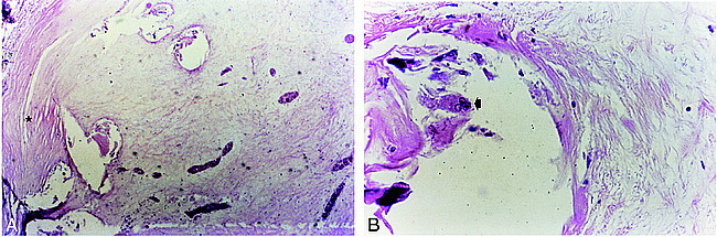
Pathologic findings of MCA aneurysm fundus. The cast of the coils, removed for cutting purposes, is completely surrounded by richly vascular fibrous tissue more cellular at the periphery of the fundus (star) and around the platinum coils (Hematoxilin and eosin stain; magnification ×200) (A). Many multinucleated foreign body giant cells are stretched along the coil casts, and some were released in the lumen of the coil casts (arrow), probably during coil removal for cutting purposes (Hematoxilin and eosin stain; magnification ×400) (B).
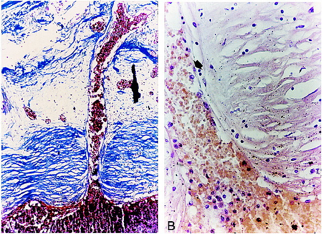
Histologic study of the aneurysm neck. A, Capillary growth into the aneurysm neck proceeded from the lumen of the ICA (P) via the neck. A thick and dense layer of collagen tissue aligned parallel to the long axis of the parent vessel, covering ostium (C) (Trichrome stain; magnification ×200). B, Transition zone between the endothelial cell lining of neovessels and endothelial cells line the surface of the collagen tissue covering the neck. The neointima is organized in two layers. The most superficial layer comprises endothelium continuous with neovessels proceding from the lumen of the parent vessel (arrow). The deepest layer of neointima consited of dense vascular and collagenous fibrous tissue (C) (Hematoxilin and eosin stain; magnification ×400).
Comment in
-
Tissue response to Guglielmi detachable coils: present implications and future developments.AJNR Am J Neuroradiol. 1999 Apr;20(4):533-4. AJNR Am J Neuroradiol. 1999. PMID: 10319951 Free PMC article. No abstract available.
References
-
- Guglielmi G, Viñuela F, Sepetka I, Macellari V. Electrothrombosis of saccular aneurysms via endovascular approach. I: electrochemical basis, technique and experimental results. J Neurosurg 1991;75:1-7 - PubMed
-
- Molyneux A, Ellison D, Morris J, Byrne J. Histological findings in giant aneurysms treated with Guglielmi detachable coils. J Neurosurg 1995;83:129-132 - PubMed
-
- Mizoi K, Yoshimoto T, Takahashi A, Nagmine Y. A Pitfall in the surgery of a recurrent aneurysm after coil embolization and its histological observation: technical case report. Neurosurgery 1996;39:165-168 - PubMed
Publication types
MeSH terms
Substances
LinkOut - more resources
Full Text Sources
Other Literature Sources
Medical