

Comparison of MR imaging with PET and ictal SPECT in 118 patients with intractable epilepsy
- PMID: 10319968
- PMCID: PMC7056008
Comparison of MR imaging with PET and ictal SPECT in 118 patients with intractable epilepsy
Abstract
Background and purpose: MR imaging, PET, and ictal SPECT have been studied extensively as individual techniques in the localization of epileptogenic foci, but only a few comparative studies have been done. We evaluated the concordance rates of ictal video/EEG, MR imaging, PET, and ictal SPECT to compare the sensitivities of these imaging methods in the lateralization of epileptogenic foci.
Methods: The study included 118 consecutive patients who underwent surgery for medically intractable epilepsy and who were followed up for 12 months or more. MR imaging was compared retrospectively with ictal video/EEG, FDG-PET, ictal 99mTc-HMPAO SPECT, and invasive EEG as to their ability to localize the epileptogenic focus; the pathologic findings served as the standard of reference.
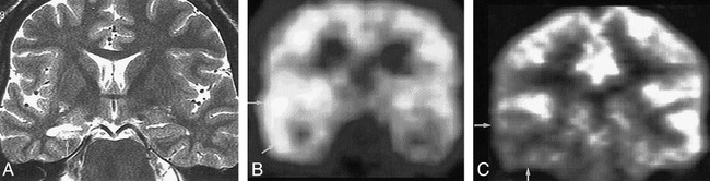
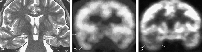
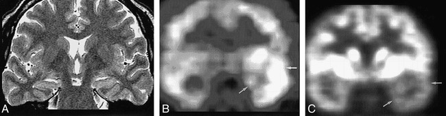
Results: MR imaging was concordant with video/EEG, PET, and ictal SPECT in 58%, 68%, and 58% of patients, respectively. With the pathologic diagnosis as the standard of reference, MR imaging, PET, and ictal SPECT correctly lateralized the lesion in 72%, 85%, and 73% of patients, respectively. Of the patients with good outcomes, MR imaging, PET, and ictal SPECT were correct in 77%, 86%, and 78%, respectively. In the good outcome group, MR imaging was concordant with PET and ictal SPECT in 73% and 62% of patients, respectively. Of 45 patients who underwent invasive EEG, MR imaging was concordant with the invasive study in 47%; PET in 58%; and ictal SPECT in 56%. Of 26 patients with normal MR findings, PET and ictal SPECT correctly lateralized the lesion in 80% and 55%, respectively.
Conclusion: Overall concordance among the techniques is approximately two thirds or less in lateralizing epileptogenic foci. PET is the most sensitive, even though it provides a broad approximate nature of the epileptogenic zone, which is not adequate for precise surgical localization of epilepsy. PET and/or ictal SPECT may be used as complementary tools in cases of inconclusive lateralization with ictal video/EEG and MR imaging.
Figures
Comment in
-
Imaging intractable epilepsy: how many tests are enough?AJNR Am J Neuroradiol. 1999 Apr;20(4):534-5. AJNR Am J Neuroradiol. 1999. PMID: 10319952 Free PMC article. No abstract available.
References
-
- Hauser WA, Hesdorffer DC. Epilepsy: Frequency, Causes and Consequences.. New York: Demos; 1990;1–51
-
- Rasmussen TB. Surgical treatment of mesiobasal limbic epilepsy. Surg Neurol 1982;17:445-457 - PubMed
-
- Schomer DJ. Current concepts in neurology: partial epilepsy. N Engl J Med 1983;309:536-539 - PubMed
-
- Stefan H, Pawlik G, Bocher-Schwarz HG, et al. Functional and morphological abnormalities in temporal lobe epilepsy: a comparison of interictal and ictal EEG, CT SPECT and PET. J Neurol 1987;234:377-384 - PubMed
-
- Coubes P, Awad IA, Antar M, et al. Comparison and spatial correlation of interictal HMPAO-SPECT and FDG-PET in intractable temporal lobe epilepsy. Neurol Res 1993;15:160-167 - PubMed
Publication types
MeSH terms
Substances
LinkOut - more resources
Full Text Sources
Medical