

Case Reports
Decreased BOLD functional MR activation of the motor and sensory cortices adjacent to a glioblastoma multiforme: implications for image-guided neurosurgery
Affiliations
- PMID: 10319970
- PMCID: PMC7056038
Item in Clipboard
Case Reports
Decreased BOLD functional MR activation of the motor and sensory cortices adjacent to a glioblastoma multiforme: implications for image-guided neurosurgery
AJNR Am J Neuroradiol.
1999 Apr.
Abstract
A patient with a glioblastoma multiforme and mild sensorimotor deficits had significantly less activation of the motor and sensory cortices on the side with the tumor than on the contralateral side on blood oxygen level-dependent (BOLD) functional MR images. This difference, which may be due to pressure effects or loss of vascular autoregulation, should be considered in preoperative planning in which BOLD functional MR imaging is used to identify eloquent cortices to be avoided during brain tumor surgery.
Figures
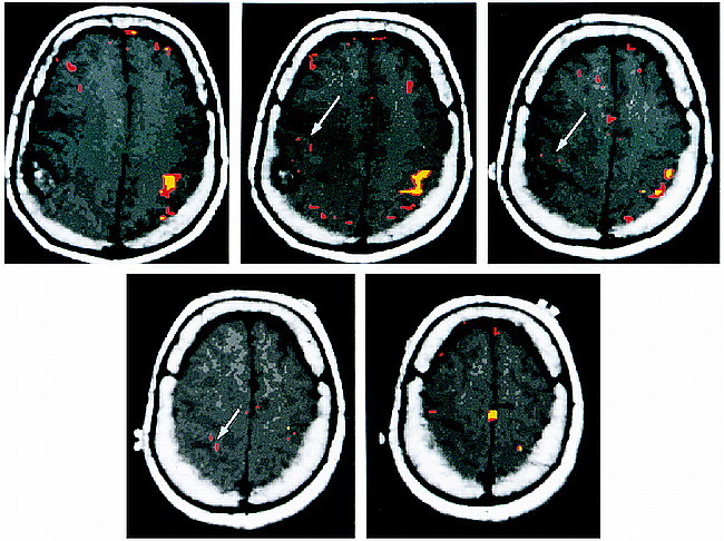
Axial T1-weighted images with coregistered functional MR images obtained during a bilateral motor paradigm show a larger volume of activation on the normal side than on the side with the tumor (arrows). This effect is seen for different correlation coefficients (r). The red areas indicate significant activation for r = .48, P < .01. The yellow areas indicate significant activation for r = .60, P < .01. Notwithstanding the difference in the volume of activation, one is still able to identify the motor cortex on the side with the tumor. The motor cortex on the right is displaced anteriorly and superiorly by the tumor mass. The accessory motor area is seen at the midline in the superiormost image (bottom right)
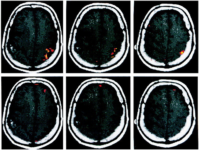
Axial T1-weighted images with coregistered functional MR images obtained during right-hand (top row, left sensory cortex) and left-hand (bottom row, right sensory cortex) sensory paradigms. The right-hand sensory paradigm shows robust activation in the left postcentral gyrus at different correlation coefficients (r). The red areas indicate activation for r = .48, P < .01. The yellow areas indicate activation for r = .60, P < .01. The left-hand sensory paradigm fails to show activation in the right postcentral gyrus (located just anterior to the tumor mass), even with P < .10
Comment in
-
The limitations of functional MR imaging: a caveat.AJNR Am J Neuroradiol. 1999 Apr;20(4):536. AJNR Am J Neuroradiol. 1999. PMID: 10319953 Free PMC article. No abstract available.
References
-
- Ammirati M, Vick N, Liao Y, et al. Effect of the extent of surgical resection on survival and quality of life in patients with supratentorial glioblastomas and anaplastic astrocytomas. Neurosurgery 1987;21:201-206 - PubMed
-
- Devaux BC, O'Fallon JR, Kelly PJ. Resection, biopsy, and survival in malignant glial neoplasms: a retrospective study of clinical parameters, therapy, and outcome. J Neurosurg 1993;78:767-75 - PubMed
-
- Maldjian JA, Schulder M, Liu WC, et al. Intraoperative functional MRI using a real-time neurosurgical navigation system. J Comput Assist Tomogr 1997;21:910-912 - PubMed
-
- Schulder M, Maldjian JA, Liu WC, et al. Functional image guided surgery of intracranial tumors located in or near the sensorimotor cortex. J Neurosurg 1998;89:412-418 - PubMed
-
- Rezai AR, Hund M, Kronberg E, et al. The interactive use of magnetoencephalography in stereotactic image-guided neurosurgery. Neurosurgery 1996;39:92-102 - PubMed
Publication types
MeSH terms
LinkOut - more resources
Full Text Sources
Medical