Histopathologic analysis of foci of signal loss on gradient-echo T2*-weighted MR images in patients with spontaneous intracerebral hemorrhage: evidence of microangiopathy-related microbleeds
- PMID: 10319975
- PMCID: PMC7056037
Histopathologic analysis of foci of signal loss on gradient-echo T2*-weighted MR images in patients with spontaneous intracerebral hemorrhage: evidence of microangiopathy-related microbleeds
Abstract
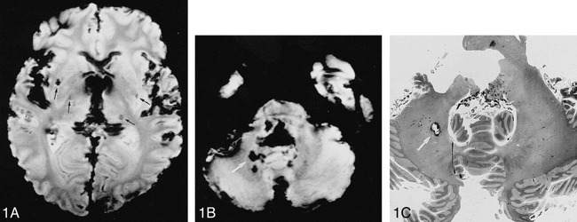
Background and purpose: Patients with spontaneous intracerebral hemorrhage (ICH) frequently have small areas of signal loss on gradient-echo T2*-weighted MR images, which have been suggested to represent remnants of previous microbleeds. Our aim was to provide histopathologic support for this assumption and to clarify whether the presence and location of microbleeds were associated with microangiopathy.
Methods: We performed MR imaging and correlative histopathologic examination in 11 formalin-fixed brains of patients who had died of an ICH (age range, 45-90 years).
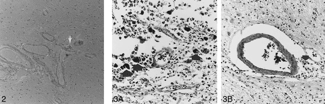
Results: Focal areas of signal loss on MR images were noted in seven brains. They were seen in a corticosubcortical location in six brains, in the basal ganglia/thalami in five, and infratentorially in three specimens. Histopathologic examination showed focal hemosiderin deposition in 21 of 34 areas of MR signal loss. No other corresponding abnormalities were found; however, hemosiderin deposits were noted without MR signal changes in two brains. All specimens with MR foci of signal loss showed moderate to severe fibrohyalinosis, and there was additional evidence of amyloid angiopathy in two of those brains.
Conclusion: Small areas of signal loss on gradient echo T2*-weighted images indicate previous extravasation of blood and are related to bleeding-prone microangiopathy of different origins.
Figures


Similar articles
-
Amyloid Angiopathy in Brain Hemorrhage: A Postmortem Neuropathological-Magnetic Resonance Imaging Study.Cerebrovasc Dis. 2018;45(3-4):124-131. doi: 10.1159/000486554. Epub 2018 Mar 20. Cerebrovasc Dis. 2018. PMID: 29558742
-
[Indications of cerebral micro-hemorrhage in MRI. Comparative histological findings and possible clinical significance].Nervenarzt. 1999 Dec;70(12):1082-7. doi: 10.1007/s001150050542. Nervenarzt. 1999. PMID: 10637813 German.
-
Cerebral microbleeds in CADASIL: a gradient-echo magnetic resonance imaging and autopsy study.Stroke. 2002 Jan;33(1):67-71. doi: 10.1161/hs0102.100885. Stroke. 2002. PMID: 11779891 Clinical Trial.
-
Magnetic resonance imaging of cerebral microbleeds.Curr Opin Neurol. 2000 Feb;13(1):69-73. doi: 10.1097/00019052-200002000-00013. Curr Opin Neurol. 2000. PMID: 10719653 Review.
-
Clinical associations of cerebral microbleeds on magnetic resonance neuroimaging.J Stroke Cerebrovasc Dis. 2014 Nov-Dec;23(10):2489-2497. doi: 10.1016/j.jstrokecerebrovasdis.2014.07.006. Epub 2014 Oct 5. J Stroke Cerebrovasc Dis. 2014. PMID: 25294059 Review.
Cited by
-
New vistas in clinical practice: susceptibility-weighted imaging.Quant Imaging Med Surg. 2015 Jun;5(3):448-52. doi: 10.3978/j.issn.2223-4292.2015.03.03. Quant Imaging Med Surg. 2015. PMID: 26029647 Free PMC article.
-
Cerebral microbleeds are associated with worse cognitive function in the nondemented elderly with small vessel disease.Cerebrovasc Dis Extra. 2014 Dec 9;4(3):212-220. doi: 10.1159/000369294. eCollection 2014 Sep-Dec. Cerebrovasc Dis Extra. 2014. PMID: 25598771 Free PMC article.
-
Blood in the Brain on Susceptibility-Weighted Imaging.Indian J Radiol Imaging. 2022 Dec 7;33(1):89-97. doi: 10.1055/s-0042-1758880. eCollection 2023 Jan. Indian J Radiol Imaging. 2022. PMID: 36855723 Free PMC article. Review.
-
Diffuse axonal injury associated with chronic traumatic brain injury: evidence from T2*-weighted gradient-echo imaging at 3 T.AJNR Am J Neuroradiol. 2003 Jun-Jul;24(6):1049-56. AJNR Am J Neuroradiol. 2003. PMID: 12812926 Free PMC article.
-
Fat emboli and critical illness-associated cerebral microbleeds (CICMs) in a patient with sickle cell disease: Do these 2 entities coexist?Radiol Case Rep. 2023 Mar 20;18(5):1978-1981. doi: 10.1016/j.radcr.2023.02.044. eCollection 2023 May. Radiol Case Rep. 2023. PMID: 36970237 Free PMC article.
References
-
- Caplan LR. Intracerebral hemorrhage. In: Tyler HR, Dawson D, eds. Current Neurology. Boston: Houghton-Mifflin; 1979;2:185–205
-
- Brott T, Thalinger K, Hertzberg V. Hypertension as a risk factor for spontaneous intracerebral hemorrhage. Stroke 1986;17:1078-1083 - PubMed
-
- Vinters HA. Cerebral amyloid angiopathy: a critical review. Stroke 1987;2:311-324 - PubMed
-
- Selekler K, Erzen C. Leukoaraiosis and intracerebral hematoma. Stroke 1989;20:1016-1020 - PubMed
MeSH terms
Substances
LinkOut - more resources
Full Text Sources
Medical