

Reduction of aneurysm clip artifacts on CT angiograms: a technical note
Affiliations
- PMID: 10319984
- PMCID: PMC7056034
Item in Clipboard
Reduction of aneurysm clip artifacts on CT angiograms: a technical note
AJNR Am J Neuroradiol.
1999 Apr.
Abstract
We describe a head tilt technique for use with CT angiography that reduces beam-hardening artifacts in patients with aneurysm clips. This simple maneuver directs the artifacts away from pertinent anatomy, thus increasing the chances for diagnostic accuracy. No significant changes in the CT angiographic protocol are required, and the maneuver can easily be combined with other artifact-minimizing strategies.
Figures
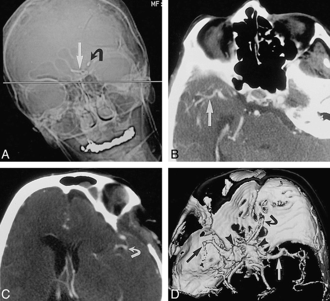
Tilted head position for intracranial CT angiography in a patient who has undergone clipping of multiple aneurysms. A, Anteroposterior scout image of the head for planning CT angiography shows plane (straight line) for axial image acquisition (straight arrow indicates aneurysm clip in anterior communicating artery; curved arrow, aneurysm clip in carotid bifurcation). Note position of head should minimize image degradation, because metal artifact will project away from vessels of interest. B and C, Axial source images from CT angiogram show right middle cerebral artery (arrow, B) and left middle cerebral artery (arrow, C) without interference from metallic beam-hardening artifact. D, Shaded-surface-display image from CT angiogram viewed from above shows left (white arrow) and right (black arrow) middle cerebral artery without interference from aneurysm clip's beam-hardening artifact (arrowheads). Owing to limited z-axis coverage, left middle cerebral artery is abbreviated because examination was targeted to suspicious right middle cerebral artery. Current CT scanner software would allow coverage of entire head, tilted or not.
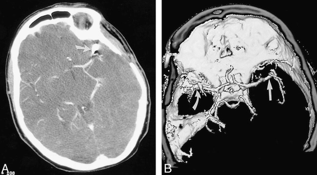
Tilted head position for intracranial CT angiography in a patient who has undergone clipping of a single aneurysm. A, Axial source image from CT angiogram shows left middle cerebral artery aneurysm clipping (arrow) with adjacent metallic beam-hardening artifact. B, Shaded-surface-display image from CT angiogram viewed from above shows right middle cerebral artery (straight arrow) without interference from aneurysm clip's beam-hardening artifact (curved arrow).
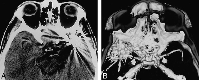
Standard head position for intracranial CT angiography. A, Axial source image from CT angiogram shows degradation of image due to beam-hardening artifacts from clips (two) on left middle cerebral artery aneurysms. B, Shaded-surface-display image from CT angiogram viewed from above shows middle cerebral arteries (arrows) and metallic beam-hardening artifact.
References
-
- Armonda RA, NehIs DG. Multiple intracranial aneurysms. In: Carter LP, Spetzler RF, eds. Neurovascular Surgery. New York: McGraw-Hill; 1994:807–814
-
- Vieco PT, Shuman WP, Alsofrom GF, et al. Detection of circle of Willis aneurysms in patients with subarachnoid hemorrhage: a comparison of CT angiography and digital subtraction angiography. AJR Am J Roentgenol 1995;165:425-430 - PubMed
-
- Klucznik RP, Carrier DA, Pyka R, Haid RW. Placement of a ferromagnetic intracerebral: aneurysm clip in a magnetic field with fatal outcome. Radiology 1993;187:855-856 - PubMed
MeSH terms
Substances
LinkOut - more resources
Full Text Sources
Other Literature Sources
Medical