

Evaluation of CSF leaks: high-resolution CT compared with contrast-enhanced CT and radionuclide cisternography
- PMID: 10319986
- PMCID: PMC7056020
Evaluation of CSF leaks: high-resolution CT compared with contrast-enhanced CT and radionuclide cisternography
Abstract
Background and purpose: Radiologic evaluation of CSF leaks is a diagnostic challenge that often involves multiple imaging studies with the associated expense and patient discomfort. We evaluated the use of screening noncontrast high-resolution CT in identifying the presence and site of CSF rhinorrhea and otorrhea and compared it with contrast-enhanced CT cisternography and radionuclide cisternography.
Methods: We retrospectively reviewed the imaging studies and medical records of all patients who were evaluated for CSF leak during a 7-year period. Forty-two patients with rhinorrhea and/or otorrhea underwent high-resolution CT of the face or temporal bone and then had CT cisternography and radionuclide cisternography via lumbar puncture. The results of the three studies were compared and correlated with the surgical findings in 21 patients.
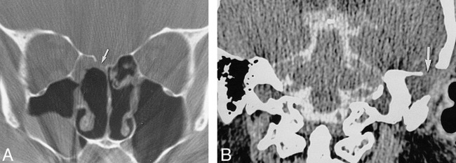
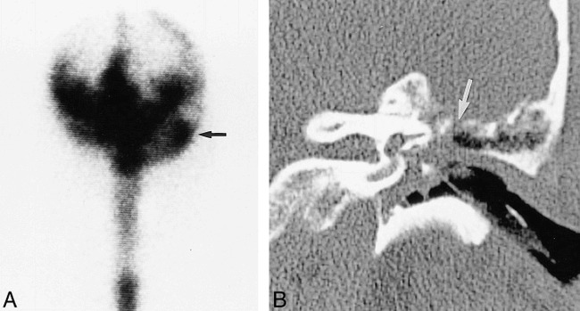
Results: High-resolution CT showed bone defects in 30 of 42 patients (71%) with CSF leak. High-resolution, radionuclide cisternography and CT cisternography did not show bone defects or CSF leak for 12 patients (29%) who had clinical evidence of CSF leak. Among the 30 patients with bone defects, 20 (66%) had positive results of their radionuclide cisternography and/or CT cisternography. For the 21 patients who underwent surgical exploration and repair, intraoperative findings correlated with the defects revealed by high-resolution CT in all cases. High-resolution CT identified significantly more patients with CSF leak than did radionuclide cisternography and CT cisternography, with a moderate degree of agreement.
Conclusion: Noncontrast high-resolution CT showed a defect in 70% of the patients with CSF leak. No radionuclide cisternography or CT cisternography study produced positive results without previous visualization of a defect on high-resolution CT. CT cisternography and radionuclide cisternography may be reserved for patients in whom initial high-resolution CT does not identify a bone defect or for patients with multiple fractures or postoperative defects.
Figures


References
-
- Zlab MK, Moore GF, Daly DT, Yonkers AJ. Cerebrospinal fluid rhinorrhea: a review of the literature. Ear Nose Throat J 1992;71:314-317 - PubMed
-
- Lloyd MNH, Kimber PM, Burrows EH. Post-traumatic cerebrospinal fluid rhinorrhoea: modern high-definition computed tomography is all that is required for the effective demonstration of the site of leakage. Clin Radiol 1994;49:100-103 - PubMed
-
- Oberascher G. A modern concept of cerebrospinal fluid diagnosis in oto- and rhinorrhea. Rhinology 1988;26:89-103 - PubMed
-
- Fagerlund M, Lilequist B. Intermittent cerebrospinal liquorrhea: cerebral computed tomography in the non-drop period. Acta Radiol 1987;28:189-192 - PubMed
Publication types
MeSH terms
Substances
LinkOut - more resources
Full Text Sources
Medical