. 1999 May;181(10):3039-50.
doi: 10.1128/JB.181.10.3039-3050.1999.

Identification and transcriptional control of the genes encoding the Caulobacter crescentus ClpXP protease

Affiliations

Identification and transcriptional control of the genes encoding the Caulobacter crescentus ClpXP protease

M Osterås et al. J Bacteriol. 1999 May.

Abstract

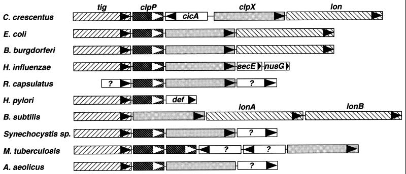
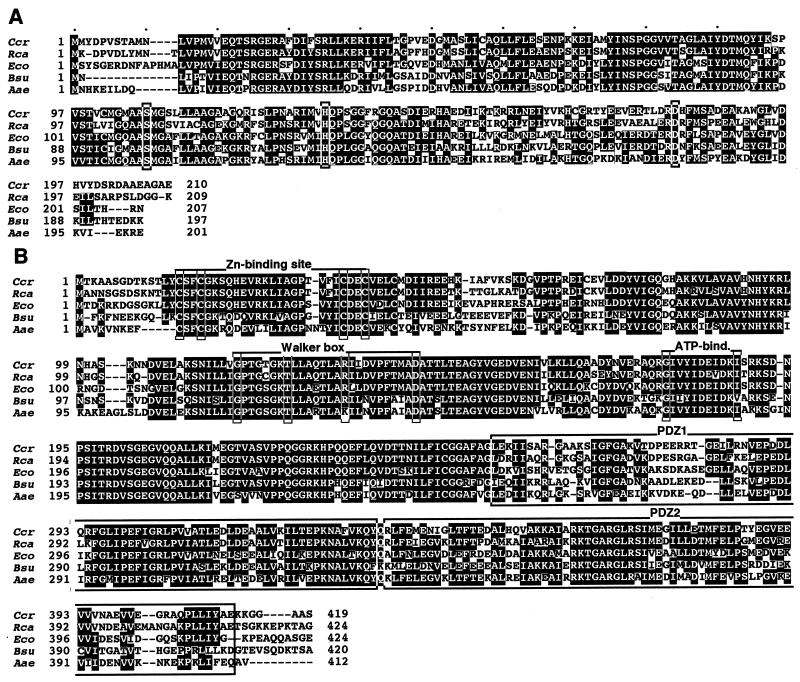
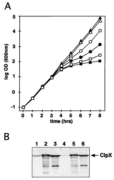
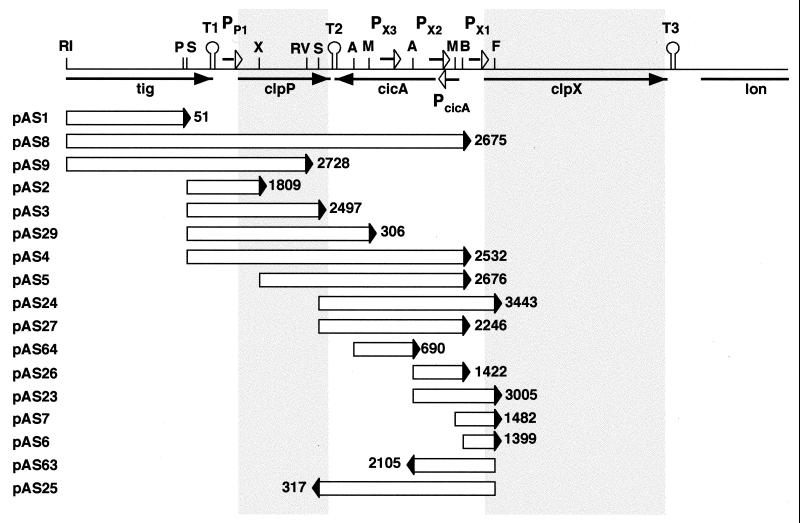
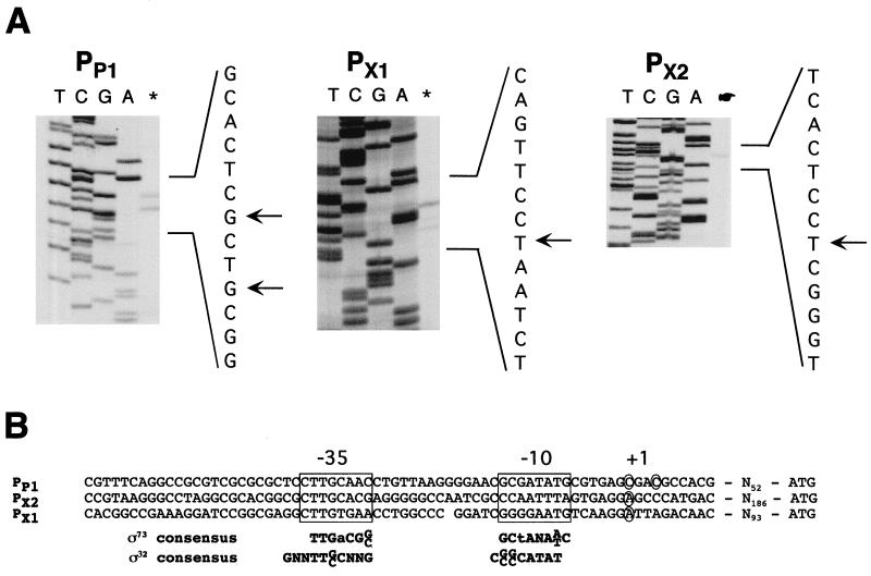
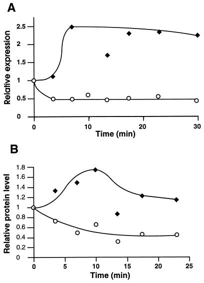
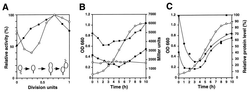
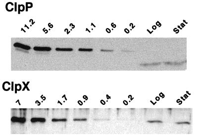
The region of the Caulobacter crescentus chromosome harboring the genes for the ClpXP protease was isolated and characterized. Comparison of the deduced amino acid sequences of the C. crescentus ClpP and ClpX proteins with those of their homologues from several gram-positive and gram-negative bacteria revealed stronger conservation for the ATPase regulatory subunit (ClpX) than for the peptidase subunit (ClpP). The C. crescentus clpX gene was shown by complementation analysis to be functional in Escherichia coli. However, clpX from E. coli was not able to substitute for the essential nature of the clpX gene in C. crescentus. The clpP and clpX genes are separated on the C. crescentus chromosome by an open reading frame pointing in the opposite direction from the clp genes, and transcription of clpP and clpX was found to be uncoupled. clpP is transcribed as a monocistronic unit with a promoter (PP1) located immediately upstream of the 5' end of the gene and a terminator structure following its 3' end. PP1 is under heat shock control and is induced upon entry of the cells into the stationary phase. At least three promoters for clpX (PX1, PX2, and PX3) were mapped in the clpP-clpX intergenic region. In contrast to PP1, the clpX promoters were found to be downregulated after heat shock but were also subject to growth phase control. In addition, the clpP and clpX promoters showed different activity patterns during the cell cycle. Together, these results demonstrate that the genes coding for the peptidase and the regulatory subunits of the ClpXP protease are under independent transcriptional control in C. crescentus. Determination of the numbers of ClpP and ClpX molecules per cell suggested that ClpX is the limiting component compared with ClpP.


Figures

FIG. 1
FIG. 1
Schematic diagram of the clpP-clpX regions of gram-positive and gram-negative bacteria. The genes coding for the trigger factor (tig), the ClpP peptidase subunit (clpP), the ClpX ATPase subunit (clpX), and the Lon protease (lon) are indicated. In most organisms analyzed so far, the gene order tig-clpP-clpX is conserved, with the lon gene located immediately downstream. In C. crescentus, the clp genes are separated by cicA, a gene of unknown function; in Haemophilus influenzae, the lon gene is not linked to the clp genes, which are followed by the secE (preprotein translocase subunit) and nusG (transcription antiterminator protein) genes (9); in R. capsulatus, neither tig nor lon is linked to the clp genes, which are flanked by genes of unknown function (?) (35a); in H. pylori, the clpX and lon genes, although linked, are not found at the same location as the tig and clpP genes, which are followed by the def (polypeptide deformylase) gene (45); and in B. subtilis, clpP is not linked to the tig, clpX, and lon genes. The lon gene has undergone duplication in B. subtilis (23); in Synechocystis and Mycobacterium tuberculosis, no gene coding for a homolog of the Lon protease is found. However, two clpP genes are present in M. tuberculosis, and four clpP genes are found in Synechocystis, one of them linked to the tig and clpX genes (19). Two genes are present between the clpP and clpX genes of M. tuberculosis, and the deduced sequences of their products are similar to those of a methyltransferase and a drug efflux protein (4). In Aquifex aeolicus, the lon gene is not linked to the tig, clpP, and clpX genes and is found elsewhere on the chromosome (5).
FIG. 2
FIG. 2
Sequence alignment of ClpP (A) and ClpX (B) from C. crescentus (Ccr) with homologous sequences from R. capsulatus (Rca) (35a), E. coli (Eco) (11, 30), B. subtilis (Bsu) (23), and A. aeolicus (Aae) (5). The amino acid residues identical to those of ClpP or ClpX from C. crescentus are indicated by white letters on a black background. A putative Zn finger-binding site, the ATP-binding (Walker box and ATP-bind.) motifs, and the proposed PDZ-like domains of ClpX (27) are marked. The serine, histidine, and aspartate residues involved in ClpP activity (31, 49) are boxed. Gaps are represented by dashes and were introduced to maximize the alignment. The alignment was generated with the Megalignment program of the DNAstar program package (DNAstar, Madison, Wis.).
FIG. 3
FIG. 3
clpXCc is able to complement an E. coli clpX mutant. (A) The activity of ClpX was monitored by its ability to degrade, together with ClpP, the antidote protein Phd of the P1 plasmid addiction module Phd-Doc (26). Loss of the phd gene results in Doc-dependent cell killing if the Phd protein is degraded by the ClpXP protease. Growth of cultures containing the phd and doc genes on a plasmid with a temperature-sensitive replicon (pGB2ts::phd-doc) was monitored after a shift to the nonpermissive temperature. Growth is shown as the log OD600. The time after the temperature increase is indicated in hours. Cessation of growth 3 to 5 h after the temperature shift was an indicator of the rapid disappearance of the antidote protein Phd and thus of ClpX activity. The following plasmids were used: plasmid pGB2ts is temperature sensitive for replication; pGB2ts::phd-doc is identical to pGB2ts except that it contains the plasmid addiction genes phd and doc; pSSN6 contains clpXCc; and pSSN3 carries clpXEc. Growth of the following E. coli strains was monitored: W3110/pGB2ts/pSSN6 plus isopropyl-β-d-thiogalactopyranoside (IPTG) (▴; negative control); W3110/pGB2ts::phd-doc/pSSN6 plus IPTG (■; positive control); SSN1/pGB2ts/pSSN6 plus IPTG (▵); SSN1/pGB2ts::phd-doc/pSSN3 plus IPTG (□); SSN1/pGB2ts::phd-doc/pSSN6 plus IPTG (●); and SSN1/pGB2ts::phd-doc/pSSN6 (○). (B) Immunoblot analysis with an anti-ClpX serum and extracts of E. coli strains expressing ClpXCc and ClpXEc. Equal amounts of total protein from the following strains were analyzed: W3110/pMR20 (lane 1); W3110/pSSN3 (lane 2); W3110/pSSN6 (lane 3); SSN1/pMR20 (lane 4); SSN1/pSSN3 (lane 5); and SSN1/pSSN6 (lane 6). The band corresponding to ClpX is marked by an arrow.
FIG. 4
FIG. 4
Identification of promoter regions in the C. crescentus clp locus. A schematic of the chromosomal clp region is shown at the top. The tig (trigger factor), clpP (ClpP peptidase), cicA (unknown function), clpX (ClpX ATPase), and lon (Lon protease) genes are indicated by black arrows. The approximate locations and orientations of the promoters identified (PP1, PX1, PX2, and PX3) are marked by short open arrows, and putative transcriptional terminator structures (T1, T2, and T3) are indicated as stem-loop outlines. Fragments that were cloned into the lacZ reporter plasmid pRKlac290 (see Materials and Methods) are indicated as open bars below the schematic, with the filled triangles marking the location and orientation of the lacZ reporter gene. The names of the corresponding constructs are on the left, and the number on the right indicates the β-galactosidase activity (Miller units) generated by each fusion construct. All measurements were determined in triplicate, and average numbers are presented. The following abbreviations are used for recognition sites of restriction enzymes: RI, EcoRI; P, PstI; S, SalI; X, XhoI; RV, EcoRV; A, ApaI; M, MluI; B, BamHI; and F, FspI.
FIG. 5
FIG. 5
Promoter analysis of the C. crescentus clpP and clpX genes. (A) Primer extension products obtained with the clpX- and clpX-specific primers are shown in the rightmost lanes. Sequencing reactions generated with the same primers are shown in lanes T, C, G, and A. The relevant sequence of the coding strand is shown to the right of each gel, and the positions of the major extension products are indicated by arrows. (B) Sequence alignment of PP1, PX1, and PX2. The −35 and −10 (boxed) and +1 (circled) regions are indicated, and the distance between the transcriptional start site and the presumed translational start codon is shown (Nx). The C. crescentus consensus sequences for ς73-dependent (29) and ς32-dependent (37, 53) promoters are shown in boldface below the three clp promoters.
FIG. 6
FIG. 6
Heat shock control of ClpP and ClpX expression. (A) The transcription of clpP (⧫) and clpX (○) was determined with cultures of NA1000/pAS2 (clpP::lacZ) and NA1000/pAS24 (clpX::lacZ) by [35S]methionine labeling, β-galactosidase immunoprecipitation, and quantitation at different times after a shift in the temperature from 30 to 42°C. (B) Cellular concentrations of ClpP (⧫) and ClpX (○) after the temperature shift from 30 to 42°C, as determined by immunoblot analysis for the same samples as those analyzed in panel A. The values are relative to the level of transcription or protein at 30°C.
FIG. 7
FIG. 7
Cell cycle- and growth phase-dependent expression of clpP and clpX. (A) The relative activities of PX1 (○) and PX3 (●) were determined during the cell cycle by pulse labeling of synchronized cultures of strain NA1000 containing plasmids pAS6 (PX1) and pAS64 (PX3), respectively, with [35S]methionine for 5 min at different intervals and determining β-galactosidase synthesis during the pulse time by immunoprecipitation (see Materials and Methods). The promoter activity is shown relative to maximal activity. Progression of the cell cycle is indicated schematically at the bottom. (B and C) Growth phase-dependent expression (B) and cellular levels (C) of ClpP and ClpX. (B) Stationary-phase overnight cultures were diluted in fresh PYE complex medium (NA1000, NA1000/pAS2, and NA1000/pAS24) or PYE complex medium plus 0.2% xylose (NA1000/pCS225), and growth was monitored by determining the OD660 (○). At different intervals, samples were removed from the cultures, and β-galactosidase activity (Miller units) was determined as described in Materials and Methods for the following strains: NA1000/pAS2 (PP1) (●), NA1000/pAS24 (PX1, PX2, and PX3) (■), and NA1000/pCS225 (xylX promoter) (□). (C) Relative cellular levels of ClpP (●) and ClpX (■) in wild-type strain NA1000 were determined as a function of the growth phase by immunoblot analysis. ○, OD660.
FIG. 8
FIG. 8
Estimation of the number of ClpP and ClpX molecules per cell. Immunoblot analysis was done with anti-ClpP and anti-ClpX sera and with specific amounts (picomoles) of purified His-tagged ClpP and ClpX proteins, respectively, and total proteins from NA1000. The band intensities of total proteins correspond to the amounts of ClpP and ClpX present in 2 × 10⁷ cells and 1 × 10⁸ cells, respectively, during exponential growth (Log) and in 1 × 10⁷ cells and 5 × 10⁷ cells, respectively, during the stationary phase (Stat).

References

    1. Alley M R K, Maddock J R, Shapiro L. Requirement of the carboxyl terminus of the bacterial chemoreceptor for its targeted proteolysis. Science. 1993;259:1754–1757. - PubMed
    1. Ausubel F M, Brent R, Kingston R E, Moore D D, Seidman J G, Smith J A, Struhl K. Current protocols in molecular biology. New York, N.Y: John Wiley & Sons, Inc.; 1992.
    1. Baldini R L, Avedissian M, Gomes S L. The CIRCE element and its putative repressor control cell cycle expression of the Caulobacter crescentus groESL operon. J Bacteriol. 1998;180:1632–1641. - PMC - PubMed
    1. Cole S T, Brosch R, Parkhill J, Garnier T, Churcher C, Harris D, Gordon S V, Eiglmeier K, Gas S, Barry III C E, Tekaia F, Badcock K, Basham D, Brown D, Chillingworth T, Connor R, Davies R, Devlin K, Feltwell T, Gentles S, Hamlin N, Holroyd S, Hornsby T, Jagels K, Barrell B G, et al. Deciphering the biology of Mycobacterium tuberculosis from the complete genome sequence. Nature. 1998;393:537–544. - PubMed
    1. Deckert G, Warren P V, Gaasterland T, Young W G, Lenox A L, Graham D E, Overbeek R, Snead M A, Keller M, Aujay M, Huber R, Feldman R A, Short J M, Olsen G J, Swanson R V. The complete genome of the hyperthermophilic bacterium Aquifex aeolicus. Nature. 1998;392:353–358. - PubMed

Publication types

MeSH terms

Associated data