. 1999 May;181(10):3087-95.
doi: 10.1128/JB.181.10.3087-3095.1999.

Cell wall-anchored CshA polypeptide (259 kilodaltons) in Streptococcus gordonii forms surface fibrils that confer hydrophobic and adhesive properties

Affiliations

Cell wall-anchored CshA polypeptide (259 kilodaltons) in Streptococcus gordonii forms surface fibrils that confer hydrophobic and adhesive properties

R McNab et al. J Bacteriol. 1999 May.

Abstract

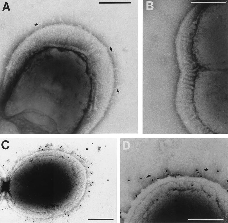
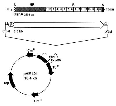
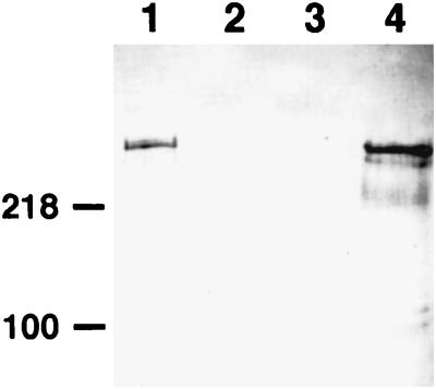
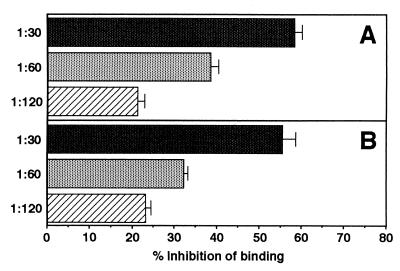
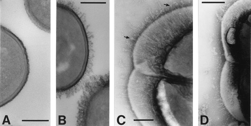
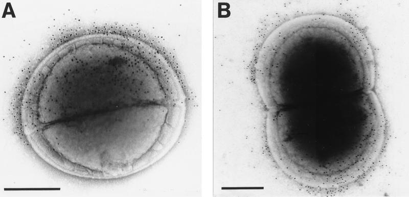
It has been shown previously that inactivation of the cshA gene, encoding a major cell surface polypeptide (259 kDa) in the oral bacterium Streptococcus gordonii, generates mutants that are markedly reduced in hydrophobicity, deficient in binding to oral Actinomyces species and to human fibronectin, and unable to colonize the oral cavities of mice. We now show further that surface fibrils 60.7 +/- 14.5 nm long, which are present on wild-type S. gordonii DL1 (Challis) cells, bind CshA-specific antibodies and are absent from the cell surfaces of cshA mutants. To more precisely determine the structural and functional properties of CshA, already inferred from insertional-mutagenesis experiments, we have cloned the entire cshA gene into the replicative plasmid pAM401 and expressed full-length CshA polypeptide on the cell surface of heterologous Enterococcus faecalis JH2-2. Enterococci expressing CshA exhibited a 30-fold increase in cell surface hydrophobicity over E. faecalis JH2-2 carrying the pAM401 vector alone and 2.4-fold-increased adhesion to human fibronectin. CshA expression in E. faecalis also promoted cell-cell aggregation and increased the ability of enterococci to bind Actinomyces naeslundii cells. Electron micrographs of negatively stained E. faecalis cells expressing CshA showed peritrichous surface fibrils 70.3 +/- 9.1 nm long that were absent from control E. faecalis JH2-2(pAM401) cells. The fibrils bound CshA-specific antibodies, as detected by immunoelectron microscopy, and the antibodies inhibited the adhesion of E. faecalis cells to fibronectin. The results demonstrate that the CshA polypeptide is the structural and functional component of S. gordonii adhesive fibrils, and they provide a molecular basis for past correlations of surface fibril production, cell surface hydrophobicity, and adhesion in species of oral "sanguis-like" streptococci.


Figures

FIG. 1
FIG. 1
Electron micrographs of S. gordonii CshA fibrils negatively stained with 1% (wt/vol) methylamine tungstate (A and B) or reacted with CshA-specific antibodies and 10-nm gold particle-conjugated secondary antibody (C and D). Fibrils of 60.7 ± 14.5 nm in length are present on the wild-type strain DL1 (indicated by arrows in panel A) but are absent from the surfaces of OB277 cshA31 cshB2 mutant cells (B). (C and D) S. gordonii DL1 cells labeled with immunogold. Gold particles demonstrate a bias for “older wall” (see Discussion) and are located up to 61.3 ± 19.2 nm from the cell surface. In panel D, fibrils may be seen at one end of the cell (arrows). In all images of negatively stained cells, some plasmolysis was observed, with the cytoplasmic membrane pulled away to some degree from the cell wall. Bars, 200 nm.
FIG. 2
FIG. 2
Construction of plasmid pAMCshA. The cshA gene and flanking regions were PCR amplified from S. gordonii DL1 DNA, and the SmaI-XbaI segment (8.1 kb) was ligated into the E. coli-E. faecalis shuttle vector pAM401 digested with EcoRV and XbaI as detailed in Materials and Methods. The structural features of the CshA polypeptide are shown as follows: L, leader peptide (41 amino acid residues); NR, nonrepetitive region (residues 42 to 878); R, amino acid repeat block region (residues 879 to 2417); A, cell wall anchor (residues 2418 to 2508); aa, amino acids; P, promoter; Ω, rho-independent transcriptional terminator.
FIG. 3
FIG. 3
Western immunoblot detection of CshA expression in S. gordonii or E. faecalis strains. Surface proteins were extracted from cells in the early-stationary phase of growth following incubation with the murolytic enzyme mutanolysin or lysozyme (see Materials and Methods). Extracts were subjected to SDS-PAGE, proteins were electroblotted onto nitrocellulose membranes, and blots were probed with polyclonal antibodies raised to the N-terminal region of recombinant CshA diluted 1:1,000. Lanes: 1, S. gordonii DL1 (wild type); 2, S. gordonii OB235 cshA3; 3, E. faecalis OB513(pAM401); 4, E. faecalis OB516(pAMCshA). Note the lack of reactivity of antibodies in lane 2, demonstrating their specificity for CshA polypeptide. Protein loadings were equalized (10 μg per lane). The numbers on the left indicate the positions of molecular mass markers (in kilodaltons).
FIG. 4
FIG. 4
Inhibition by antibodies to N-terminal CshA of bacterial-cell adhesion to fibronectin. Radioactively labeled cells of E. faecalis OB516 (A) or S. gordonii DL1 (B) were mixed with immune or preimmune antiserum diluted appropriately, and portions (2 × 10⁷ per well) were applied to microtiter plate wells coated with human fibronectin (1 μg per well) or to wells blocked with bovine albumin. The numbers of cells that adhered were measured as described elsewhere (31). Data are presented as percent inhibition of adhesion in the presence of immune serum compared with adhesion in the presence of preimmune serum. The results for CshA antibody inhibition of S. gordonii cells binding to fibronectin are as previously reported (31).
FIG. 5
FIG. 5
Electron microscopy of E. faecalis JH2-2 cells carrying pAM401 or pAMCshA. (A and B) Thin-section micrographs stained with RR and osmium tetroxide; (C and D) negatively stained cells. (A) E. faecalis OB513(pAM401) control cells are surrounded by a compact and densely stained layer covering a less-densely stained cell wall layer. (B) E. faecalis OB516(pAMCshA) cells expressing CshA polypeptide show a densely stained fibrillar fringe. (C) E. faecalis OB516 cells show peritrichous fibrils 70.3 ± 9.1 nm long that are more sparsely located in the region of the septum, and some fibrils have globular ends (arrows). (D) E. faecalis OB516 cells showing the absence of fibrils from the septal region and their expression restricted to the older ends of the cells. Bars, 200 nm.
FIG. 6
FIG. 6
Immunogold-labeled E. faecalis OB516(pAMCshA) cells. Cells from the early-stationary phase of growth were incubated with N-terminus-specific CshA antibodies and a gold-conjugated secondary antibody. (A) A newly-divided cell binds gold label that is restricted to one-half of the cell as defined by the nascent septum; (B) sparse deposition of gold particles in the septal region of the cell in the process of cell wall synthesis prior to cell division. Gold particles were located 61.7 ± 12.2 nm from the cell surface. Bars, 200 nm.

References

    1. Boyer H W, Roulland-Dussoix D. A complementation analysis of the restriction and modification of DNA in Escherichia coli. J Mol Biol. 1969;41:459–472. - PubMed
    1. Cruz-Rodz A L, Gilmore M S. High-efficiency introduction of plasmid DNA into glycine-treated Enterococcus faecalis by electroporation. Mol Gen Genet. 1990;224:152–154. - PubMed
    1. Demuth D R, Berthold P, Leboy P S, Golub E E, Davis C A, Malamud D. Saliva-mediated aggregation of Enterococcus faecalis transformed with a Streptococcus sanguis gene encoding the SSP-5 surface antigen. Infect Immun. 1989;57:1470–1475. - PMC - PubMed
    1. Demuth D R, Duan Y, Brooks W, Holmes A R, McNab R, Jenkinson H F. Tandem genes encode cell-surface polypeptides SspA and SspB which mediate adhesion of the oral bacterium Streptococcus gordonii to human and bacterial receptors. Mol Microbiol. 1996;20:403–413. - PubMed
    1. Fachon-Kalweit S, Elder B L, Fives-Taylor P. Antibodies that bind to fimbriae block adhesion of Streptococcus sanguis to saliva-coated hydroxyapatite. Infect Immun. 1985;48:617–624. - PMC - PubMed

Publication types

MeSH terms

LinkOut - more resources