

Hematuria: an unusual presentation for mucocele of the appendix. Case report and review of the literature
- PMID: 10323174
- PMCID: PMC3015341
Hematuria: an unusual presentation for mucocele of the appendix. Case report and review of the literature
Abstract
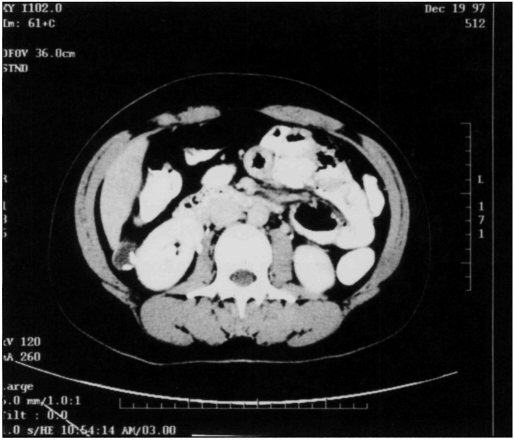
Mucocele of the appendix is a nonspecific term that is used to describe an appendix abnormally distended with mucus. This may be the result of either neoplastic or non-neopleastic causes and may present like most appendiceal pathology with either mild abdominal pain or life-threatening peritonitis. Urologic manifestations of mucocele of the appendix have rarely been reported. Laparoscopy can be used as a diagnostic tool in equivocal cases. Conversion to laparotomy may be indicated if there is a special concern for the ability to remove the appendix intact or if more extensive resection is warranted, as in malignancy. We here report our experience with a woman presenting with hematuria whose ultimate diagnosis was mucocele of the appendix, and we review the appropriate literature. This case highlights the mucocele as a consideration in the differential diagnosis of appendiceal pathology and serves to remind the surgeon of the importance for careful intact removal of the diseased appendix.
Figures


References
-
- Landen S, Bertrand C, Maddern GJ, et al. Appendiceal mucoceles and pseudomyxoma peritonei. Surg Gynecol Obstet. 1992;175:401–404 - PubMed
-
- Sivam NS, Ananthakrishnan N, Kate V. A case of appendicular diverticulitis leading to mucocele of the appendix. Trop Gastroenterology. 1997;18:36–37 - PubMed
-
- Baskin LS, Stoller ML. Unusual appendiceal pathology presenting as urologie disease. Urology. 1991;38:432–436 - PubMed
-
- Vale J, Kirby RS. Hematuria due to mucocele of the appendix. Br J Urology. 1989;63:218–219 - PubMed
Publication types
MeSH terms
LinkOut - more resources
Full Text Sources