. 1999 May 1;13(9):1140-55.
doi: 10.1101/gad.13.9.1140.

A putative protein complex consisting of Ctf19, Mcm21, and Okp1 represents a missing link in the budding yeast kinetochore

Affiliations

A putative protein complex consisting of Ctf19, Mcm21, and Okp1 represents a missing link in the budding yeast kinetochore

J Ortiz et al. Genes Dev. .

Abstract

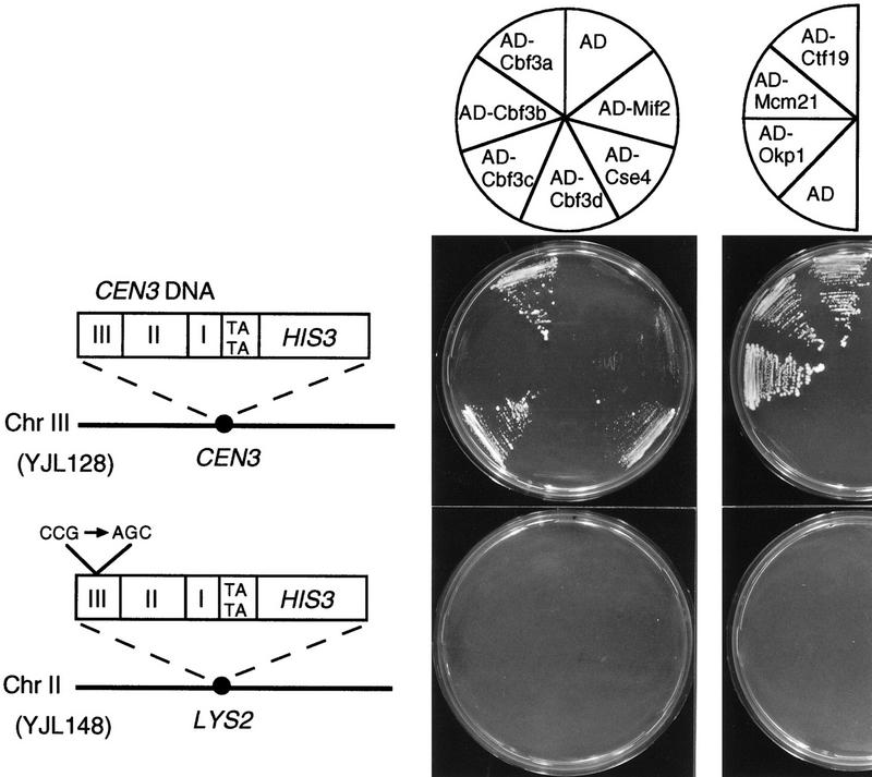
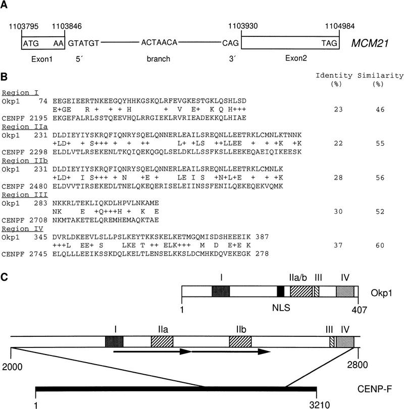
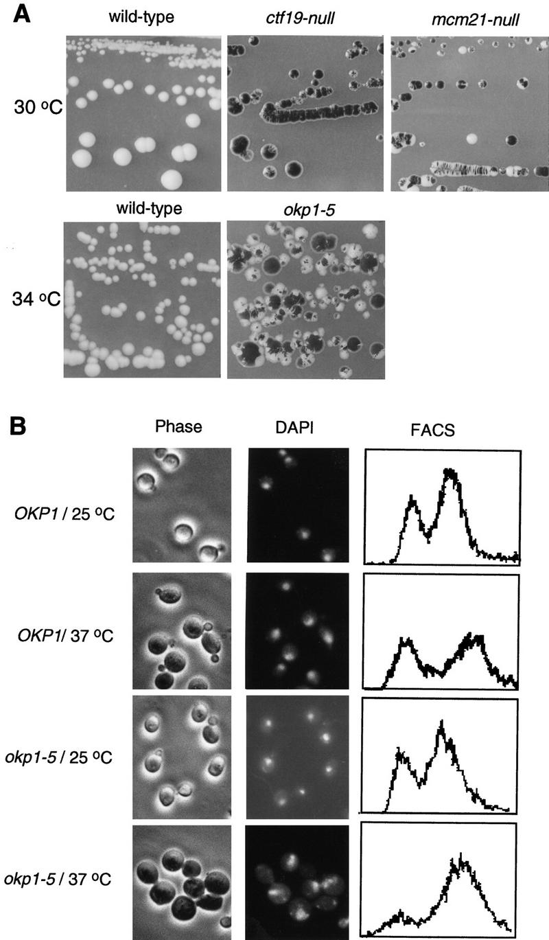
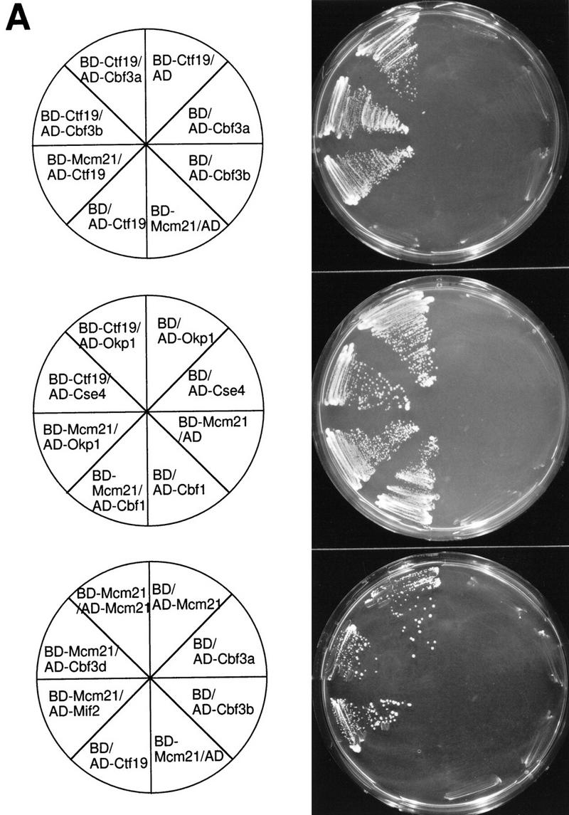
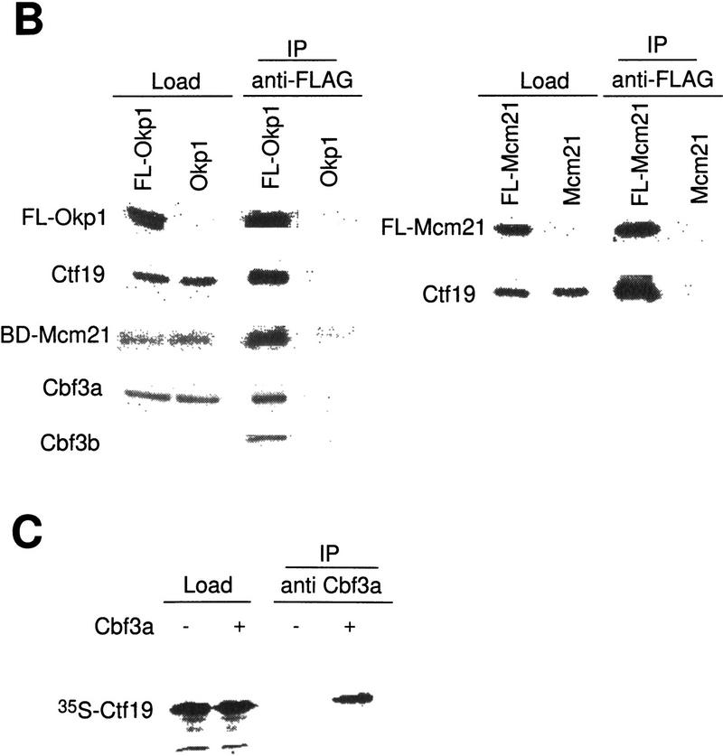
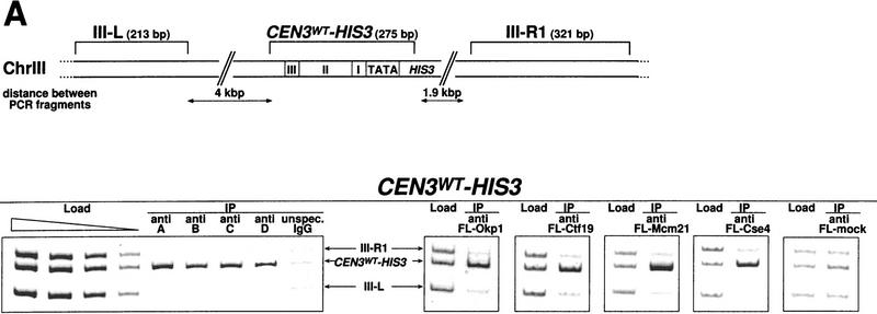
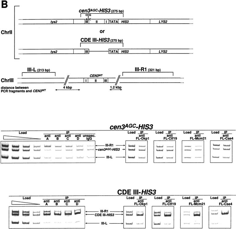
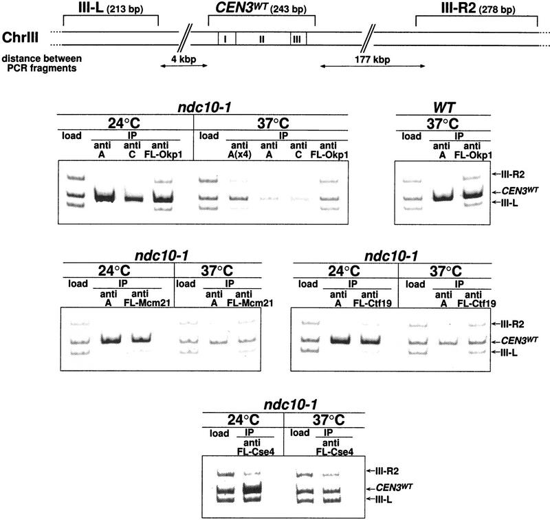
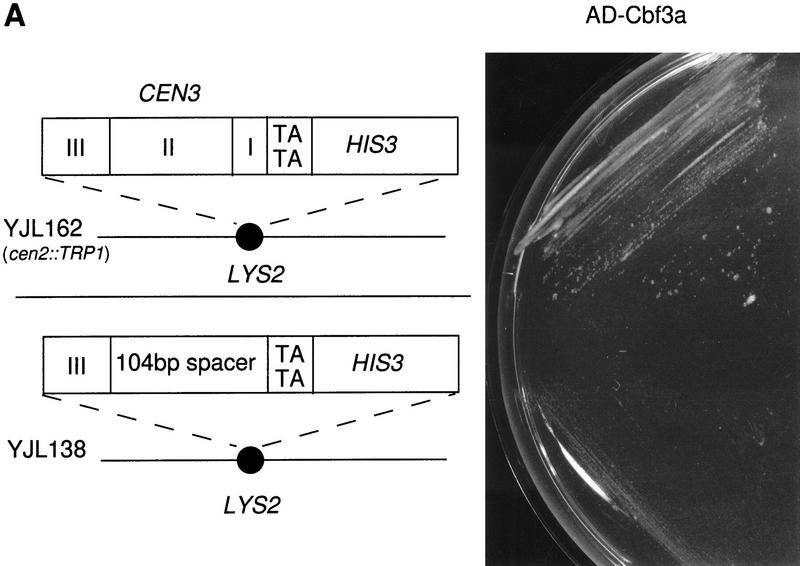
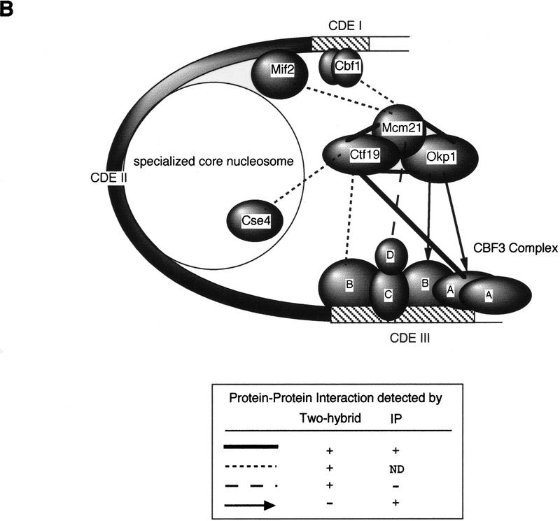
We have established a one-hybrid screen that allows the in vivo localization of proteins at a functional Saccharomyces cerevisiae centromere. Applying this screen we have identified three proteins-Ctf19, Mcm21, and the product of an unspecified open reading frame that we named Okp1-as components of the budding yeast centromere. Ctf19, Mcm21, and Okp1 most likely form a protein complex that links CBF3, a protein complex directly associated with the CDE III element of the centromere DNA, with further components of the budding yeast centromere, Cbf1, Mif2, and Cse4. We demonstrate that the CDE III element is essential and sufficient to localize the established protein network to the centromere and propose that the interaction of the CDE II element with the CDE III localized protein complex facilitates a protein-DNA conformation that evokes the active centromere.


Figures

Figure 1
Figure 1
Cbf3a, Cbf3c, Cse4, Ctf19, Mcm21, and Okp1 fused to the transcriptional activation domain of Gal4 (AD) stimulate the expression of HIS3 only when the reporter construct contains a functional CEN DNA. The reporter construct of strain YJL128 (cen3::CEN3–HIS3) containing wild-type CEN3 DNA and strain YJL148 (LYS2::cen3AGC–HIS3) containing a nonfunctional CEN3 DNA with a CCG to AGC mutation are presented. Growth of YJL128 (cen3::CEN3–HIS3) and YJL148 (LYS2::cen3AGC–HIS3) that express isolated AD (from pACT2) or the indicated AD fusion proteins (see Materials and Methods) on SD/His medium containing 5 mm 3-AT is shown. AD–Ctf19, AD–Mcm21, and AD–Okp1 were identified by expressing AD fusion libraries in YJL128 (cen3::CEN3–HIS3) as described in the text.
Figure 2
Figure 2
(A) Intron–exon structure of MCM21. 5′- and 3′-splice sites and a putative branch point are indicated. Arabic numerals refer to positions in Chromosome IV. (B) Regions of homology (I–IV) between Okp1 and CENP-F. (+) A conservative amino acid change. (C) Schematic representation of homology regions. Arrows indicate a direct repeat in CENP-F, and NLS denotes a putative nuclear localization signal. Roman numerals indicate regions of homology, as shown in B. Arabic numerals refer to amino acid positions.
Figure 3
Figure 3
(A) ctf19, mcm21, and okp1 mutants exhibit elevated chromosome loss. Colony sectoring of wild-type or mutant strains harboring an artificial test chromosome on SD medium containing reduced amounts of adenine (Koshland and Hieter 1987) is shown. The temperature-sensitive okp1-5 mutant and a corresponding control strain were grown for 2 days at 34°C and then shifted to 28°C. (B) okp1-5 terminal phenotype. Strains as indicated were grown at 25°C to early logarithmic stage and than shifted to 37°C or kept at 25°C for 4 hr. Cellular morphology (phase contrast), nuclear staining (DAPI), and DNA content (FACS) are shown. okp1-5 arrests at the nonpermissive temperature predominantly as large budded cells with one nucleus and replicated DNA.
Figure 4
Figure 4
Protein–protein interactions involving Ctf19, Mcm21, Okp1, and other components of the S. cerevisiae centromere. (A) Two-hybrid analysis. The indicated fusion constructs containing the DNA-binding domain of GAL4 (BD) or the transcriptional activation domain of Gal4 (AD) were coexpressed in HF7c (for plasmid construction see Materials and Methods); growth on SD/His medium containing 15 mm 3-AT is shown. As controls, coexpressions of corresponding fusion proteins with the appropriate two-hybrid vector containing no insert are shown. (B) For coimmunoprecipitation of Cbf3a, Cbf3b, and Ctf19 with Flag-tagged Okp1 (FL-Okp1), extracts from strain YJL132 (okp1::TRP1) harboring pJL471 (Flag–OKP1) were prepared. As a control, extracts from YJL19 were used that did not contain Flag-tagged Okp1. For coimmunoprecipitation of Mcm21 fused to the DNA-binding domain of Gal4 (BD-Mcm21) with Flag-tagged Okp1, extracts from YPH499 (Sikorski and Hieter 1992) harboring pJL471 (Flag–OKP1) and pJO508 (BD–MCM21) were prepared and as a control, extracts from YPH499 harboring only pJO508 (BD–MCM21) was used. For coimmunoprecipitation of Ctf19 with Flag-tagged Mcm21 (FL-Mcm21) extracts from YJL146 (mcm21::TRP1) harboring pJL505 (Flag–MCM21) were prepared and as a control, extracts from YJL19 were used. The extracts were immunoprecipitated with anti-Flag M2 agarose. Aliquots (0.5%–1.5% of the total load) of the extracts (Load) and aliquots (5% or 25% of the total load for the analysis of Flag-tagged proteins or coprecipitated proteins, respectively) of the immunoprecipitations (IPs) were subjected to Western analysis. Anti-Cbf3a, anti-Cbf3b, and anti-Ctf19 antibodies were used to detect corresponding proteins. Anti-HA antibody was used to detect BD–Mcm21, which contains an HA tag. The FL-Okp1 shown for Load and IP fractions corresponds to an experiment where the coprecipitation of Ctf19 and Cbf3a was revealed. For BD-Mcm21 or Cbf3b coimmunoprecipitation, the amount of FL-Okp1 detected in the Load and IP fractions was similar. Note that Cbf3b was undetectable in the loads. However, the same amount of total protein from the extract containing FL-Okp1 and of the control extract was subjected to immunoprecipitation. (C) 35S-Labeled Ctf19, synthesized in vitro, was incubated with or without 1 μg of purified Cbf3a–His6 expressed in Pichia pastoris (Stemmann and Lechner 1996). Subsequently, the samples were immunoprecipitated with anti Cbf3a antibody. Aliquots of the load (5% of the total) and IP fraction (30% of the total load) were subjected to SDS-PAGE and autoradiography.
Figure 4
Figure 4
Protein–protein interactions involving Ctf19, Mcm21, Okp1, and other components of the S. cerevisiae centromere. (A) Two-hybrid analysis. The indicated fusion constructs containing the DNA-binding domain of GAL4 (BD) or the transcriptional activation domain of Gal4 (AD) were coexpressed in HF7c (for plasmid construction see Materials and Methods); growth on SD/His medium containing 15 mm 3-AT is shown. As controls, coexpressions of corresponding fusion proteins with the appropriate two-hybrid vector containing no insert are shown. (B) For coimmunoprecipitation of Cbf3a, Cbf3b, and Ctf19 with Flag-tagged Okp1 (FL-Okp1), extracts from strain YJL132 (okp1::TRP1) harboring pJL471 (Flag–OKP1) were prepared. As a control, extracts from YJL19 were used that did not contain Flag-tagged Okp1. For coimmunoprecipitation of Mcm21 fused to the DNA-binding domain of Gal4 (BD-Mcm21) with Flag-tagged Okp1, extracts from YPH499 (Sikorski and Hieter 1992) harboring pJL471 (Flag–OKP1) and pJO508 (BD–MCM21) were prepared and as a control, extracts from YPH499 harboring only pJO508 (BD–MCM21) was used. For coimmunoprecipitation of Ctf19 with Flag-tagged Mcm21 (FL-Mcm21) extracts from YJL146 (mcm21::TRP1) harboring pJL505 (Flag–MCM21) were prepared and as a control, extracts from YJL19 were used. The extracts were immunoprecipitated with anti-Flag M2 agarose. Aliquots (0.5%–1.5% of the total load) of the extracts (Load) and aliquots (5% or 25% of the total load for the analysis of Flag-tagged proteins or coprecipitated proteins, respectively) of the immunoprecipitations (IPs) were subjected to Western analysis. Anti-Cbf3a, anti-Cbf3b, and anti-Ctf19 antibodies were used to detect corresponding proteins. Anti-HA antibody was used to detect BD–Mcm21, which contains an HA tag. The FL-Okp1 shown for Load and IP fractions corresponds to an experiment where the coprecipitation of Ctf19 and Cbf3a was revealed. For BD-Mcm21 or Cbf3b coimmunoprecipitation, the amount of FL-Okp1 detected in the Load and IP fractions was similar. Note that Cbf3b was undetectable in the loads. However, the same amount of total protein from the extract containing FL-Okp1 and of the control extract was subjected to immunoprecipitation. (C) 35S-Labeled Ctf19, synthesized in vitro, was incubated with or without 1 μg of purified Cbf3a–His6 expressed in Pichia pastoris (Stemmann and Lechner 1996). Subsequently, the samples were immunoprecipitated with anti Cbf3a antibody. Aliquots of the load (5% of the total) and IP fraction (30% of the total load) were subjected to SDS-PAGE and autoradiography.
Figure 5
Figure 5
CDE III is essential and sufficient to localize Cbf3a, Cbf3b, Cbf3c, Cbf3d, Okp1, Ctf19, Mcm21, and Cse4 to the CEN DNA in vivo as shown by Chip assays. (A) Association with wild-type CEN DNA, represented by CEN3WT–HIS3. Centromeric and noncentromeric loci analyzed are illustrated with corresponding sizes. Following in vivo cross-linking, extracts from strain YJL128 (cen3::CEN3–HIS3) without plasmids or YJL128 (cen3::CEN3–HIS3) harboring plasmids (see Materials and Methods) that expressed Flag-tagged versions of Okp1, Ctf19, Mcm21, and Cse4 were immunoprecipitated with anti CBF3 antibodies, unspecific IgG or anti Flag M2 agarose as indicated. (Anti Fl-mock) An extract from YJL128 (cen3::CEN3–HIS3) expressing no Flag-tagged proteins was immunoprecipitated with anti-Flag agarose. To test for specific copurification of CEN DNA, aliquots (∼0.016 μl of chromatin solution) of the extracts before immunoprecipitation (Load) and aliquots (∼5 μl of chromatin solution) of the immunoprecipitates (IPs) were analyzed by PCR. In the load dilution series, 0.1, 0.04, 0.016, and 0.0064 μl of chromatin solution were used. PCR mixtures were designed (see Table 2 for primers) to amplify the CEN DNA fragment in addition to two noncentromeric control fragments from chromosome III as indicated. (B) Association with nonfunctional CEN DNA, as represented by CEN3AGC–HIS3 and CDE III–HIS3. Centromeric and noncentromeric loci analyzed are illustrated with corresponding sizes. After in vivo cross-linking, extracts from strains YJL148 (LYS2::cen3AGC–HIS3), YJL138 (LYS2::CDE III–HIS3), or corresponding strains harboring plasmids, which express Flag-tagged versions of Okp1, Ctf19, Mcm21, and Cse4, were immunoprecipi tated as described in A. To test for specific copurification of cen3AGC DNA (inactive CDE III element) or the isolated CDE III DNA, aliquots (0.016 μl of chromatin solution) of the extracts before immunoprecipitation (Load) and aliquots (5 μl chromatin solution) of the immunoprecipitates were analyzed by PCR. In the load dilution series, 0.1, 0.04, 0.016, and 0.0064 μl of chromatin solution (for cen3AGC–HIS3) or 0.1, 0.03, and 0.008 μl of chromatin solution (for CDE III–HIS3) were used. PCR mixtures were designed (see Table 2 for primers) to amplify the cen3AGC or CDE III fragment in addition to two noncentromeric control fragments as indicated.
Figure 5
Figure 5
CDE III is essential and sufficient to localize Cbf3a, Cbf3b, Cbf3c, Cbf3d, Okp1, Ctf19, Mcm21, and Cse4 to the CEN DNA in vivo as shown by Chip assays. (A) Association with wild-type CEN DNA, represented by CEN3WT–HIS3. Centromeric and noncentromeric loci analyzed are illustrated with corresponding sizes. Following in vivo cross-linking, extracts from strain YJL128 (cen3::CEN3–HIS3) without plasmids or YJL128 (cen3::CEN3–HIS3) harboring plasmids (see Materials and Methods) that expressed Flag-tagged versions of Okp1, Ctf19, Mcm21, and Cse4 were immunoprecipitated with anti CBF3 antibodies, unspecific IgG or anti Flag M2 agarose as indicated. (Anti Fl-mock) An extract from YJL128 (cen3::CEN3–HIS3) expressing no Flag-tagged proteins was immunoprecipitated with anti-Flag agarose. To test for specific copurification of CEN DNA, aliquots (∼0.016 μl of chromatin solution) of the extracts before immunoprecipitation (Load) and aliquots (∼5 μl of chromatin solution) of the immunoprecipitates (IPs) were analyzed by PCR. In the load dilution series, 0.1, 0.04, 0.016, and 0.0064 μl of chromatin solution were used. PCR mixtures were designed (see Table 2 for primers) to amplify the CEN DNA fragment in addition to two noncentromeric control fragments from chromosome III as indicated. (B) Association with nonfunctional CEN DNA, as represented by CEN3AGC–HIS3 and CDE III–HIS3. Centromeric and noncentromeric loci analyzed are illustrated with corresponding sizes. After in vivo cross-linking, extracts from strains YJL148 (LYS2::cen3AGC–HIS3), YJL138 (LYS2::CDE III–HIS3), or corresponding strains harboring plasmids, which express Flag-tagged versions of Okp1, Ctf19, Mcm21, and Cse4, were immunoprecipi tated as described in A. To test for specific copurification of cen3AGC DNA (inactive CDE III element) or the isolated CDE III DNA, aliquots (0.016 μl of chromatin solution) of the extracts before immunoprecipitation (Load) and aliquots (5 μl chromatin solution) of the immunoprecipitates were analyzed by PCR. In the load dilution series, 0.1, 0.04, 0.016, and 0.0064 μl of chromatin solution (for cen3AGC–HIS3) or 0.1, 0.03, and 0.008 μl of chromatin solution (for CDE III–HIS3) were used. PCR mixtures were designed (see Table 2 for primers) to amplify the cen3AGC or CDE III fragment in addition to two noncentromeric control fragments as indicated.
Figure 6
Figure 6
Localization of Cse4, Okp1, Ctf19 and Mcm21 to CEN DNA in vivo requires an intact CBF3 complex as shown by Chip. Centromeric and noncentromeric loci analyzed are illustrated with corresponding sizes. The ndc10-1 strain harboring a temperature-sensitive Cbf3a mutation, with or without plasmids that expressed Flag-tagged versions of Cse4, Okp1, Ctf19, and Mcm21, was grown at 24°C to early logarithmic stage before it was shifted to 37°C or kept at 24°C for 3 additional hr. A wild-type (WT) control strain (YPH499) was grown at 24°C and shifted to 37°C in the same way. After in vivo cross-linking, extracts of these strains were immunoprecipitated with anti-Cbf3a and anti-Cbf3c antibodies or with anti Flag M2 agarose as indicated. To test for the specific copurification of CEN DNA, aliquots of the extracts (Load; 0.016 μl of chromatin solution, with the exception of Fl-Cse4 load that contained 0.4 μl) and the immunoprecipitates [5 μl of chromatin solution; for A(x4), 20 μl] were analyzed by PCR. PCR mixtures were designed (see Table 2 for primers) to amplify the CEN DNA fragment in addition to two noncentromeric control fragments from chromosome III as indicated.
Figure 7
Figure 7
(A) Substitution of CDE I+II in a CEN3–HIS3 reporter construct interferes with AD–Cbf3a-promoted activation of HIS3 transcription. The reporter constructs of strain YJL162 (cen2::TRP1, LYS2::CEN3–HIS3) containing wild-type CEN3 DNA and strain YJL138 (LYS2::CDE III–HIS3) are presented. Growth of strains YJL162 (cen2::TRP1, LYS2::CEN3–HIS3) and YJL138 (LYS2::CDE III–HIS3) expressing AD–Cbf3a (pJO196) on SD/His medium containing 5 mm 3-AT is shown. (B) S. cerevisiae kinetochore model. Solid lines, broken lines, and arrows represent protein–protein interactions revealed by experimental methods as indicated. For further explanation of the model, see text.
Figure 7
Figure 7
(A) Substitution of CDE I+II in a CEN3–HIS3 reporter construct interferes with AD–Cbf3a-promoted activation of HIS3 transcription. The reporter constructs of strain YJL162 (cen2::TRP1, LYS2::CEN3–HIS3) containing wild-type CEN3 DNA and strain YJL138 (LYS2::CDE III–HIS3) are presented. Growth of strains YJL162 (cen2::TRP1, LYS2::CEN3–HIS3) and YJL138 (LYS2::CDE III–HIS3) expressing AD–Cbf3a (pJO196) on SD/His medium containing 5 mm 3-AT is shown. (B) S. cerevisiae kinetochore model. Solid lines, broken lines, and arrows represent protein–protein interactions revealed by experimental methods as indicated. For further explanation of the model, see text.

References

    1. Bai C, Sen P, Hofmann K, Ma L, Goebl M, Harper JW, Elledge J. SKP1 connects cell cycle regulators to the ubiquitin proteolysis machinery through a novel motif, the F-box. Cell. 1996;86:265–274. - PubMed
    1. Berben G, Dumont J, Gilliquet V, Bolle P, Hilger F. The YDp plasmids: A uniform set of vectors bearing versatile gene disruption cassettes for Saccharomyces cerevisiae. Yeast. 1991;7:475–477. - PubMed
    1. Bourns BD, Alexander MK, Smith AM, Zakian VA. Sir proteins, Rif proteins, and Cdc13p bind Saccharomyces telomeres in vivo. Mol Cell Biol. 1998;18:5600–5608. - PMC - PubMed
    1. Brown MT. Sequence similarities between the yeast chromosome segregation protein Mif2 and the mammalian centromere protein CENP-C. Gene. 1995;160:111–116. - PubMed
    1. Brown MT, Goetsch L, Hartwell LH. Mif2 is required for mitotic spindle integrity during anaphase spindle elongation in Saccharomyces cerevisiae. J Cell Biol. 1993;123:387–403. - PMC - PubMed

Publication types

MeSH terms