. 1999 May 1;13(9):1181-9.
doi: 10.1101/gad.13.9.1181.

Ubiquitination of p27 is regulated by Cdk-dependent phosphorylation and trimeric complex formation

Affiliations

Ubiquitination of p27 is regulated by Cdk-dependent phosphorylation and trimeric complex formation

A Montagnoli et al. Genes Dev. .

Abstract

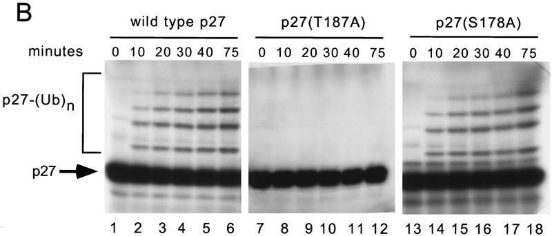
The cellular abundance of the cyclin-dependent kinase (Cdk) inhibitor p27 is regulated by the ubiquitin-proteasome system. Activation of p27 degradation is seen in proliferating cells and in many types of aggressive human carcinomas. p27 can be phosphorylated on threonine 187 by Cdks, and cyclin E/Cdk2 overexpression can stimulate the degradation of wild-type p27, but not of a threonine 187-to-alanine p27 mutant [p27(T187A)]. However, whether threonine 187 phosphorylation stimulates p27 degradation through the ubiquitin-proteasome system or an alternative pathway is still not known. Here, we demonstrate that p27 ubiquitination (as assayed in vivo and in an in vitro reconstituted system) is cell-cycle regulated and that Cdk activity is required for the in vitro ubiquitination of p27. Furthermore, ubiquitination of wild-type p27, but not of p27(T187A), can occur in G1-enriched extracts only upon addition of cyclin E/Cdk2 or cyclin A/Cdk2. Using a phosphothreonine 187 site-specific antibody for p27, we show that threonine 187 phosphorylation of p27 is also cell-cycle dependent, being present in proliferating cells but undetectable in G1 cells. Finally, we show that in addition to threonine 187 phosphorylation, efficient p27 ubiquitination requires formation of a trimeric complex with the cyclin and Cdk subunits. In fact, cyclin B/Cdk1 which can phosphorylate p27 efficiently, but cannot form a stable complex with it, is unable to stimulate p27 ubiquitination by G1 extracts. Furthermore, another p27 mutant [p27(CK-)] that can be phosphorylated by cyclin E/Cdk2 but cannot bind this kinase complex, is refractory to ubiquitination. Thus throughout the cell cycle, both phosphorylation and trimeric complex formation act as signals for the ubiquitination of a Cdk inhibitor.


Figures

Figure 1
Figure 1
p27 polyubiquitination depends on Cdk-dependent phosphorylation on T187. (A) Human p27 cDNA was transcribed and translated in vitro in the presence of [35S] methionine and subjected to a ubiquitination reaction with a HeLa extract, as described in the Materials and Methods. Samples shown in lanes 2–4 were incubated with hexokinase and deoxyglucose to deplete extract from ATP (ATP depl.). Then, AMP–PNP (lane 3) or ATP and an ATP regeneration system (lane 4) were added. Sample in lane 5 was incubated with staurosporine (Stauro.). Samples in lanes 7 and 8 were incubated with the indicated concentrations of flavopiridol (Flav.), used as described in Materials and Methods. Samples in the last three lanes were depleted with control beads (lane 9) or p13 beads (lanes 10,11). Then, purified recombinant cyclin E/Cdk2 complex was added (lane 11). The reaction products were analyzed by SDS-PAGE and autoradiography. The bracket (left) marks a ladder of bands >27,000 corresponding to polyubiquitinated p27. (B) In vitro-translated wild-type p27 (lanes 1–6), p27(T187A) (lanes 7–12), or p27(S178A) (lanes 13–18) was incubated for the indicated times in the presence of a HeLa extract. The reaction products were analyzed by SDS-PAGE and autoradiography. The bracket (left) marks a ladder of bands >27,000 corresponding to polyubiquitinated p27.
Figure 1
Figure 1
p27 polyubiquitination depends on Cdk-dependent phosphorylation on T187. (A) Human p27 cDNA was transcribed and translated in vitro in the presence of [35S] methionine and subjected to a ubiquitination reaction with a HeLa extract, as described in the Materials and Methods. Samples shown in lanes 2–4 were incubated with hexokinase and deoxyglucose to deplete extract from ATP (ATP depl.). Then, AMP–PNP (lane 3) or ATP and an ATP regeneration system (lane 4) were added. Sample in lane 5 was incubated with staurosporine (Stauro.). Samples in lanes 7 and 8 were incubated with the indicated concentrations of flavopiridol (Flav.), used as described in Materials and Methods. Samples in the last three lanes were depleted with control beads (lane 9) or p13 beads (lanes 10,11). Then, purified recombinant cyclin E/Cdk2 complex was added (lane 11). The reaction products were analyzed by SDS-PAGE and autoradiography. The bracket (left) marks a ladder of bands >27,000 corresponding to polyubiquitinated p27. (B) In vitro-translated wild-type p27 (lanes 1–6), p27(T187A) (lanes 7–12), or p27(S178A) (lanes 13–18) was incubated for the indicated times in the presence of a HeLa extract. The reaction products were analyzed by SDS-PAGE and autoradiography. The bracket (left) marks a ladder of bands >27,000 corresponding to polyubiquitinated p27.
Figure 2
Figure 2
p27 ubiquitinating activity is cell-cycle regulated. In vitro-translated wild-type p27 (lanes 1–5 and 10–13), p27(T187A) (lanes 6,7), or 125I-labeled cyclin B (lanes 8,9) were incubated in the presence of extracts (ext.) from proliferating HeLa (lanes 1,8), G1 HeLa (lanes 2–7,9), G1 IMR-90 (lanes 10,12), or G0 IMR-90 cells (lanes 11,13). Purified recombinant cyclin E/Cdk2 complex (80 ng) was added to the samples contained in lanes 3–5, 7, 12, and 13. Samples in lanes 4 and 5 were incubated in the presence of staurosporine (Stauro.) added to the ubiquitination reaction either before (lane 4) or after p27 incubation with the cyclin E/Cdk2 complex (lane 5). The reaction products were analyzed by SDS-PAGE and autoradiography. The bracket (left) marks a ladder of bands >27,000 corresponding to polyubiquitinated p27.
Figure 3
Figure 3
Phosphorylation on T187 and ubiquitination of cellular p27 are cell-cycle regulated. (A) Recombinant purified p27 incubated in a kinase reaction with either recombinant purified cyclin E/Cdk2 (lane 1) or cyclin E/Cdk2m (lane 2). Sample in lane 3 is from G1-enriched cells (75% in G1, 6% in S, and 19% in G2/M); sample in lane 4 is from proliferating cells (58% in G1, 21% in S, and 21% in G2/M); and sample in lane 5 is from G1-depleted cells (4% in G1, 35% in S, and 61% in G2/M). Lanes 3–5 represent immunoprecipitations with a mouse anti-p27 monoclonal antibody. (Top) samples were immunoblotted with a rabbit phospho-T187 site-specific p27 antibody (P-T187 p27); (bottom) samples were immunoblotted with a goat anti-p27 antibody. (B) Samples in lanes 1 and 4 are from G1-depleted cells; sample in lane 2 is from G1-enriched cells; and sample in lane 3 is from proliferating cells. (Lane 1) Immunoprecipitation with purified rabbit IgG (IgG); (lanes 2–4) immunoprecipitations with a mixture of a rabbit phospho-T187 site-specific p27 antibody and a rabbit anti-p27 antibody. Samples were then immunoblotted with a mouse monoclonal antibody to ubiquitin. The bracket (left) marks a ladder of bands corresponding to polyubiquitinated p27; (→) the IgG heavy chains (Ig).
Figure 3
Figure 3
Phosphorylation on T187 and ubiquitination of cellular p27 are cell-cycle regulated. (A) Recombinant purified p27 incubated in a kinase reaction with either recombinant purified cyclin E/Cdk2 (lane 1) or cyclin E/Cdk2m (lane 2). Sample in lane 3 is from G1-enriched cells (75% in G1, 6% in S, and 19% in G2/M); sample in lane 4 is from proliferating cells (58% in G1, 21% in S, and 21% in G2/M); and sample in lane 5 is from G1-depleted cells (4% in G1, 35% in S, and 61% in G2/M). Lanes 3–5 represent immunoprecipitations with a mouse anti-p27 monoclonal antibody. (Top) samples were immunoblotted with a rabbit phospho-T187 site-specific p27 antibody (P-T187 p27); (bottom) samples were immunoblotted with a goat anti-p27 antibody. (B) Samples in lanes 1 and 4 are from G1-depleted cells; sample in lane 2 is from G1-enriched cells; and sample in lane 3 is from proliferating cells. (Lane 1) Immunoprecipitation with purified rabbit IgG (IgG); (lanes 2–4) immunoprecipitations with a mixture of a rabbit phospho-T187 site-specific p27 antibody and a rabbit anti-p27 antibody. Samples were then immunoblotted with a mouse monoclonal antibody to ubiquitin. The bracket (left) marks a ladder of bands corresponding to polyubiquitinated p27; (→) the IgG heavy chains (Ig).
Figure 4
Figure 4
p27 polyubiquitination requires its stable binding to a cyclin/Cdk complex. (A) In vitro-translated p27 was incubated in the presence of extracts from G1 HeLa cells (G1 ext.) and 80 ng of purified recombinant cyclin E/Cdk2 complex (lane 2), 80 ng of purified recombinant cyclin E in complex with catalytic inactive Cdk2 mutant (Cdk2m) (lanes 3,4,6–8), 80 ng of purified recombinant cyclin E alone (lane 5), or buffer (lane 1). Sample in lane 4 lacked okadaic acid (−OA); samples in lanes 7 and 8 were incubated in the presence of the indicated concentrations of flavopiridol (Flav.). The reaction products were analyzed by SDS-PAGE and autoradiography. The bracket (left) marks a ladder of bands >27,000 corresponding to polyubiquitinated p27. (B) Comparison of the abilities of cyclin E/Cdk2, cyclin E/Cdk2m, cyclin B/Cdk1, and cyclin A/Cdk2 to phosphorylate, bind, and stimulate the ubiquitination of p27. The amounts of purified cyclin/HA-tagged Cdk complexes were measured by colorimetric methods and confirmed with an anti-HA antibody (HA-Cdk). (Lane 1) Cyclin B/Cdk1 (200 ng); (lane 2) cyclin E/Cdk2 (200 ng); (lane 3) cyclin B/Cdk1 (50 ng); (lane 4) cyclin E/Cdk2 (50 ng); (lane 5) cyclin E/Cdk2m (200 ng); (lane 6) no kinase added; (lane 7) cyclin E/Cdk2 (200 ng); (lane 8) cyclin A/Cdk2 (200 ng); (lane 9) cyclin A/Cdk2 (50 ng). Lanes 1–5 and 6–9 are from two separate experiments. The phosphorylation of p27 by different amounts of cyclin/Cdk complexes was performed as described in Materials and Methods. Phosphorylated p27 was detected by autoradiography (32P p27). The binding of p27 to different cyclin/Cdks was assayed by incubating in vitro-translated p27 with His-tagged cyclin/Cdk complexes and then purifying them with nickel-agarose. Bound p27 was then detected by immunoblot with an anti-p27 monoclonal antibody (Cdk-Bound p27).The stimulation of p27 ubiquitination by different amount of cyclin/Cdk complexes was assayed by incubating in vitro-translated p27 with these complexes and extracts from G1 HeLa cells. The reaction products were analyzed by SDS-PAGE and autoradiography. The bracket (left) marks a ladder of bands >27,000 corresponding to polyubiquitinated p27. (C) Phosphorylation, binding, and ubiquitination of wild-type p27 (WT p27) and p27(CK) in the presence (lanes 2,4) or in the absence (lanes 1,3) of 200 ng of cyclin E/Cdk2. In vitro-translated wild-type p27 (lanes 1,2) and p27(CK) (lanes 3,4) were immunoprecipitated with an anti-p27 antibody and then subjected to a phosphorylation reaction in the presence or in the absence of cyclin E/Cdk2. Phosphorylated p27 was detected by autoradiography with a cellophane screen that blocked the 35S but not 32P (32P p27) (top). The binding to cyclin E/Cdk2 was assayed by incubating in vitro-translated wild-type p27 and p27(CK) with His-tagged cyclin E/Cdk2 complex and then purifying the complex with nickel–agarose. Bound p27 was then detected by autoradiography (Cdk-Bound p27) (middle). The ubiquitination reaction was performed by incubating in vitro-translated wild-type p27 and p27(CK) with extracts from G1 HeLa cells in the presence and in the absence of cyclin E/Cdk2 (bottom). The reaction products were analyzed by SDS-PAGE and autoradiography. The bracket (left) marks a ladder of bands >27,000 corresponding to polyubiquitinated p27.
Figure 4
Figure 4
p27 polyubiquitination requires its stable binding to a cyclin/Cdk complex. (A) In vitro-translated p27 was incubated in the presence of extracts from G1 HeLa cells (G1 ext.) and 80 ng of purified recombinant cyclin E/Cdk2 complex (lane 2), 80 ng of purified recombinant cyclin E in complex with catalytic inactive Cdk2 mutant (Cdk2m) (lanes 3,4,6–8), 80 ng of purified recombinant cyclin E alone (lane 5), or buffer (lane 1). Sample in lane 4 lacked okadaic acid (−OA); samples in lanes 7 and 8 were incubated in the presence of the indicated concentrations of flavopiridol (Flav.). The reaction products were analyzed by SDS-PAGE and autoradiography. The bracket (left) marks a ladder of bands >27,000 corresponding to polyubiquitinated p27. (B) Comparison of the abilities of cyclin E/Cdk2, cyclin E/Cdk2m, cyclin B/Cdk1, and cyclin A/Cdk2 to phosphorylate, bind, and stimulate the ubiquitination of p27. The amounts of purified cyclin/HA-tagged Cdk complexes were measured by colorimetric methods and confirmed with an anti-HA antibody (HA-Cdk). (Lane 1) Cyclin B/Cdk1 (200 ng); (lane 2) cyclin E/Cdk2 (200 ng); (lane 3) cyclin B/Cdk1 (50 ng); (lane 4) cyclin E/Cdk2 (50 ng); (lane 5) cyclin E/Cdk2m (200 ng); (lane 6) no kinase added; (lane 7) cyclin E/Cdk2 (200 ng); (lane 8) cyclin A/Cdk2 (200 ng); (lane 9) cyclin A/Cdk2 (50 ng). Lanes 1–5 and 6–9 are from two separate experiments. The phosphorylation of p27 by different amounts of cyclin/Cdk complexes was performed as described in Materials and Methods. Phosphorylated p27 was detected by autoradiography (32P p27). The binding of p27 to different cyclin/Cdks was assayed by incubating in vitro-translated p27 with His-tagged cyclin/Cdk complexes and then purifying them with nickel-agarose. Bound p27 was then detected by immunoblot with an anti-p27 monoclonal antibody (Cdk-Bound p27).The stimulation of p27 ubiquitination by different amount of cyclin/Cdk complexes was assayed by incubating in vitro-translated p27 with these complexes and extracts from G1 HeLa cells. The reaction products were analyzed by SDS-PAGE and autoradiography. The bracket (left) marks a ladder of bands >27,000 corresponding to polyubiquitinated p27. (C) Phosphorylation, binding, and ubiquitination of wild-type p27 (WT p27) and p27(CK) in the presence (lanes 2,4) or in the absence (lanes 1,3) of 200 ng of cyclin E/Cdk2. In vitro-translated wild-type p27 (lanes 1,2) and p27(CK) (lanes 3,4) were immunoprecipitated with an anti-p27 antibody and then subjected to a phosphorylation reaction in the presence or in the absence of cyclin E/Cdk2. Phosphorylated p27 was detected by autoradiography with a cellophane screen that blocked the 35S but not 32P (32P p27) (top). The binding to cyclin E/Cdk2 was assayed by incubating in vitro-translated wild-type p27 and p27(CK) with His-tagged cyclin E/Cdk2 complex and then purifying the complex with nickel–agarose. Bound p27 was then detected by autoradiography (Cdk-Bound p27) (middle). The ubiquitination reaction was performed by incubating in vitro-translated wild-type p27 and p27(CK) with extracts from G1 HeLa cells in the presence and in the absence of cyclin E/Cdk2 (bottom). The reaction products were analyzed by SDS-PAGE and autoradiography. The bracket (left) marks a ladder of bands >27,000 corresponding to polyubiquitinated p27.

References

    1. Alessandrini A, Chiaur DS, Pagano M. Regulation of the cyclin-dependent kinase inhibitor p27 by degradation and phosphorylation. Leukemia. 1997;11:342–345. - PubMed
    1. Amon A, Irniger S, Nasmyth K. Closing the cell cycle circle in yeast: G2 cyclin proteolysis initiated at mitosis persists until the activation of G1 cyclins in the next cycle. Cell. 1994;77:1037–1050. - PubMed
    1. Blain SW, Montalvo E, Massague J. Differential interaction of the cyclin-dependent kinase (Cdk) inhibitor p27Kip1 with cyclin A/Cdk2 and cyclin D2/Cdk4. J Biol Chem. 1997;272:25863–25872. - PubMed
    1. Brancolini C, Lazarevic D, Rodriguez J, Schneider C. Dismantling cell–cell contacts during apoptosis is coupled to a caspase-dependent proteolytic cleavage of beta-catenin. J Cell Biol. 1997;139:759–771. - PMC - PubMed
    1. Brandeis M, Hunt T. The proteolysis of mitotic cyclins in mammalian cells persists from the end of mitosis until the onset of S phase. EMBO J. 1996;15:5280–5289. - PMC - PubMed

Publication types

MeSH terms