. 1999 May 14;84(9):1020-31.
doi: 10.1161/01.res.84.9.1020.

Activation of distinct cAMP-dependent and cGMP-dependent pathways by nitric oxide in cardiac myocytes

Affiliations

Activation of distinct cAMP-dependent and cGMP-dependent pathways by nitric oxide in cardiac myocytes

M G Vila-Petroff et al. Circ Res. .

Abstract

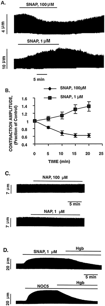
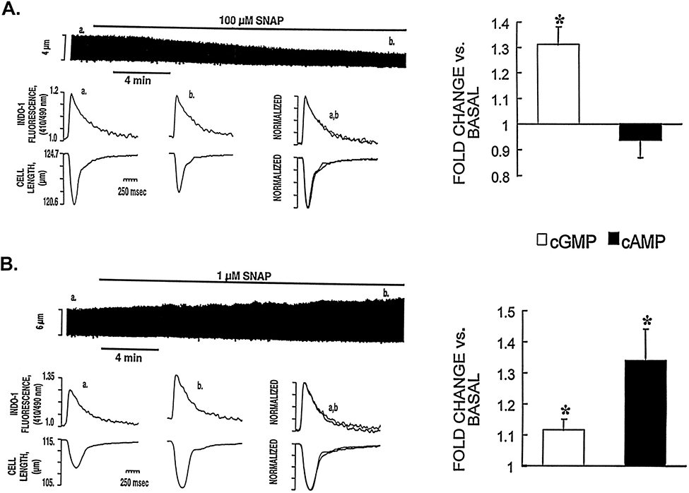
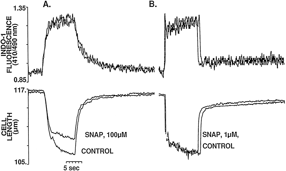
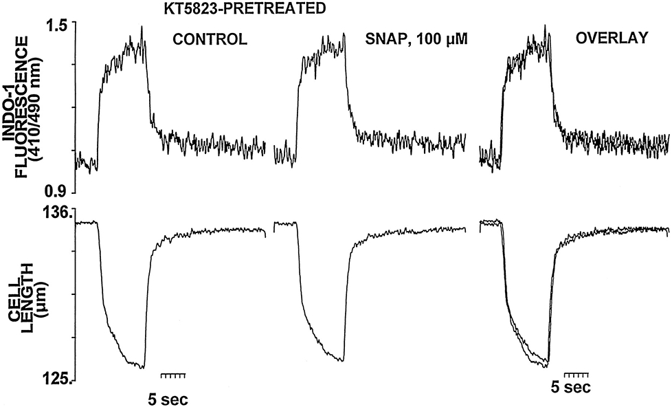
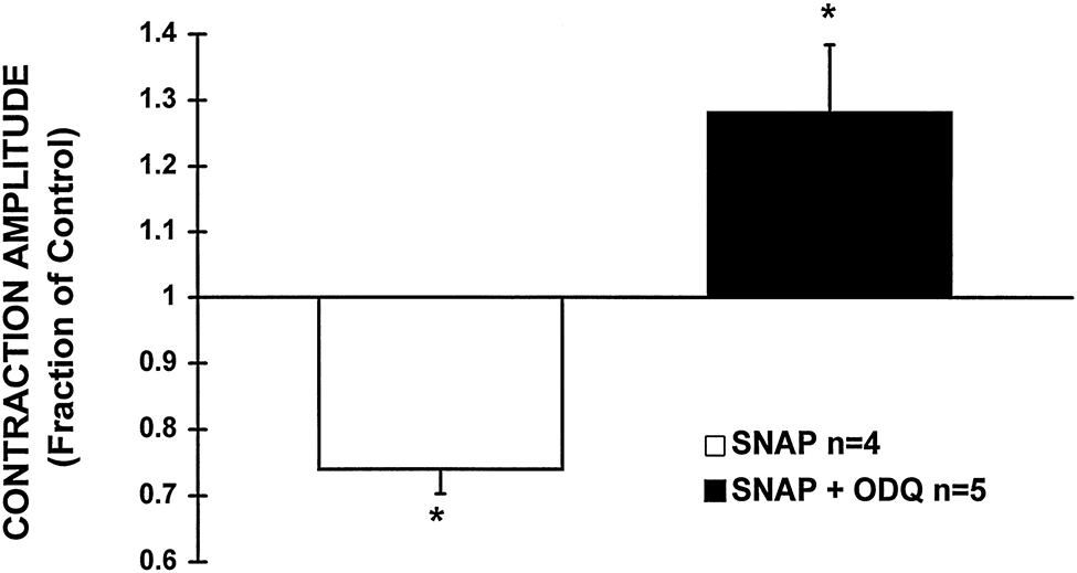
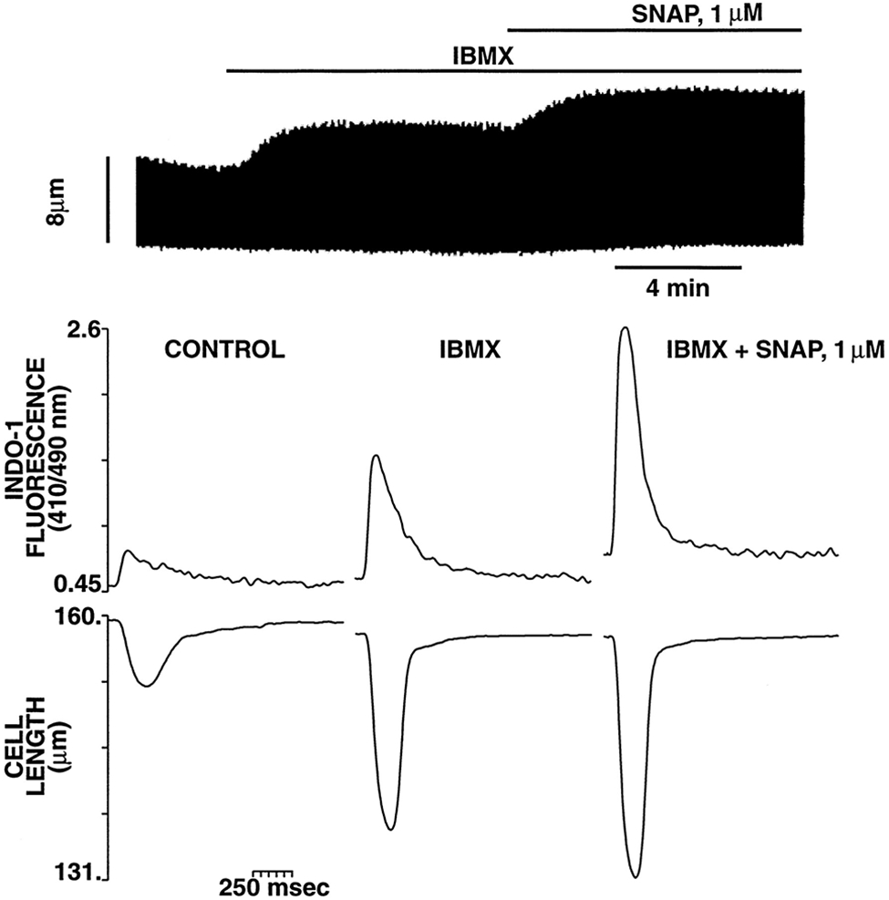
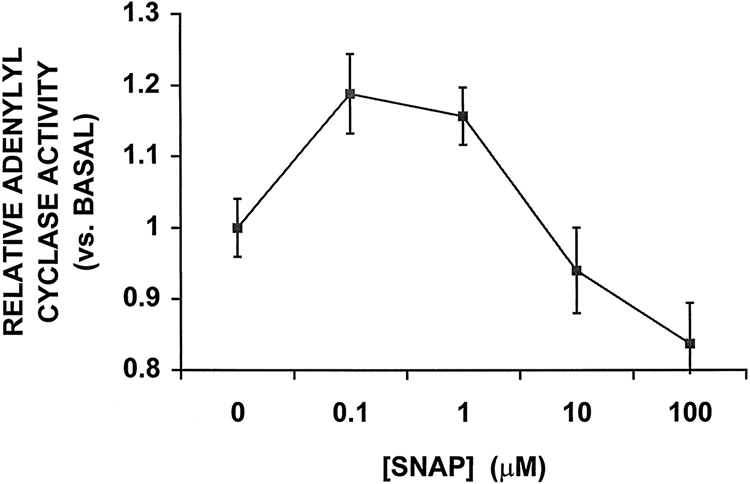
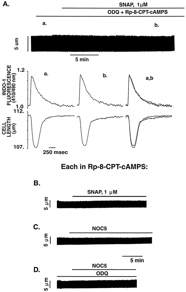
Nitric oxide (NO) donors were recently shown to produce biphasic contractile effects in cardiac tissue, with augmentation at low NO levels and depression at high NO levels. We examined the subcellular mechanisms involved in the opposing effects of NO on cardiac contraction and investigated whether NO modulates contraction exclusively via guanylyl cyclase (GC) activation or whether some contribution occurs via cGMP/PKG-independent mechanisms, in indo 1-loaded adult cardiac myocytes. Whereas a high concentration of the NO donor S-nitroso-N-acetylpenicillamine (SNAP, 100 micromol/L) significantly attenuated contraction amplitude by 24.4+/-4.5% (without changing the Ca2+ transient or total cAMP), a low concentration of SNAP (1 micromol/L) significantly increased contraction amplitude (38+/-10%), Ca2+ transient (26+/-10%), and cAMP levels (from 6.2 to 8.5 pmol/mg of protein). The negative contractile response of 100 micromol/L SNAP was completely abolished in the presence of the specific blocker of PKG KT 5823 (1 micromol/L); the positive contractile response of 1 micromol/L SNAP persisted, despite the presence of the selective inhibitor of GC 1H-[1,2,4]oxadiazolo[4,3-a]quinoxalin-1-one (ODQ, 10 micromol/L) alone, but was completely abolished in the presence of ODQ plus the specific inhibitory cAMP analog Rp-8-CPT-cAMPS (100 micromol/L), as well as by the NO scavenger oxyhemoglobin. Parallel experiments in cell suspensions showed significant increases in adenylyl cyclase (AC) activity at low concentrations (0.1 to 1 micromol/L) of SNAP (AC, 18% to 20% above basal activity). We conclude that NO can regulate both AC and GC in cardiac myocytes. High levels of NO induce large increases in cGMP and a negative inotropic effect mediated by a PKG-dependent reduction in myofilament responsiveness to Ca2+. Low levels of NO increase cAMP, at least in part, by a novel cGMP-independent activation of AC and induce a positive contractile response.


Figures

Figure 1.
Figure 1.
Effect of high (100 μmol/L) and low (1 μmol/L) concentrations of SNAP on the electrically stimulated myocyte contraction. A, Representative examples of the opposing contractile effects induced by high and low concentrations of SNAP from 2 different cardiac myocytes. The continuous chart recording of cell length (top) shows that a high concentration of SNAP induced a progressive decrease in twitch amplitude, whereas the bottom tracing shows that a low concentration of SNAP induced a progressive increase in contraction amplitude. Both negative and positive contractile effects of SNAP were entirely reversible on washout. B, Overall data of the time course of the contractile effects induced by high and low concentrations of SNAP. Data are mean±SE; n=5 cells per group. C, Representative examples of the lack of contractile effects of exposure to NAP 100 μmol/L and 1 μmol/L (the byproduct of SNAP after release of NO). D, Representative examples demonstrating that the positive contractile effects of both SNAP (1 μmol/L) and the unrelated NO donor NOC5 (1 μmol/L) were completely abolished by coincubation with the NO scavenger oxyhemoglobin (Hgb, 0.1 mmol/L).
Figure 2.
Figure 2.
Effect of high (100 μmol/L) and low (1 μmol/L) concentrations of SNAP on [Ca2+]i, cGMP and cAMP levels, and contraction. A, Representative example of simultaneously recorded Cai and contraction traces showing the effects induced by a high concentration of SNAP in an indo 1-AM–loaded myocyte. The chart recording (top) shows that SNAP (100 μmol/L) induced a progressive decrease in twitch amplitude. The bottom tracings show, on an expanded time scale, the [Ca2+]i transients and cell length at control (a) and 15 to 20 minutes after addition of SNAP (b). The superimposed tracing (overlay of data from a and b; scales normalized to peak heights) shows that SNAP (100 μmol/L) reduced twitch duration and twitch amplitude without affecting the Cai. The right panel shows the effect of SNAP 100 μmol/L on cGMP and cAMP levels. The results are presented as fractions of the basal values of cGMP and cAMP (4.2±0.2 and 6.2±0.4 pmol/mg of protein, respectively). Each bar represents the mean±SEM of 3 independent experiments performed in triplicate. Using the same presentation scheme, Figure 2B shows the effect of low concentrations of SNAP (1 μmol/L) on [Ca2+]i, cGMP and cAMP levels, and contraction. Note that this concentration of SNAP was associated with a large increase in the Cai, cAMP levels, and contraction. *P<0.05 vs basal level.
Figure 3.
Figure 3.
Representative example of the effects of SNAP 100 μmol/L (A) and 1 μmol/L (B) on the steady-state myofilament Ca2+ response during tetanic contraction (10 Hz) of myocytes pretreated with thapsigargin (0.2 μmol/L). The top tracings correspond to the superimposed Ca2+ at control and after 20 minutes of exposure to the indicated concentration of SNAP; immediately below are the superimposed tetanic contractions. Although SNAP had no effect on peak tetanic Ca2+ at either of the concentrations studied, the amplitude of the tetanic contraction is significantly decreased after exposure to high concentrations of SNAP but unchanged at the low concentration. It should be noted that the [Ca2+]i achieved during tetanization protocols is <1 μmol/L (ie, on the order of that achieved during peak systole), and, moreover, that Ca2+–indo 1 binding is far from saturation, even at the plateau of the tetanus.
Figure 4.
Figure 4.
PKG inhibition with KT 5823 (1 μmol/L) prevents the negative myofilament effect of SNAP 100 μmol/L. Representative example of the effects of SNAP 100 μmol/L on the steady-state Cai and contraction during tetanic contractions of a myocyte pretreated and continuously perfused with KT 5823 1 μmol/L. The superimposed tracing shows the failure of SNAP to decrease the amplitude of the tetanic contraction in the presence of KT 5823.
Figure 5.
Figure 5.
Failure of ODQ 10 μmol/L to block the positive contractile response of SNAP 1 μmol/L. A, Typical continuous chart recording of contraction amplitude in response to ODQ (10 μmol/L) alone (top tracing) and SNAP (1 μmol/L) in the continued presence of ODQ (10 μmol/L) (bottom tracing). The superimposed tracings of cell length on the right show the lack of effect of ODQ (10 μmol/L) on baseline contraction (top) and the increase of contraction amplitude in response to a low concentration of SNAP (1 μmol/L) in the continued presence of ODQ (10 μmol/L) (bottom). B, Parallel measurements of cGMP and cAMP in response to SNAP (1 μmol/L) in the presence and absence of ODQ. The results are presented as fractions of the basal values of cGMP and cAMP, 4.2±0.2 and 6.2±0.4 pmol/mg of protein, respectively. ODQ (10 μmol/L) alone did not affect basal cGMP or cAMP levels (4.1±0.2 and 5.2±0.7 pmol/mg of protein, respectively). ODQ completely abolished the SNAP-induced increase in cGMP but had no effect on increased cAMP levels. *P<0.05 vs basal level. C, Representative example of the lack of effect of SNAP (1 μmol/L) in the presence of ODQ (10 μmol/L) on the steady-state myofilament response to Ca2+ of a myocyte during tetanic contractions (10 Hz). This indicates that the positive contractile effect seen in panel A is purely the result of the increase in magnitude of the Cai.
Figure 6.
Figure 6.
Inhibition of GC with ODQ (10 μmol/L) converts a negative contractile response of SNAP (10 μmol/L) to a sustained positive response. The bar graph depicts the average change in contraction amplitude, expressed as a fraction of the basal contraction, after 20 minutes of exposure of cells to SNAP (10 μmol/L) either in the absence or presence of ODQ (10 μmol/L). SNAP alone significantly attenuated contraction amplitude by 26±4%, whereas in the presence of ODQ, SNAP induced a pronounced increase in contraction, 28±10% above baseline. *P<0.05 vs basal level.
Figure 7.
Figure 7.
Tonic PDE inhibition with IBMX does not prevent the positive contractile response of SNAP. Top, Typical continuous chart recording of contraction amplitude in response to IBMX (0.1 mmol/L) followed by SNAP (1 μmol/L) in the continued presence of IBMX. Bottom, Representative example of the effect of IBMX and SNAP (same protocol as in the top panel) on contraction and [Ca2+]i from a different myocyte. Each tracing is taken at steady state under the condition listed. Note that even in the presence of IBMX, SNAP can still exert a positive inotropic effect via an increase of the Cai.
Figure 8.
Figure 8.
Effects of SNAP on AC activity in rat cardiac sarcolemmal membranes. AC activity of membranes prepared from rat heart was determined under basal conditions and in the presence of different concentrations of SNAP. SNAP produced a biphasic response in AC activity in a concentration-dependent fashion. Low concentrations of SNAP (0.1 to 1 μmol/L) were associated with significant increases in AC activity (P<0.05 vs control), whereas higher concentrations of SNAP resulted in either no change (10 μmol/L) or a small decrease (100 μmol/L) in AC activity. Average AC activity is expressed as a fraction of the basal activity (92±6 pmol/mg of protein per minute). Each point represents the mean±SEM of 9 independent experiments performed in triplicate. The increases in AC activity at 0.1 and 1 μmol/L SNAP were 40% and 34% of maximal Mn2+-stimulated AC activity, respectively.
Figure 9.
Figure 9.
PKA inhibition with Rp-8-CPT-cAMPS (100 μmol/L) completely abolishes the positive contractile responses of SNAP and NOC5. A, Representative example of the effect of SNAP (1 μmol/L) in the continued presence of inhibitors of GC (ODQ 10 μmol/L) and PKA (Rp-8-CPT-cAMPS 100 μmol/L) on myocyte contraction and [Ca2+]i. The continuous chart recording shows the failure of SNAP (1 μmol/L) to increase the isotonic twitch contraction in a cardiac myocyte pretreated and continuously perfused with ODQ and Rp-8-CPT-cAMPS. The bottom tracings show, on an expanded time scale, [Ca2+]i and cell length at the times indicated (a, baseline; b, 10 minutes after addition of SNAP; and c, 15 to 20 minutes after SNAP). Under these conditions, SNAP had no significant effect on either contraction amplitude or Cai amplitude. Representative examples of the lack of effect on myocyte contraction of SNAP (1 μmol/L) (B), NOC5 (1 μmol/L) (C), and NOC5 (1 μmol/L) pretreated with the GC inhibitor ODQ (10 μmol/L) (D), each in the continued presence of the inhibitor of PKA (Rp-8-CPT-cAMPS 100 μmol/L).

References

    1. Moncada S, Palmer RMJ, Higgs EA. Nitric oxide: physiology, pathology, and pharmacology. Pharmacol Rev. 1991;43:109–142. - PubMed
    1. Ignarro LJ, Kadoitz PJ. The pharmacological role of cyclic GMP in vascular smooth muscle relaxation. Annu Rev Pharmacol Toxicol. 1985;25:171–191. - PubMed
    1. Brady AJB, Warren JB, Poole-Wilson PA, Williams TJ, Harding SE. Nitric oxide attenuates cardiac myocyte contraction. Am J Physiol. 1993;265:H176–H182. - PubMed
    1. Meulemans AL, Sipido KR, Sys SU, Brutsaert DL. Atriopeptin III induces early relaxation of ventricular cardiac muscle. Circ Res. 1988;62:1171–1174. - PubMed
    1. Smith JA, Shah AM, Lewis MJ. Factors released from endocardium of the ferret and pig modulate myocardial contraction. J Physiol (Lond). 1991;439:1–14. - PMC - PubMed

MeSH terms