. 1999 May;154(5):1345-52.
doi: 10.1016/S0002-9440(10)65388-6.

Identification of a glioblastoma-associated tenascin-C isoform by a high affinity recombinant antibody

Affiliations

Identification of a glioblastoma-associated tenascin-C isoform by a high affinity recombinant antibody

B Carnemolla et al. Am J Pathol. 1999 May.

Abstract

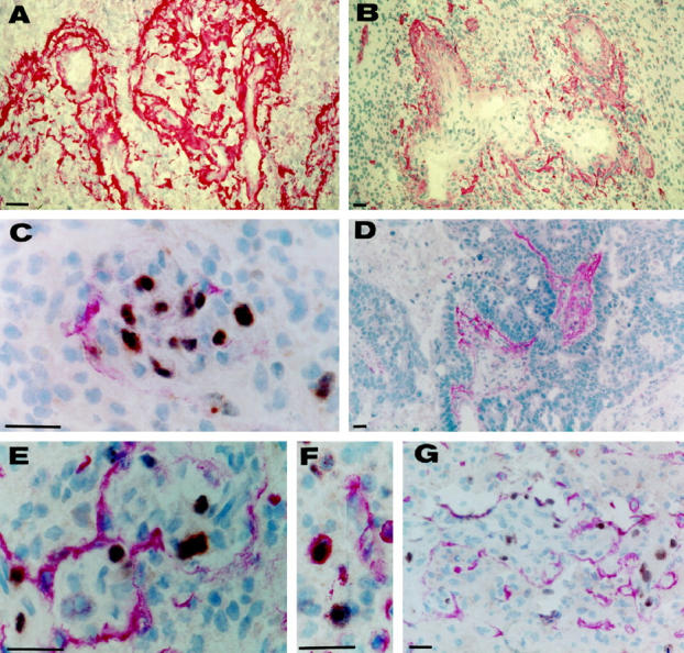
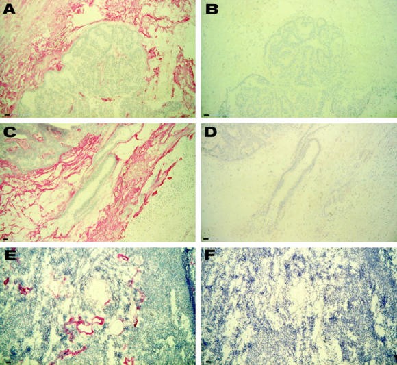
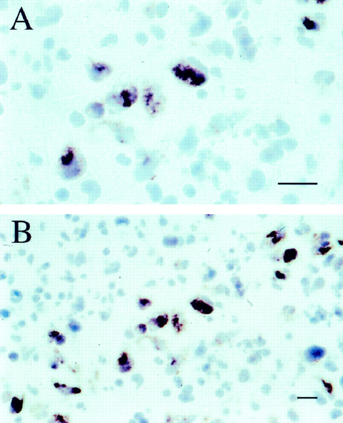
Tenascin-C exists in several polymorphic isoforms due to alternative splicing of nine fibronectin-like type III repeats. Large Tenascin-C isoforms are present in almost all normal adult tissues but are upregulated in fetal, regenerating, and neoplastic tissues. Here, we report a human antibody fragment, TN11, derived from a phage library with high affinity for the spliced repeat C and demonstrate that this repeat is undetectable in normal adult tissues, barely detectable or undetectable in breast, lung and gastric carcinomas, meningioma, and low grade astrocytoma, but extremely abundant in high grade astrocytoma (grade III and glioblastoma), especially around vascular structures and proliferating cells. The antibody appears to have potential for development of a therapeutic agent for patients with high grade astrocytoma.


Figures

Figure 1.
Figure 1.
Confirmation by Southern blot analysis of the specificity of the cRNA probe used for in situ hybridization experiments. Bottom: ethidium bromide staining of the agarose gel. Lane 1: TNfnALL (including human tenascin DNA from type III repeat 2 to type III repeat 7, including all the alternatively spliced type III repeats). Lane 2: TNfn 1-8 (same sequence as TNfnALL but lacking all the alternatively spliced repeats). Lane 3: All TNegf like repeats. Lane 4: Alternatively spliced type III repeat D. Lane 5: Alternatively spliced type III repeat C. Lane 6: type III repeat 1. Lane 7: TNegf like repeats from 8 to 10. Lane 8: 1-Kb standard. Above: Southern blot showing DIG-hybrids detection. The numbers on the right are expressed in kilobases.
Figure 2.
Figure 2.
A: Model of the domain structure of a human TN-C subunit. The ovals and the squares represent the EGF-like and FN-like repeats, respectively. The globular N-terminal knob and the fibrinogen-like C-terminal domain are also depicted. The FN-like repeats A1 to D, whose expression is regulated by the alternative splicing of the pre-mRNA, are shaded. The upper part of the figure also shows the TN-C-β-galactosidase fusion proteins or recombinant proteins used. The arrows show the sequence in which each epitope of recombinant or monoclonal antibodies was localised. ∧ indicates contiguity. B: Sodium dodecyl sulfate polyacryl-amide gel electro-phoresis (4-18%) of recombinant Large TN-C (containing repeats from A1 to D) and Small TN-C (without repeats from A1 to D) stained with Coomassie blue and immunoblots stained with scFv TN11 and TN12. C: Immunoblots of different fusion and recombinant proteins (A) using the scFv TN11 and TN12. Values on the left indicate molecular masses (in kilodaltons) of the standards.
Figure 3.
Figure 3.
Northern blots of poly(A)-rich RNA from human adult heart (1), brain (2), placenta (3), lung (4), liver (5), skeletal muscle (6), kidney (7), and pancreas (8) tissues and fetal brain (1), lung (2), liver (3), and kidney (4) tissues using the cDNA probe (see Materials and Methods) specific for c-TN-C isoform, the HT11 probe that recognizes all TN-C isoforms, and the human G3PDH cDNA to normalize the blots (see Materials and Methods). Numbers on the left are the size, in kb, of the standards.
Figure 4.
Figure 4.
Immunohistochemical experiments on sections of glioblastoma stained using scFv TN11 (A and B) and double-stained using scFv TN11 (red) and mAbKI67 (brown) (C, E, F, and G); a section of a brain metastasis from lung carcinoma stained using scFv TN11 (D). Scale bar, 10 μm.
Figure 5.
Figure 5.
Immunohistochemical experiments on serial sections of invasive ductal breast carcinoma stained using scFv TN12 (A and C) and scFv TN11 (B and D) and on serial sections of meningioma stained using scFv TN12 (E) and scFv TN11 (F). Scale bar, 10 μm.
Figure 6.
Figure 6.
Two different magnifications of an in situ hybridization experiment using human glioblastoma cryostat sections with the DIG-labeled cRNA repeat C probe (see Material and Methods section). Positive signal was visible only in some tumoral cells with large nuclei.

Similar articles

Cited by

References

    1. Folkman J: Angiogenesis in cancer, vascular, rheumathoid and other diseases. Nat Med 1995, 1:27-31 - PubMed
    1. Van den Hoff A: Stromal involvement in malignant growth. Adv Cancer Res 1988, 50:159-196 - PubMed
    1. Risau W, Lemmon V: Changes in the vascular extracellular matrix during embryonic vasculogenesis and angiogenesis. Devel Biol 1988, 125:441-450 - PubMed
    1. Ingber D, Folkman J: How does extracellular matrix control capillary morphogenesis? Cell 1989, 58:803-805 - PubMed
    1. Borsi L, Carnemolla B, Nicoló G, Spina B, Tanara G, Zardi L: Expression of different tenascin isoforms in normal, hyperplastic and neoplastic human breast tissues. Int J Cancer 1992, 52:688-692 - PubMed

Publication types

LinkOut - more resources