

Parkinson's disease is associated with oxidative damage to cytoplasmic DNA and RNA in substantia nigra neurons
- PMID: 10329595
- PMCID: PMC1866598
- DOI: 10.1016/S0002-9440(10)65396-5
Parkinson's disease is associated with oxidative damage to cytoplasmic DNA and RNA in substantia nigra neurons
Abstract
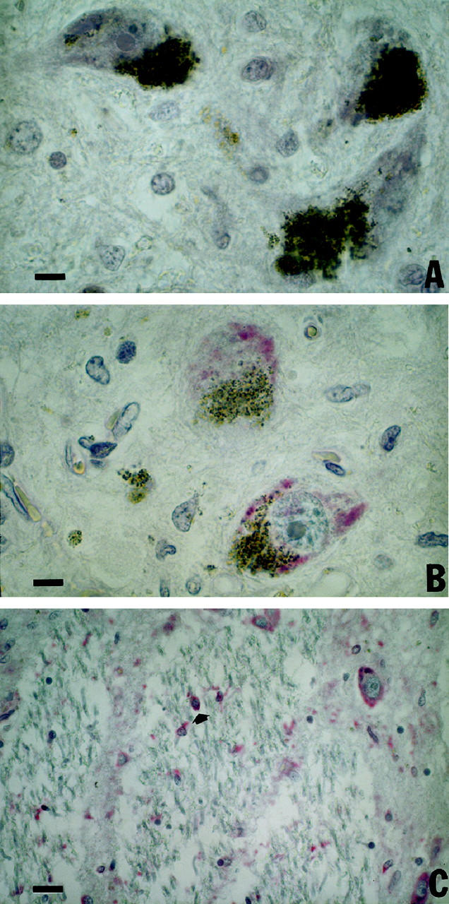
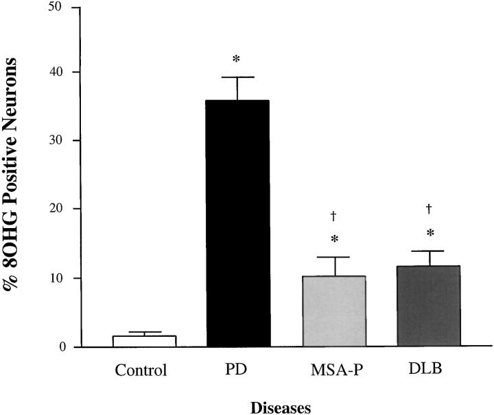
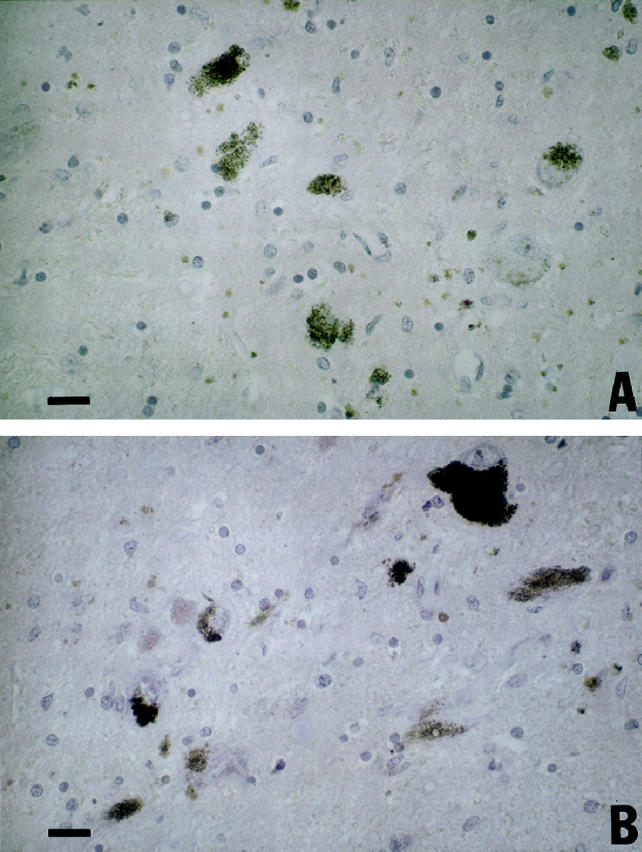
Oxidative damage, including modification of nucleic acids, may contribute to dopaminergic neurodegeneration in the substantia nigra (SN) of patients with Parkinson's disease (PD). To investigate the extent and distribution of nucleic acid oxidative damage in these vulnerable dopaminergic neurons, we immunohistochemically characterized a common product of nucleic acid oxidation, 8-hydroxyguanosine (8OHG). In PD patients, cytoplasmic 8OHG immunoreactivity was intense in neurons of the SN, and present to a lesser extent in neurons of the nucleus raphe dorsalis and oculomotor nucleus, and occasionally in glia. The proportion of 8OHG immunoreactive SN neurons was significantly greater in PD patients compared to age-matched controls. Midbrain sections from patients with multiple system atrophy-Parkinsonian type (MSA-P) and dementia with Lewy bodies (DLB) also were examined. These showed increased cytoplasmic 8OHG immunoreactivity in SN neurons in both MSA-P and DLB compared to controls; however, the proportion of positive neurons was significantly less than in PD patients. The regional distribution of 8OHG immunoreactive neurons within the SN corresponded to the distribution of neurodegeneration for these three diseases. Nuclear 8OHG immunoreactivity was not observed in any individual. The type of cytoplasmic nucleic acid responsible for 8OHG immunoreactivity was analyzed by preincubating midbrain sections from PD patients with RNase, DNase, or both enzymes. 8OHG immunoreactivity was substantially diminished by either RNase or DNase, and completely ablated by both enzymes. These results suggest that oxidative damage to cytoplasmic nucleic acid is selectively increased in midbrain, especially the SN, of PD patients and much less so in MSA-P and DLB patients. Moreover, oxidative damage to nucleic acid is largely restricted to cytoplasm with both RNA and mitochondrial DNA as targets.
Figures
Similar articles
-
RNA oxidation is a prominent feature of vulnerable neurons in Alzheimer's disease.J Neurosci. 1999 Mar 15;19(6):1959-64. doi: 10.1523/JNEUROSCI.19-06-01959.1999. J Neurosci. 1999. PMID: 10066249 Free PMC article.
-
Isofurans, but not F2-isoprostanes, are increased in the substantia nigra of patients with Parkinson's disease and with dementia with Lewy body disease.J Neurochem. 2003 May;85(3):645-50. doi: 10.1046/j.1471-4159.2003.01709.x. J Neurochem. 2003. PMID: 12694390
-
A comparison of changes in proteasomal subunit expression in the substantia nigra in Parkinson's disease, multiple system atrophy and progressive supranuclear palsy.Brain Res. 2010 Apr 22;1326:174-83. doi: 10.1016/j.brainres.2010.02.045. Epub 2010 Feb 20. Brain Res. 2010. PMID: 20176003
-
Interaction of alpha-synuclein and dopamine metabolites in the pathogenesis of Parkinson's disease: a case for the selective vulnerability of the substantia nigra.Acta Neuropathol. 2006 Aug;112(2):115-26. doi: 10.1007/s00401-006-0096-2. Epub 2006 Jun 22. Acta Neuropathol. 2006. PMID: 16791599 Review.
-
Glial reactions in Parkinson's disease.Mov Disord. 2008 Mar 15;23(4):474-83. doi: 10.1002/mds.21751. Mov Disord. 2008. PMID: 18044695 Review.
Cited by
-
Oxidative damage to RNA in aging and neurodegenerative disorders.Neurotox Res. 2012 Oct;22(3):231-48. doi: 10.1007/s12640-012-9331-x. Epub 2012 Jun 6. Neurotox Res. 2012. PMID: 22669748 Review.
-
Parkinson's disease brain mitochondria have impaired respirasome assembly, age-related increases in distribution of oxidative damage to mtDNA and no differences in heteroplasmic mtDNA mutation abundance.Mol Neurodegener. 2009 Sep 23;4:37. doi: 10.1186/1750-1326-4-37. Mol Neurodegener. 2009. PMID: 19775436 Free PMC article.
-
The Impact of Oxidative Stress on Ribosomes: From Injury to Regulation.Cells. 2019 Nov 2;8(11):1379. doi: 10.3390/cells8111379. Cells. 2019. PMID: 31684095 Free PMC article. Review.
-
Hydrogen sulfide induces oxidative damage to RNA and DNA in a sulfide-tolerant marine invertebrate.Physiol Biochem Zool. 2010 Mar-Apr;83(2):356-65. doi: 10.1086/597529. Physiol Biochem Zool. 2010. PMID: 19327040 Free PMC article.
-
Mutational Spectrum Analysis of Neurodegenerative Diseases and Its Pathogenic Implication.Int J Mol Sci. 2015 Oct 14;16(10):24295-301. doi: 10.3390/ijms161024295. Int J Mol Sci. 2015. PMID: 26473852 Free PMC article.
References
-
- Lowe J, Graham L, Leigh PN: Disorders of movement and system degeneration. Graham DI Lantos PL eds. Greenfield’s Neuropathology. 1997, :pp 281-366 Arnold, London
-
- Dexter DT, Wells FR, Lees AJ, Agid F, Agid Y, Jenner P, Marsden CD: Increased nigral iron content and alterations in other metal ions occurring in brain in Parkinson’s disease. J Neurochem 1989, 52:1830-1836 - PubMed
-
- Saggu H, Cooksey J, Dexter D, Wells FR, Lees A, Jenner P, Marsden CD: A selective increase in particulate superoxide dismutase activity in Parkinsonian substantia nigra. J Neurochem 1989, 53:692-697 - PubMed
-
- Perry TL, Godin DV, Hansen S: Parkinson’s disease: a disorder due to nigral glutathione deficiency? Neurosci Lett 1982, 33:305-310 - PubMed
Publication types
MeSH terms
Substances
Grants and funding
LinkOut - more resources
Full Text Sources
Other Literature Sources
Medical
