

Molecular genetic alterations in radiation-induced astrocytomas
- PMID: 10329596
- PMCID: PMC1866591
- DOI: 10.1016/S0002-9440(10)65397-7
Molecular genetic alterations in radiation-induced astrocytomas
Abstract
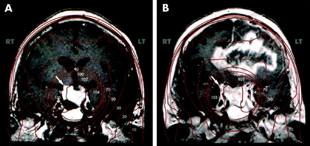
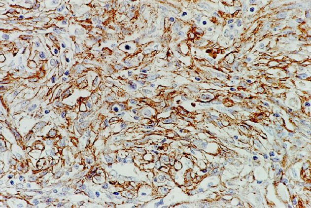
Astrocytic tumors occasionally arise in the central nervous system following radiotherapy. It is not clear if these gliomas represent a unique molecular genetic subset. We identified nine cases in which an astrocytoma arose within ports of previous radiation therapy, with total doses ranging from 2400 to 5500 cGy. Irradiated primary lesions included craniopharyngioma, pituitary adenoma, Hodgkin's lymphoma, ependymoma, pineal neoplasm, rhabdomyosarcoma, and three cases of lymphoblastic malignancies. Patients ranged from 9 to 60 years of age and developed secondary tumors 5 to 23 years after radiotherapy. The 9 postradiation neoplasms presented as either anaplastic astrocytoma (3 cases) or glioblastoma multiforme (6 cases). Two of the latter contained malignant mesenchymal components. We performed DNA sequence analysis, differential polymerase chain reaction (PCR), and quantitative PCR on DNA from formalin-fixed, paraffin-embedded tumors to evaluate possible alterations of p53, PTEN, K-ras, EGFR, MTAP, and p16 (MTS1/CDKN2) genes. By quantitative PCR, we found EGFR gene amplification in 2 of 8 tumors. One of these demonstrated strong immunoreactivity for EGFR. Quantitative PCR showed chromosome 9p deletions including p16 tumor suppressor gene (2 of 7 tumors) and MTAP gene (3 of 7). Five of 9 tumors demonstrated diffuse nuclear immunoreactivity for p53 protein. Sequencing of the p53 gene in these 9 cases revealed a mutation in only one of these cases, a G-to-A substitution in codon 285 (exon 8). Somewhat unexpectedly, no mutations were identified in PTEN, a commonly altered tumor suppressor gene in de novo glioblastoma multiformes. Unlike some radiation-induced tumors, no activating point mutations of the K-ras proto-oncogene or base pair deletions of tumor suppressor genes were noted. These radiation-induced tumors are distinctive in their high histological grade at clinical presentation. The spectrum of molecular genetic alterations appears to be similar to that described in spontaneous high grade astrocytomas, especially those of the de novo type.
Figures

References
-
- Harrison MJ, Wolfe DE, Lau T, Mitnick RJ, Sachdev VP: Radiation-induced meningiomas: experience at the Mount Sinai Hospital and review of the literature. J Neurosurg 1991, 75:564-574 - PubMed
-
- Kaschten B, Flandroy P, Reznik M, Hainaut H, Stevenaert A: Radiation-induced gliosarcoma: case report and review of the literature. J Neurosurg 1995, 83:154-162 - PubMed
-
- Simmons NE, Laws ER, Jr: Glioma occurrence after sellar irradiation: case report and review. Neurosurgery 1998, 42:172-178 - PubMed
-
- Ron E, Modan B, Boice JD, Jr., Alfandary E, Stovall M, Chetrit A, Katz L: Tumors of the brain and nervous system after radiotherapy in childhood. N Engl J Med 1988, 319:1033-1039 - PubMed
MeSH terms
Substances
LinkOut - more resources
Full Text Sources
Other Literature Sources
Medical
Research Materials
Miscellaneous
