Clinical Trial
. 1999 May;154(5):1453-64.
doi: 10.1016/S0002-9440(10)65400-4.

Pediatric AIDS-associated lymphocytic interstitial pneumonia and pulmonary arterio-occlusive disease: role of VCAM-1/VLA-4 adhesion pathway and human herpesviruses

Affiliations
Clinical Trial

Pediatric AIDS-associated lymphocytic interstitial pneumonia and pulmonary arterio-occlusive disease: role of VCAM-1/VLA-4 adhesion pathway and human herpesviruses

S J Brodie et al. Am J Pathol. 1999 May.

Abstract

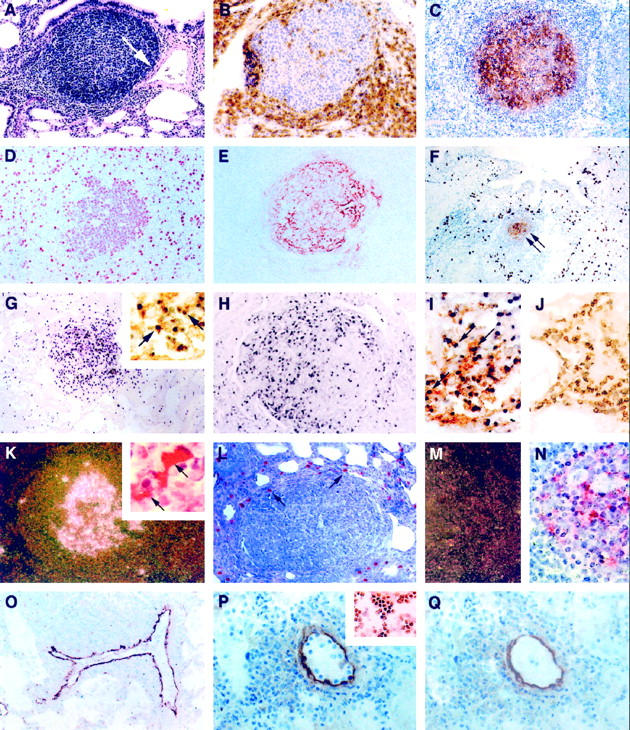
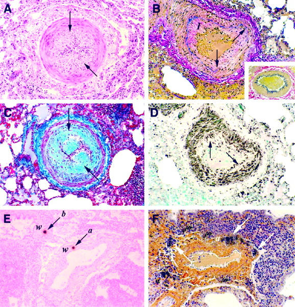
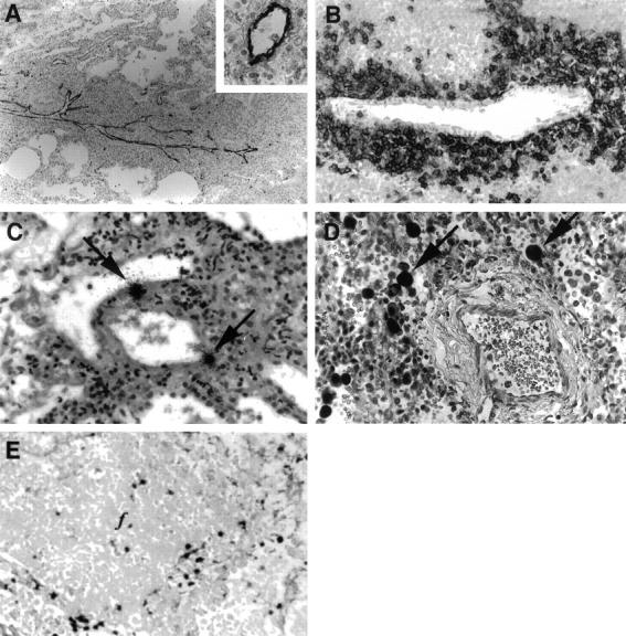
Because the mechanisms of lymphocyte accumulation in the lungs of children with AIDS-associated lymphocytic interstitial pneumonia (LIP) are unknown, we studied the relative contributions of known adhesion pathways in mediating lymphocyte adherence to endothelium and the potential role of human herpesviruses in the expansion of these lesions. LIP was characterized by lymphoid hyperplasia of the bronchus-associated lymphoid tissue (BALT) and infiltration of the pulmonary interstitium with CD8(+) T lymphocytes. In some individuals there was expansion of the alveolar septae with dense aggregates of B lymphocytes, many containing the Epstein-Barr viral (EBV) genome. Patients with concurrent EBV infection also demonstrated large-vessel arteriopathy characterized by thickening of the intimae with collagen and smooth muscle. Venular endothelium from the lung of children with LIP, but not uninflamed lung from other children with AIDS or lung from children with nonspecific pneumonitis, expressed high levels of vascular cell adhesion molecule-1 (VCAM-1) protein. In turn, inflammatory cells expressing very late activation antigen-4 (VLA-4), the leukocyte ligand for VCAM-1, were the predominant perivascular infiltrate associated with vessels expressing VCAM-1. Expression of other endothelial adhesion molecules, including intracellular adhesion molecule-1 and E-selectin, was not uniformly associated with LIP. Using a tissue adhesion assay combined with immunohistochemistry for VCAM-1, we show that CD8(+) T cell clones that express VLA-4 bind preferentially to pulmonary vessels in sites of LIP: vessels that expressed high levels of VCAM-1. When tissues and cells were pretreated with antibodies to VCAM-1 or VLA-4, respectively, adhesion was inhibited by >/=80%. Thus, infiltration of alveolar septae with CD8(+) T cells was highly correlative with VCAM-1/VLA-4 adhesive interactions, and focal expansion of B cells was coincidental to co-infection with EBV.


Figures

Figure 1.
Figure 1.
Infiltrative and proliferative lesions associated with pediatric lymphocytic interstitial pneumonia. Immunoperoxidase (IP) procedures used 3,3′-diaminobenzidine as the chromogen, and in situ hybridization (ISH) procedures used anti-digoxigenin-alkaline phosphatase (αDIG-AP) or 125I-labeled CTP as an indicator. A: Widespread thickening of the pulmonary interstitium with mononuclear leukocytes and a single peribronchiole and perivascular aggregate of lymphocytes. Note that the expansion of lymphocytes is associated with constriction of a medium-sized artery (arrow) H&E, magnification, ×100. B: Perifollicular aggregation of CD8+ T lymphocytes. IP, CD8 (clone C8/144B, DAKO); magnification, ×250. C: Lymphoid aggregate showing a predominance of immature B cells within the germinal center. IP, CD19 (clone 4G7/2E, Novocastra, Newcastle, UK); magnification, ×100. D to F: Most germinal center lymphoblasts were mitotically active (D; IP, PCNA (clone PC10, DAKO); magnification, ×250) and surrounded well developed networks of FDC (E and F, arrows; IP, CD21 (clone 1F8, DAKO); magnification, ×100). F: Alveolar and interstitial macrophages were found in high numbers on the periphery of lymphoid follicles. IP, CD68 (KP1, DAKO); magnification, ×100. G: A high percentage of cells (>20%) within the follicular germinal center of lymphoid aggregates contained high copy numbers of EBV EBER-1 RNA. ISH αDIG-AP for EBER-1; magnification, ×100. Most of these EBV RNA-positive cells were of B cell lineage (G, inset; combined ISH αDIG-AP for EBER-1 and IP for CD20; magnification, ×400), and many expressed EBV latent membrane proteins (H, IP for EBV LMP (clone CS1–4, DAKO); magnification, ×200). I: EBV also co-localized to follicular dendritic cells (arrows; combined ISH αDIG-AP for EBV RNA and IP for CD21 FDC; magnification, ×400), but not to interstitial T lymphocytes J; combined ISH αDIG-AP for EBV RNA and IP for CD3; magnification, ×400). K: HIV mRNA was observed within follicular germinal centers (125I ISH for HIV-1 mRNA, dark-field; magnification, ×40) in association with FDC (combined 125I ISH for HIV-1 mRNA and IP for CD21 FDC; inset, ×1000); however, cells that expressed viral antigens were found mostly on the periphery of lymphoid aggregates in areas normally occupied by macrophages (L, arrows; IP for HIV gag p24 (clone Kal-1, DAKO; magnification, ×200). M and N: Serial tissue sections hybridized with noncomplementary HIV RNA probes transcribed in sense-strand orientation did not form hybrids (M, 125I ISH for HIV-1 RNA, dark-field; magnification, ×40; N, 125I ISH for HIV-1 RNA; magnification, ×500). O: VCAM-1 expression was heightened in vessels with perivascular inflammatory foci. IP, CD106 (clone 51–10C9, PharMingen); magnification, ×100. P: Tissue adhesion assay demonstrating that CD8+ T cell clones that co-express the VLA-4 ligand (inset, IP for VLA-4α4/β1; magnification, ×200) selectively adhere to venular endothelium in the lung from a child with severe LIP (IP for VCAM-1; magnification, ×400). Q: Pretreatment of sequential tissue sections (shown in P) with blocking antibodies to VCAM-1 and incubation of CD8+ T cell clones with blocking antibodies to VLA-4α4/β1 inhibited >80% of this adhesion. After the adhesion assay, the tissues were stained with antibody to VCAM-1 to demonstrate presence of VCAM-1 expression. IP for VCAM-1; magnification, ×400.
Figure 2.
Figure 2.
Histochemical and immunochemical characterization of pulmonary arterial lesions. A: Marked intimal thickening (arrows) with luminal narrowing. H&E; magnification, ×100. B: Disruption of the internal elastic laminae (arrows) due to expansion of the tunica intimae (I). Weigert’s resorcin fuchsin; magnification, ×100. Vessel showing normal intimae and normal elastic laminae. Weigert’s resorcin fuchsin; magnification, ×200 (inset). C and D: Advanced narrowing of the vascular lumen (arrows) attributable to intimal fibrous (C; Masson’s trichrome; magnification, ×100) and smooth muscle cell proliferation (D; immunoperoxidase for α-actin (clone 1A4, DAKO); magnification, ×100. E: Localization of cells harboring high copy number HIV gag mRNA (arrows) within the wall (w) of a medium-sized artery (a) and bronchiole (b). 125I ISH; magnification, ×100. F: Mineral deposition surrounding the tunica adventitia (a) of a medium-sized artery. Von Kossa’s; magnification, ×100.
Figure 3.
Figure 3.
Mechanisms of lymphocyte infiltration and localization of virus in pediatric lymphocytic interstitial pneumonia. A: Longitudinal section of a vein showing VCAM-1 expression and associated mononuclear cell infiltrates. IP for VCAM-1 (clone 51–10C9, PharMingen); magnification, ×100. The inset shows a transverse section of a venule and demonstrates the uniform expression of endothelial VCAM-1. IP for VCAM-1; magnification, ×400. B: Perivenular aggregation of CD8+ T lymphocytes in areas of interstitial collapse. IP for CD8 (clone C8/144B, DAKO); magnification, ×400. C: Localization of cells harboring HIV mRNA (arrows) within a thin-walled vein. 125I ISH for HIV-1 mRNA; magnification, ×100. D: Expression of CMV early gene RNA in cytomegaloid cells (arrows) adjacent to a vein and in a region of interstitial collapse and lymphocyte infiltration. ISH αDIG-AP for EBV EBER-1 RNA; magnification, ×400. E: Expression of KSHV latent gene transcripts in cells associated with follicular aggregation. ISH αDIG-AP for KSHV T0.7 RNA; magnification, ×400.

Comment in

  • Findings of misconduct in science.
    [No authors listed] [No authors listed] NIH Guide Grants Contracts (Bethesda). 2010 May 14:NOT-OD-10-095. NIH Guide Grants Contracts (Bethesda). 2010. PMID: 20486276 Free PMC article. No abstract available.
  • Note of concern.
    [No authors listed] [No authors listed] Am J Pathol. 2010 Oct;177(4):2147. doi: 10.2353/ajpath.2010.100773. Am J Pathol. 2010. PMID: 20884964 Free PMC article. No abstract available.

References

    1. Bye MR, Cairns-Bazarian AM, Ewig JM: Markedly reduced mortality associated with corticosteroid therapy of Pneumocystis carinii pneumonia in children with acquired immune deficiency syndrome. Arch Pediatr Adolesc Med 1995, 148:638-641 - PubMed
    1. Blanche S, Newell ML, Mayaux MJ, Dunn DT, Teglas JP, Rouzioux C, Peckham CS: Morbidity and mortality in European children vertically infected with HIV-1: the French Pediatric HIV Infection Study Group and European Collaborative Study. J Acquir Immune Defic Syndr Hum Retrovirol 1997, 14:442-450 - PubMed
    1. Scott GB, Hutto C, Makuch RW, Mastrucci MT, O’Connor T, Mitchell CD, Trapido EJ, Parks WP: Survival in children with perinatally acquired immunodeficiency virus type 1 infection. N Engl J Med 1989, 321:1791-1796 - PubMed
    1. Marolda J, Pace B, Bonforte RJ, Kotin NM, Rabinowitz J, Kattan M: Pulmonary manifestations of HIV infection in children. Pediatr Pulmonol 1991, 10:231-235 - PubMed
    1. Moran CA, Suster S, Pavlova Z, Mullick FG, Koss MN: The spectrum of pathological changes in the lung in children with the acquired immunodeficiency syndrome: an autopsy study of 36 cases. Hum Pathol 1994, 25:877-882 - PubMed

Publication types

MeSH terms

Substances