. 1999 May;154(5):1479-87.
doi: 10.1016/S0002-9440(10)65402-8.

Keratinocyte growth factor protects alveolar epithelium and endothelium from oxygen-induced injury in mice

Affiliations

Keratinocyte growth factor protects alveolar epithelium and endothelium from oxygen-induced injury in mice

C Barazzone et al. Am J Pathol. 1999 May.

Abstract

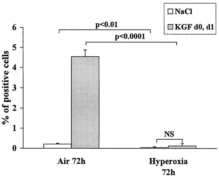
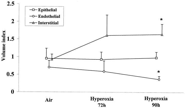
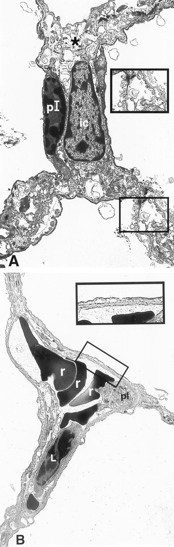
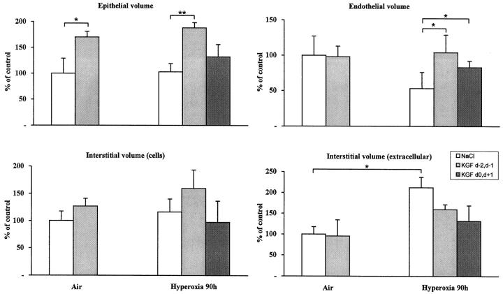
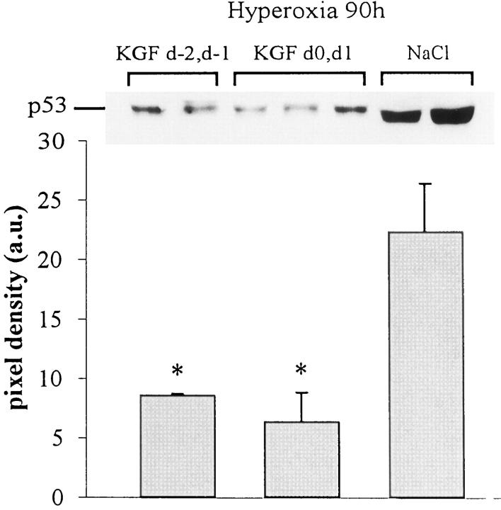
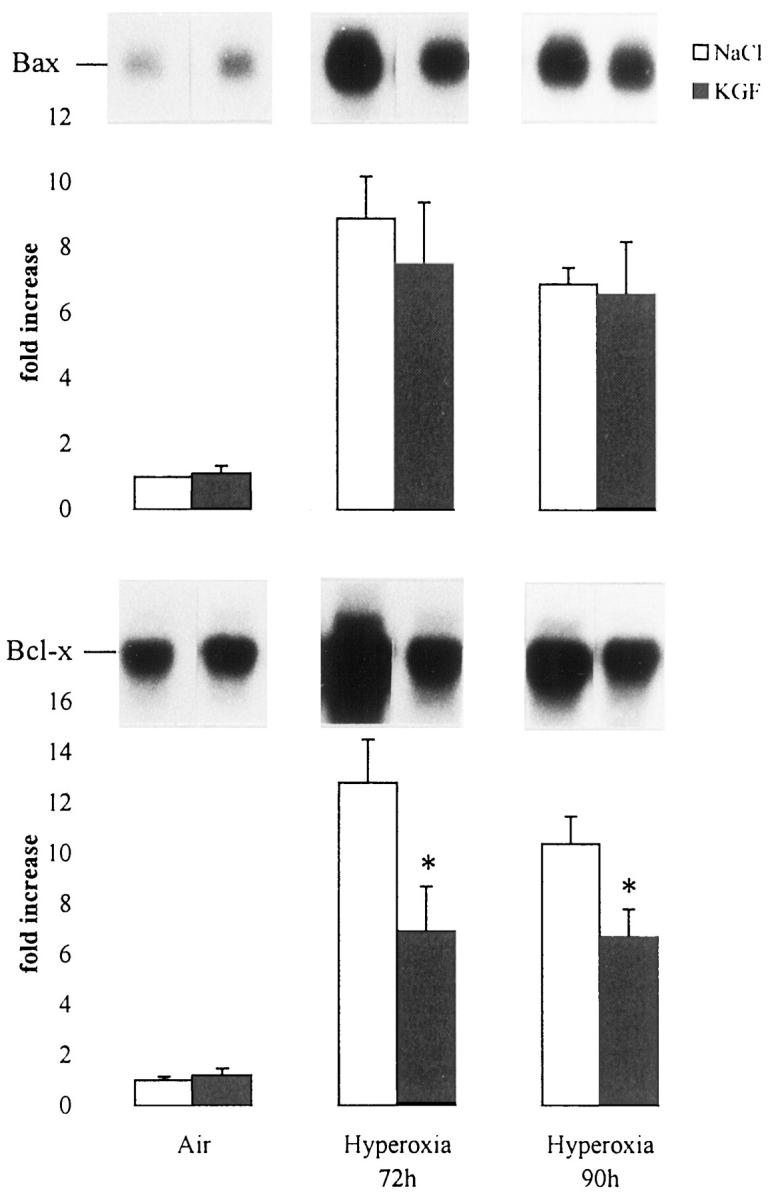
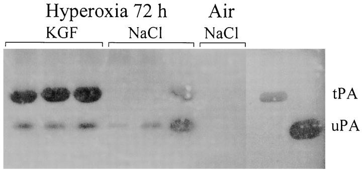
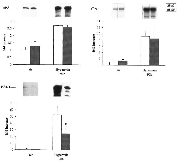
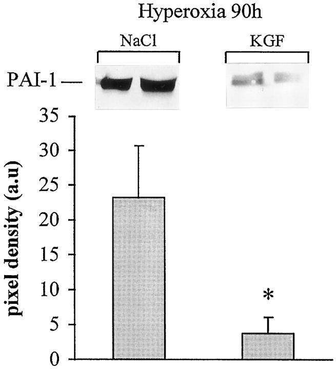
Keratinocyte growth factor (KGF) has been used successfully to prevent alveolar damage induced by oxygen exposure in rodents. However, this treatment was used intratracheally and before oxygen exposure, which limited its clinical application. In the present study, mice were treated with the recombinant human KGF intravenously before (days -2 and -1) or during (days 0 and +1) oxygen exposure. In both cases, lung damage was attenuated. KGF increased the number of cells incorporating bromodeoxyuridine (BrdU) in the septa and in bronchial epithelium of air-breathing mice but not of oxygen-exposed mice, indicating that the protective effect of KGF is not necessarily associated with proliferation. Oxygen-induced damage of alveolar epithelium and, unexpectedly, of endothelium was prevented by KGF treatment as seen by electron microscopy. We investigated the effect of KGF on different mechanisms known to be involved in oxygen toxicity. The induction of p53, Bax, and Bcl-x mRNAs during hyperoxia was to a large extent prevented by KGF. Surfactant proteins A and B mRNAs were not markedly modified by KGF. The anti-fibrinolytic activity observed in the alveoli during hyperoxia was to a large extent prevented by KGF, most probably by suppressing the expression of plasminogen activator inhibitor-1 (PAI-1) mRNA and protein. As PAI-1 -/- mice are more resistant to hyperoxia, KGF might act, at least in part, by decreasing the expression of this protease inhibitor and by restoring the fibrinolytic activity into the lungs.


Figures

Figure 1.
Figure 1.
Mice were injected intraperitoneally with 100 mg/kg BrdU 2 hours and 15 minutes before sacrifice. BrdU uptake of the nuclei was revealed by immunohistochemistry, and quantification was performed. The whole section was analyzed (20 fields) for the BrdU staining and the same number in the adjacent section for the control staining. Three to four different mice were analyzed for each condition, and the percentage of positive cells was calculated for each condition. The results are the mean ± SD of three to four mice for each condition. KGF treatment given 72 hours before sacrifice increased significantly the number of positive cells in air-breathing mice. When mice were exposed to oxygen, the number of positive cells was diminished compared with air-breathing mice with (P < 0.0001) or without KGF treatment (P < 0.01).
Figure 2.
Figure 2.
Morphometric analysis of lung volumes during hyperoxia. Results are the mean ± SD of five to six mice. Endothelial volume decreased and total interstitial volume increased (cellular and extracellular) significantly at 90 hours of hyperoxia (*P < 0.05), whereas epithelial volume did not change.
Figure 3.
Figure 3.
Alveolar septa from mice exposed to oxygen for 90 hours in a saline-treated mouse (A) and in a KGF-treated mouse (B). KGF was injected on day 0 and day 1 of oxygen exposure. A: Note the very significant interstitial edema and the condensed chromatin of the nucleus of the type I epithelial cell. Higher magnification of the damaged epithelium and endothelium. B: Presence of red blood cells and probably of a leukocyte within the capillary. Note the minimal changes of the epithelium and endothelium (original magnification, ×2800 and ×5200, respectively). pI, type I epithelial cell; ic, interstitial cell; r, red blood cell; L, leukocyte; pl, platelet.
Figure 4.
Figure 4.
Effect of KGF treatment on different lung volumes. Results are the mean ± SD of five to six mice. *P < 0.05; **P < 0.001.
Figure 5.
Figure 5.
Western blot analysis for the detection of p53 in lung extracts. p53 was undetectable in control lungs treated with saline (NaCl) or with KGF and was strongly expressed in hyperoxia-exposed lungs. KGF treatment before or concomitantly to oxygen exposure decreased significantly the expression of p53. The film was exposed for 30 seconds. Quantification of three different samples for each condition was performed by scanning densitometry. Values represent the mean ± SD of pixel density (*P < 0.05 compared with NaCl-treated group).
Figure 6.
Figure 6.
Effect of rhKGF (day −2, day −1) treatment on bax and bcl-x in murine lung during hyperoxia. Mice were exposed to air or to hyperoxia for the indicated times. Total lung RNA was isolated, and 10 μg was electrophoresed, transferred onto nitrocellulose membrane, and hybridized with 32P-labeled RNA probes. Quantification of the samples was performed with a Molecular Dynamics PhosphorImager and adjusted to the quantity of 18 S rRNA detected with methylene blue staining. Values represent means ± SD of three animals and are expressed as fold increases over air-breathing animals set as 1. KGF treatment did not change the basal level of bax and bcl-x in air-breathing animals, whereas it decreased significantly hyperoxia-induced bcl-x (P < 0.05). This effect was not significant with bax.
Figure 7.
Figure 7.
Zymographic analysis of PA activities in BAL. A total of 20 μg of protein recovered in BAL from individual mice was loaded onto each lane. Mice were exposed to hyperoxia for 72 hours and treated with KGF (lanes 1 to 3) or with NaCl (lanes 4 to 6). Lanes 7 to 8, air-breathing mice. The migration of tPA and uPA was determined with purified standard proteins electrophoresed in adjacent lanes. Note the marked induction of tPA-mediated activity in KGF-treated mice compared with NaCl-treated mice. The photograph was taken after 48 hours of incubation at 37°C.
Figure 8.
Figure 8.
Quantification of uPA, tPA, and PAI-1 in murine lungs. Mice were exposed to air or to hyperoxia for the indicated time and treated with saline (NaCl) or rhKGF on days 0 and 1, and 5 μg of total lung RNA was hybridized to 32P-labeled UTP cRNA probes and RNAse-resistant hybrids analyzed after separation in urea/polyacrylamide gels. Quantification of the samples was performed as described. Values represent means ± SD of three animals and are expressed as fold increase compared with NaCl controls set as 1. uPA mRNA level increased two to threefold in NaCl- or in KGF-treated mice, tPA mRNA increased eight- to ninefold, whereas PAI-1 mRNA increase was significantly less in KGF-treated mice exposed to hyperoxia. *P < 0.05 versus NaCl-treated mice exposed to hyperoxia.
Figure 9.
Figure 9.
Western blot analysis for the detection of PAI-1 in lung tissue extracts during hyperoxic injury. PAI-1 was almost undetectable in the lung of an air-exposed animal (not shown). KGF treatment before or concomitantly to oxygen exposure decreased significantly the expression of PAI-1. The film was exposed for 30 seconds. Quantification of three different samples for each condition was performed by scanning densitometry. Values represent the mean ± SD of pixel density (*P < 0.05 compared with NaCl-treated group).

References

    1. Crapo JD, Barry BE, Foscue A, Shelburne J: Structural and biochemical changes in rat lung during exposure to lethal and adaptative doses of oxygen. Am Rev Respir Dis 1980, 122:123-143 - PubMed
    1. Ulich TR, Yi ES, Longmuir K, Yin S, R. B, Morris CF, Housley RM, Pierce GF: Keratinocyte growth factor is a growth factor for type II pneumocytes in vivo. J Clin Invest 1994, 93:1298–1236 - PMC - PubMed
    1. Yano T, Deterding RR, Scott Simonet W, Shannon J, Mason R: Keratinocyte growth factor reduces lung damage due to acid instillation in rats. Am J Respir Cell Mol Biol 1996, 15:433-442 - PubMed
    1. Panos RJ, Bak PM, Scott Simonet W, Rubin JS, Smith LJ: Intratracheal instillation of keratinocyte growth factor decreases hyperoxia-induced mortality in rats. J Clin Invest 1995, 96:2026-2033 - PMC - PubMed
    1. Yi ES, Williams ST, Lee H, Malicki D, Chin EM, Yin S, Tarpley J, Ulich T: Keratinocyte growth factor ameliorates radiation- and bleomycin-induced lung injury. Am J Pathol 1996, 149:1963-1970 - PMC - PubMed

Publication types

MeSH terms