

The development of M cells in Peyer's patches is restricted to specialized dome-associated crypts
- PMID: 10329609
- PMCID: PMC1866609
- DOI: 10.1016/S0002-9440(10)65410-7
The development of M cells in Peyer's patches is restricted to specialized dome-associated crypts
Abstract
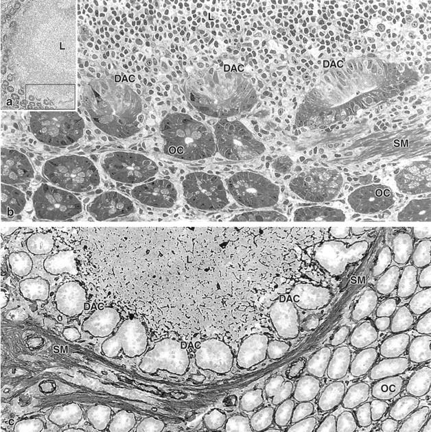
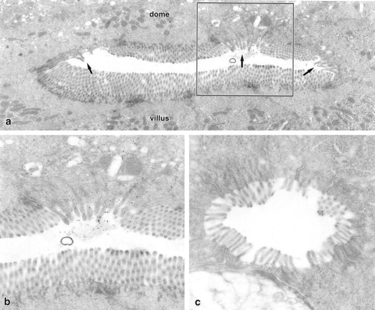
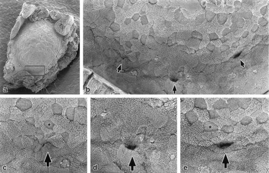
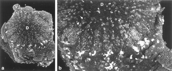
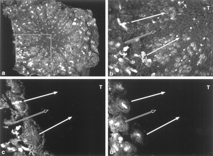
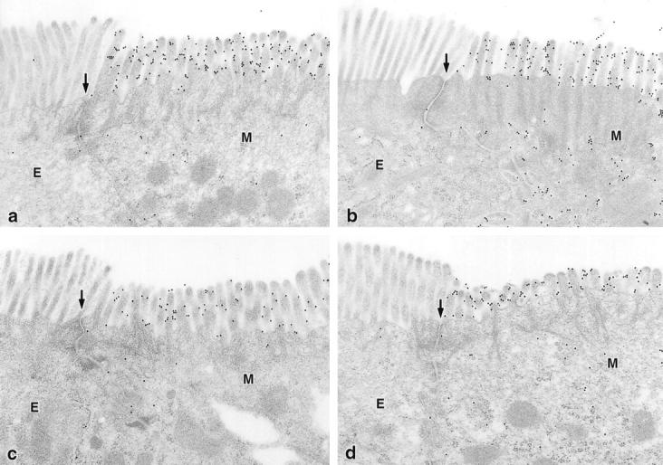
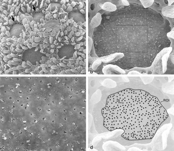
It is controversial whether the membranous (M) cells of the Peyer's patches represent a separate cell line or develop from enterocytes under the influence of lymphocytes on the domes. To answer this question, the crypts that produce the dome epithelial cells were studied and the distribution of M cells over the domes was determined in mice. The Ulex europaeus agglutinin was used to detect M cells in mouse Peyer's patches. Confocal microscopy with lectin-gold labeling on ultrathin sections, scanning electron microscopy, and laminin immuno-histochemistry were combined to characterize the cellular composition and the structure of the dome-associated crypts and the dome epithelium. In addition, the sites of lymphocyte invasion into the dome epithelium were studied after removal of the epithelium using scanning electron microscopy. The domes of Peyer's patches were supplied with epithelial cells that derived from two types of crypt: specialized dome-associated crypts and ordinary crypts differing not only in shape, size, and cellular composition but also in the presence of M cell precursors. When epithelial cells derived from ordinary crypts entered the domes, they formed converging radial strips devoid of M cells. In contrast to the M cells, the sites where lymphocytes invaded the dome epithelium were not arranged in radial strips, but randomly distributed over the domes. M cell development is restricted to specialized dome-associated crypts. Only dome epithelial cells that derive from these specialized crypts differentiate into M cells. It is concluded that M cells represent a separate cell line that is induced in the dome-associated crypts by still unknown, probably diffusible lymphoid factors.
Figures
Similar articles
-
Differential distribution of lymphocytes and accessory cells in mouse Peyer's patches.Anat Rec. 1986 Jun;215(2):144-52. doi: 10.1002/ar.1092150208. Anat Rec. 1986. PMID: 3729011
-
M cells and granular mononuclear cells in Peyer's patch domes of mice depleted of their lymphocytes by total lymphoid irradiation.Am J Pathol. 1989 Mar;134(3):529-37. Am J Pathol. 1989. PMID: 2923183 Free PMC article.
-
Lectin histochemistry reveals the appearance of M-cells in Peyer's patches of SCID mice after syngeneic normal bone marrow transplantation.J Histochem Cytochem. 1998 Feb;46(2):143-8. doi: 10.1177/002215549804600202. J Histochem Cytochem. 1998. PMID: 9446820
-
[Structure and function of Peyer's patches in the intestines of different animal species].Schweiz Arch Tierheilkd. 1989;131(10):595-603. Schweiz Arch Tierheilkd. 1989. PMID: 2690336 Review. German.
-
Development of Peyer's patches, follicle-associated epithelium and M cell: lessons from immunodeficient and knockout mice.Semin Immunol. 1999 Jun;11(3):183-91. doi: 10.1006/smim.1999.0174. Semin Immunol. 1999. PMID: 10381864 Review.
Cited by
-
Intestinal villous M cells: an antigen entry site in the mucosal epithelium.Proc Natl Acad Sci U S A. 2004 Apr 20;101(16):6110-5. doi: 10.1073/pnas.0400969101. Epub 2004 Apr 7. Proc Natl Acad Sci U S A. 2004. PMID: 15071180 Free PMC article.
-
Unsolved mysteries of intestinal M cells.Gut. 2000 Nov;47(5):735-9. doi: 10.1136/gut.47.5.735. Gut. 2000. PMID: 11034595 Free PMC article. Review. No abstract available.
-
Infection-generated electric field in gut epithelium drives bidirectional migration of macrophages.PLoS Biol. 2019 Apr 9;17(4):e3000044. doi: 10.1371/journal.pbio.3000044. eCollection 2019 Apr. PLoS Biol. 2019. PMID: 30964858 Free PMC article.
-
Novel antigen delivery technologies: a review.Drug Deliv Transl Res. 2011 Apr;1(2):103-12. doi: 10.1007/s13346-011-0014-6. Drug Deliv Transl Res. 2011. PMID: 25788109
-
Enhancing oral vaccine potency by targeting intestinal M cells.PLoS Pathog. 2010 Nov 11;6(11):e1001147. doi: 10.1371/journal.ppat.1001147. PLoS Pathog. 2010. PMID: 21085599 Free PMC article. Review.
References
-
- Owen RL: Sequential uptake of horseradish peroxidase by lymphoid follicle epithelium of Peyer’s patches in the normal unobstructed mouse intestine: an ultrastructural study. Gastroenterology 1977, 72:440-451 - PubMed
-
- Neutra MR, Frey A, Kraehenbuhl JP: Epithelial M cells: gateways for mucosal infection and immunization. Cell 1996, 86:345-348 - PubMed
-
- Gebert A, Rothkötter HJ, Pabst R: M cells in Peyer’s patches of the intestine. Int Rev Cytol 1996, 167:91-159 - PubMed
-
- Cheng H, Leblond CP: Origin, differentiation, and renewal of the four main epithelial cell types in the mouse small intestine. V. Unitarian theory of the origin of the four epithelial cell types. Am J Anat 1974, 141:537-562 - PubMed
-
- Bhalla DK, Owen RL: Cell renewal and migration in lymphoid follicles of Peyer’s patches and cecum: an autoradiographic study in mice. Gastroenterology 1982, 82:232-242 - PubMed
Publication types
MeSH terms
Substances
LinkOut - more resources
Full Text Sources
