

7q31-32 allelic loss is a frequent finding in splenic marginal zone lymphoma
- PMID: 10329610
- PMCID: PMC1866606
- DOI: 10.1016/S0002-9440(10)65411-9
7q31-32 allelic loss is a frequent finding in splenic marginal zone lymphoma
Abstract
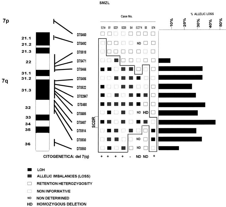
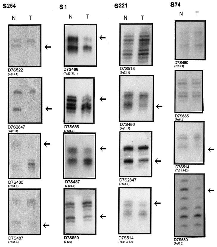
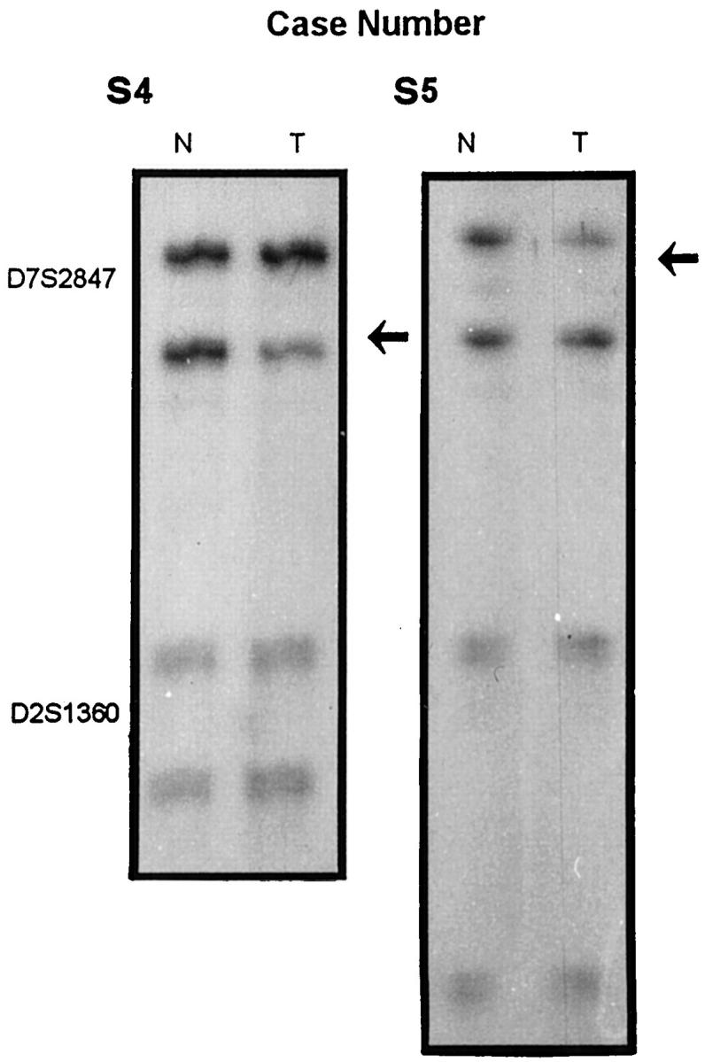
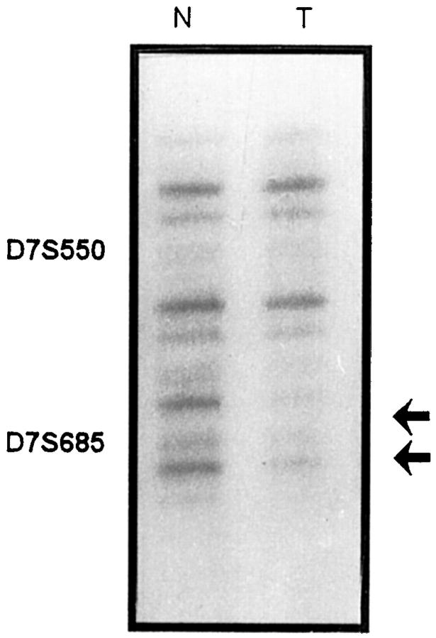
Splenic marginal zone lymphoma (SMZL) has been recognized as an entity defined on the basis of its morphological, phenotypic, and clinical characteristic features. Nevertheless, no characteristic genetic alterations have been described to date for this entity, thus making an exact diagnosis of SMZL difficult in some cases. As initial studies showed that chromosome region 7q22-32 is deleted in some of these cases, we analyzed a larger group of SMZL and other lymphoproliferative disorders that may partially overlap with it. To better define the frequency of 7q deletion in SMZL and further identify the deleted region, polymerase chain reaction analysis of 13 microsatellite loci spanning 7q21-7q36 was performed on 20 SMZL and 26 non-SMZL tissue samples. The frequency of allelic loss in SMZL (8/20; 40%) was higher than that observed in other B-cell lymphoproliferative syndromes (2/26; 7.7%). This difference was statistically significant (P < 0.05). The most frequently deleted microsatellite was D7S487 (5/11; 45% of informative cases). Surrounding this microsatellite the smallest common deleted region of 5cM has been identified, defined between D7S685 and D7S514. By comparative multiplex polymerase chain reaction analysis, we detected a homozygous deletion in the D7S685 (7q31.3) marker in one case. These results suggest that 7q31-q32 loss may be used as a genetic marker of this neoplasia, in conjunction with other morphologic, phenotypic, and clinical features. A correlation between 7q allelic loss and tumoral progression (death secondary to the tumor or large cell transformation) in SMZL showed a borderline statistical significance. The observation of a homozygous deletion in this chromosomal region may indicate that there is a tumor suppressor gene involved in the pathogenesis of this lymphoproliferative neoplasia.
Figures
References
-
- Harris NL, Jaffe NS, Stein H, Banks PM, Chan JKC, Cleary M, Delsol G, de Wolf-Peters C, Falini B, Gatter KC, Grogan TM, Isaacson PG, Knowles DM, Masson SY, Muller-Hermelink HK, Pileri SA, Piris MA, Ralfkiaer E, Warnke RA: A revised European-American classification of lymphoid neoplasms: a proposal from the International Lymphoma Study Group. Blood 1994, 84:1361-1392 - PubMed
-
- Mollejo M, Menarguez J, LLoret E, Sanchez A, Campo E, Algara P, Cristobal E, Sanchez E, Piris MA: Splenic marginal zone lymphoma, a distinctive type of low-grade B-cell lymphoma: a clinicopathological study of 13 cases. Am J Surg Pathol 1995, 19:1146-1157 - PubMed
-
- Isaacson PG, Piris MA: Splenic marginal zone lymphoma. Adv Anat Pathol 1997, 4:191-201
-
- Sole F, Woessner S, Florensa L, Espinet B, Mollejo M, Martinez P, Piris MA: Frequent involvement of chromosomes 1, 3, 7 and 8 in splenic marginal zone B-cell lymphoma. Br J Haematology 1998, 98:446-449 - PubMed
-
- Dierlam D, Pittaluga S, Wlodarska I, Stul M, Thomas J, Boogaerts M, Michaux L, Driessen A, Mecucci C, Cassiman J-J, De Wolf-Peeters, Van den Berghe H: Marginal zone B-cell lymphomas of different sites share similar cytogenetic an morphologic features. Blood 1996, 87:299–307 - PubMed
Publication types
MeSH terms
LinkOut - more resources
Full Text Sources
Other Literature Sources
Medical
Research Materials
