. 1999 May;154(5):1601-10.
doi: 10.1016/S0002-9440(10)65413-2.

HIP/PAP gene, encoding a C-type lectin overexpressed in primary liver cancer, is expressed in nervous system as well as in intestine and pancreas of the postimplantation mouse embryo

Affiliations

HIP/PAP gene, encoding a C-type lectin overexpressed in primary liver cancer, is expressed in nervous system as well as in intestine and pancreas of the postimplantation mouse embryo

C Lasserre et al. Am J Pathol. 1999 May.

Abstract

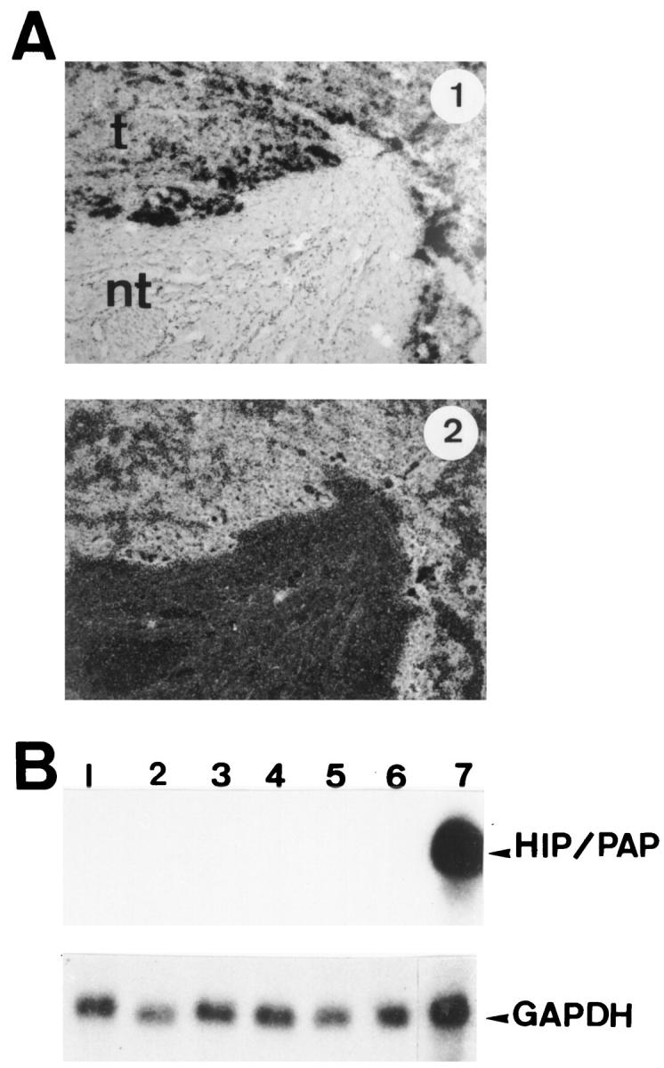
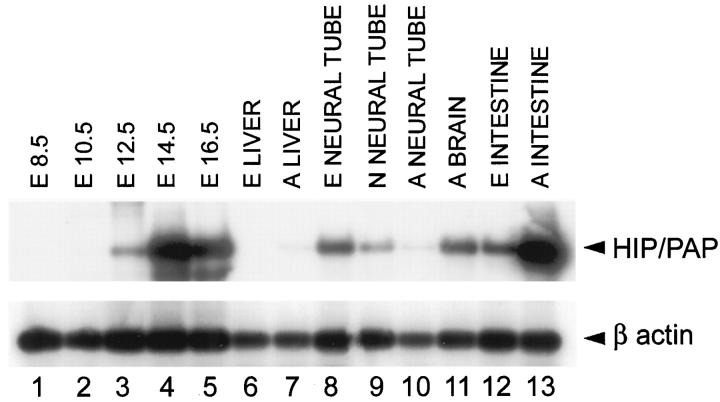
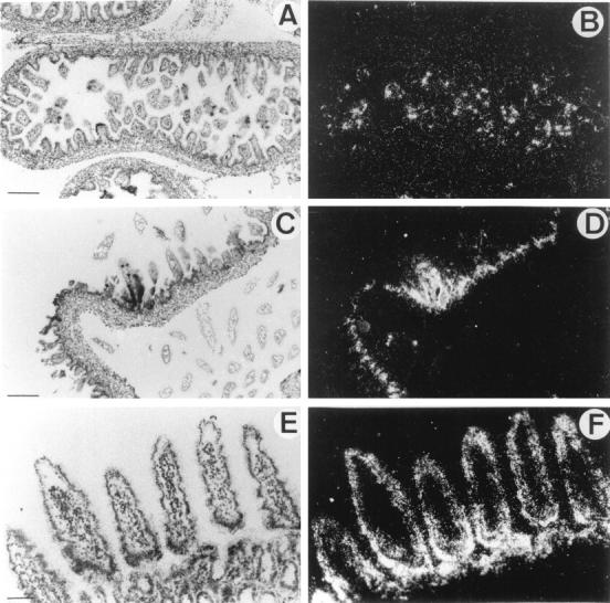
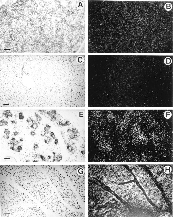
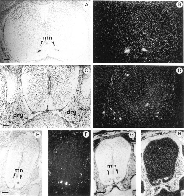
We originally isolated the HIP/PAP gene in a differential screen of a human hepatocellular carcinoma cDNA library. This gene is expressed at high levels in 25% of primary liver cancers but not in nontumorous liver. HIP/PAP belongs to the family of C-type lectins and acts as an adhesion molecule for hepatocytes. In normal adult human tissues, HIP/PAP expression is found in pancreas (exocrine and endocrine cells) and small intestine (Paneth and neuroendocrine cells). In order to gain insight into the possible role of HIP/PAP in vivo, we have investigated the pattern of HIP/PAP expression in the developing postimplantation mouse embryo by in situ hybridization. Detailed analysis of developing mouse embryos revealed that HIP/PAP gene exhibits a restricted expression pattern during development. Thus, HIP/PAP transcripts are first observed within the nervous system from day 14.5 onwards in trigeminal ganglia, dorsal root ganglia, and spinal cord where it appears to be an early specific marker of a subpopulation of motor neurons. At laster stages, HIP/PAP transcripts were detected in intestine and pancreas at day 16.5 but not in embryonic liver. This highly restricted expression pattern suggests that HIP/PAP might participate in neuronal as well as intestinal and pancreatic cell development.


Figures

Figure 1.
Figure 1.
Expression of HIP/PAP mRNA in human tissues. A: In situ hybridization of 35S-labeled antisense human HIP/PAP probe to sections of human hepatocarcinoma (HCC) under bright field and dark field illumination: t, tumor; nt, nontumoral tissue. Original magnification, ×75. Exposure time is 2 days. B: Northern blot analysis of human fetal liver mRNA. The probe was the full human HIP/PAP cDNA. Lanes 1 to 6: 10 μg of total RNA from human fetal liver (7 days exposure); lane 1: 21.5 weeks old; lane 2: 14.6 weeks old; lane 3: 11.3 weeks old; lane 4: 9.6 weeks old; lane 5: 10 weeks old; lane 6: 9 weeks old. Lane 7: human adult ileum, 20 μg of total RNA (1 day exposure). The same filter was stripped and reprobed with human glyceraldehyde-3-phosphate dehydrogenase (GAPDH) probe. For GAPDH hybridization (lower panel) exposure time is 1 day.
Figure 2.
Figure 2.
RT-PCR analysis of mouse HIP/PAP expression in embryonic and adult tissues. RNA extracted from total embryos (lanes 1 to 5) of 8.5 days old (E8.5), 10.5 days old (E10.5), 12.5 days old (E12.5), 14.5 days old (E14.5), and 16.5 days old (E16.5). RNA extracted from isolated mouse tissues (lanes 6 to 13) : liver from E16.5 embryo (E LIVER); adult liver (A LIVER); neural tube from E16.5 embryo (E NEURAL TUBE); neural tube from newborn mouse (N NEURAL TUBE); adult neural tube (A NEURAL TUBE); adult brain (A BRAIN); intestine from E16.5 embryo (E INTESTINE); adult small intestine (A INTESTINE). Reaction products were electrophoresed and an autoradiography is shown. Exposure times were 1 day for HIP/PAP and 1 hour for β actin.
Figure 3.
Figure 3.
In situ hybridization of 35S-labeled antisense mouse HIP/PAP probe to mouse intestine sections under bright-field (A,C,E) and dark field (B,D,F) illumination. A and B: Longitudinal section through small intestine of E16.5 mouse embryo. C and D: Section of mouse newborn intestine. E and F: Section of mouse adult jejunum. Scale bar, 250 μm (A,B,C,D); 100 μm (E,F).
Figure 4.
Figure 4.
In situ hybridization of 35S-labeled antisense mouse HIP/PAP probe to sections of liver and pancreas under bright field (A,C,E,G) and dark field (B,D,F,H) illumination. A and B: Section through liver of a E16.5 mouse embryo; C and D: Section of mouse adult liver; E and F: Section through pancreas of a E16.5 mouse embryo; G and H: Section of mouse adult pancreas (10-day exposure time). Note that the white specks seen in B are caused by the refringent membrane of erythrocytes and not to silver grains. Scale bar, 250 μm (A,B,C,D); 100 μm (E,F,G,H).
Figure 5.
Figure 5.
In situ hybridization of 35S-labeled antisense mouse HIP/PAP or mouse Galectin 1 probe to transverse sections through the spinal cord of E16.5 mouse embryo under bright field (A,C,E,G) and dark field (B,D,F,H) illumination. A and B: cervical region; C–H: lumbar region. For A–F the probe was mouse HIP/PAP, for G and H the probe was Galectin 1 cDNA (scale bars, 250 μm). mn, motor neuron. Note dorsal root ganglia (DRG) in C and D. DRG are aggregate of neuronal cell bodies that transmit somatic sensory information from periphery to spinal cord. Dorsal side towards the top, ventral side towards the bottom of the photographs.
Figure 6.
Figure 6.
In situ hybridization of 35S-labeled antisense mouse HIP/PAP probe to sections of E16.5 mouse embryo central nervous system under bright field (A,C,E) and dark field (B,D,F) illumination. A and B: Coronal section of the mouse head of a E16.5; C and D: Detail of the same coronal section of the mouse brain at E16.5. Note trigeminal ganglia (tr) that are aggregate of neuronal cell bodies of the primary sensory neurons of V cranial nerve; E and F: Longitudinal section through mouse brain at E16.5. mb, midbrain; lv, lateral ventricule; tv, third ventricule; tr, trigeminal ganglia; p, pituitary gland; tb, temporal bone; t, tongue; sc, spinal cord; mb, muscle blocks. Scale bar, 800 μm (A,B); 400 μm (C,D); 100 μm (E,F).

References

    1. Drickamer K, Taykor ME: Biology of animal lectins. Annu Rev Cell Biol 1993, 9:237-264 - PubMed
    1. Ochieng J, Warfield P: Galectin-3 binding potentials of mouse tumor EHS and human placental laminins. Biochem Biophys Res Commun 1995, 217:402-406 - PubMed
    1. Drickamer K: Two distinct classes of carbohydrate-recognition domain in animal lectins. J Biol Chem 1988, 263:9557-9560 - PubMed
    1. Lasserre C, Christa L, Simon M-T, Vernier P, Brechot C: A novel gene (HIP) activated in human primary liver cancer. Cancer Res 1992, 52:5089-5095 - PubMed
    1. Christa I, Felin M, Morali O, Simon M-T, Lasserre C, Brechot C, Seve A-P: The human HIP gene, overexpressed in primary liver cancer encodes a C-type carbohydrate binding protein with lactose binding activity. FEBS Lett 1994, 337:114-118 - PubMed