. 1999 May 21;274(21):14918-25.
doi: 10.1074/jbc.274.21.14918.

A human RNA viral cysteine proteinase that depends upon a unique Zn2+-binding finger connecting the two domains of a papain-like fold

Affiliations

A human RNA viral cysteine proteinase that depends upon a unique Zn2+-binding finger connecting the two domains of a papain-like fold

J Herold et al. J Biol Chem. .

Erratum in

  • J Biol Chem 1999 Jul 23;274(30):21490

Abstract

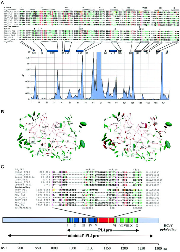
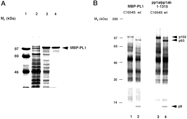
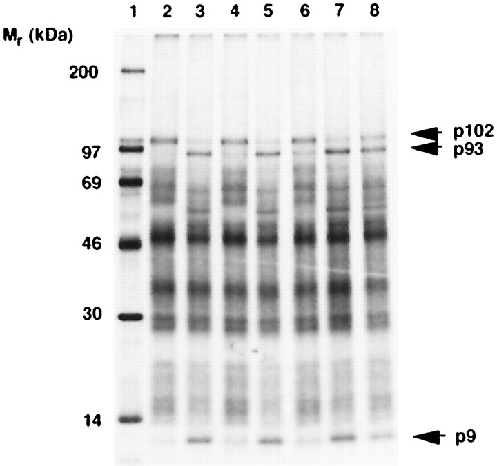
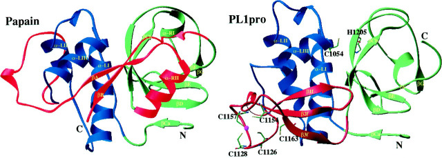
A cysteine proteinase, papain-like proteinase (PL1pro), of the human coronavirus 229E (HCoV) regulates the expression of the replicase polyproteins, pp1a and ppa1ab, by cleavage between Gly111 and Asn112, far upstream of its own catalytic residue Cys1054. In this report, using bioinformatics tools, we predict that, unlike its distant cellular homologues, HCoV PL1pro and its coronaviral relatives have a poorly conserved Zn2+ finger connecting the left and right hand domains of a papain-like fold. Optical emission spectrometry has been used to confirm the presence of Zn2+ in a purified and proteolytically active form of the HCoV PL1pro fused with the Escherichia coli maltose-binding protein. In denaturation/renaturation experiments using the recombinant protein, its activity was shown to be strongly dependent upon Zn2+, which could be partly substituted by Co2+ during renaturation. The reconstituted, Zn2+-containing PL1pro was not sensitive to 1,10-phenanthroline, and the Zn2+-depleted protein was not reactivated by adding Zn2+ after renaturation. Consistent with the proposed essential structural role of Zn2+, PL1pro was selectively inactivated by mutations in the Zn2+ finger, including replacements of any of four conserved Cys residues predicted to co-ordinate Zn2+. The unique domain organization of HCoV PL1pro provides a potential framework for regulatory processes and may be indicative of a nonproteolytic activity of this enzyme.


Figures

Figure 1
Figure 1
Fig. 1. Coronaviral and cellular PLpros: structural similarities and unique features.A, secondary structure-based sequence alignment of coronaviral and cellular PLpros. The primary structures of HCoV PL1pro and its coronaviral relatives (for accession numbers see C) were aligned using ClustalW program (43) in a stepwise manner and manually corrected with the ClustalX program (44) and the MACAW workbench (45). The main portion of this alignment is presented as 10 ungapped blocks. Only blocks II and III were statistically significant (p < 10−20 and p = 1.5−13, respectively), and blocks IV and VII, excluding MHV PL1pro, were conditionally significant, using a searching space between blocks III and VIII and between blocks IV and VIII, respectively. The validity of block VIII was previously confirmed by site-directed mutagenesis of conserved His for MHV and HCoV PL1pros and IBV PLpro (37, 41, 42). The secondary structures predicted by the PhD program are shown at the top (SS_coronaPL; A anda represent α-helix, and B and brepresent β-strand, predictions in capital letters have a reliability >5 and predictions inlowercase letters have a reliability of 5 and less (49)). The validity of this prediction was confirmed when similar secondary structure profiles were also returned for (i) the same alignment using the DSC program (50) and (ii) two automatically generated alignments containing either PL1pros or PL2pros encoded by HCoV and TGEV (not shown). The secondary structure profile of coronaviral PLpros was aligned with secondary structure elements conserved in the tertiary structure of 11 cellular PLpros (SS_celPL) (Protein Data Bank accession numbers: 1ppn, Papaya_Pap, papain (77);1gec, Papaya_Glep, glycyl endopeptidase (78); 1ppo, caricain (79);1yac, chymopapain (80); 1mem, cathepsin K (81); 1cjl, Human_CatL, cathepsin L (82); 1cte, Rat_Catb, cathepsin B (83); 2aim, trypanosoma cruzain (84); 2act, actinidin (85); 1gcb, yeast Gal6/bleomycin hydrolase (86)). The secondary structure alignment guided a sequence alignment of coronaviral and cellular proteases. A register of the alignment within each block was (arbitrarily) selected to maximize interfamily sequence similarity, although two or more poorly discriminated alignments were produced for all blocks except blocks II and VIII. When the three-dimensional structures of coronaviral PLpros become available, this alignment may need to be locally adjusted. For cellular PLpros, only a representative set of five sequences is shown. Coloring of the alignment of 12 sequences indicates the following:pink, invariant residues; red, residues conserved in >50% of the sequences; green, group of similar residues. The alignments of coronaviral and cellular PLpros highlight the active site residues of cellular proteases (66, 78). *, principal catalytic; +, “accessory” catalytic; 1, 2,3, and 4, substrate-binding pocket subsites S1, S2, S3, and S4, respectively; #, oxyanion hole-forming residue.Beneath the alignments, a plot displaying the positional structural variability (55) of cellular PLpros is shown.Above the plot, the positions of conserved secondary structure elements of cellular PLpros (66) as well as four conserved hydrogen-forming elements consisting of one residue (not marked) in the primary structure are displayed. Vertical axis, space variability at a position of the alignment; horizontal axis, numeration in the structural alignment containing only aligned residues. B, core structural residues of the cellular PLpros and residues conserved in cellular and coronaviral PLpros. Using the CORE package (55), a structural alignment of 11 cellular PLpros was converted into an average PL structure. It is characterized by the mean position of each C-α atom common in the family. The size of the ellipsoid around each of these atoms is proportional to the volume of atom variance. The two identical average PL structures, consisting of 178 atoms, are displayed in the “standard” papain orientation (66) featuring left-hand and right-hand domains as well as the interdomain active site cleft with the two catalytic residues of papain, Cys25 and His159. Conserved secondary structure elements of cellular PLpros are also marked. These structures are colored ingreen and red as follows. The left structure, the half of C-α atoms plus two atoms having the lowest space variance (91 atoms) are colored in red (core), and the remaining atoms are in green (noncore). The right structure, 109 atoms, whose residues were aligned with coronaviral PL residues in Fig. 1A, are shown in red (interfamily conserved residues), and the remaining atoms are in green. Note that the cellular PL core residues and the interfamily conserved residues are mainly from the same pool. C, A unique Zn2+finger connects the two domains of the PL fold of coronaviral PLpros. A region of the coronaviral PLpros between blocks V and VI was aligned as specified in Fig. 1A. Using the secondary structures predicted for the PLpros (SS_coronaPL) (50) and derived from the NMR structure (69) of the TFIIS Zn2+ ribbon (SS_TFI), an alignment of Zn2+ fingers of coronaviral PLpros and TFIIS was generated. The positions of these sequences in the corresponding proteins are given on the left, and accession numbers in the sequence data bases are shown on the right.Coloring of the alignment is as detailed for A. Residues involved in Zn2+ binding in TFIIS (69) are marked. A bar depicts the region of HCoV pp1a/pp1ab characterized in this study with the conserved blocks (Fig. 1A) shown. These blocks are organized in three groups colored differently.Blue, left-hand α-helix domain; green, right-hand β-sheet domain without counterparts of βA- and βB-strands; red, Zn2+ finger domain.Beneath the bar, the positions of the PL1pro domain, which is conserved among coronaviruses, and the HCoV minimal PL1pro domain determined by deletion analysis (41) are shown. The positions of mutations (Ref. and Table II) are depicted withyellow vertical lines in thebar and yellow amino acid background in the alignments in A and C.
Figure 2
Figure 2
Expression and purification of proteolytically activeHCoVPL1pro fused with the E. colimaltose-binding protein.A, purification of the fusion protein. MBP-PL1 was purified by affinity chromatography on amylose column from lysates of E. coli transformed with TB1[pMal-PL1] as described under “Experimental Procedures.” Aliquots taken from different stages of the purification were analyzed by 12.5% SDS-polyacrylamide gel electrophoresis. Lane 1, molecular mass markers; lane 2, noninduced bacterial lysate; lane 3, isopropyl-1-thio-β-d-galactopyranoside-induced bacterial lysate; lane 4, protein after amylose affinity chromatography. The position of MBP-PL1 is indicated. B, proteolytic activity of MBP-PL1. The trans-cleavage assay usingin vitro generated [35S]Met-labeled substrate was used to monitor proteolytic activity of purified MBP-PL1 (lanes 1 and 2) and in vitro generated, nonlabeled polypeptide containing PL1pro (pp1a/pp1ab-(1–1315)) and its mutated derivative (lanes 3 and 4). After immunoprecipitation of the cleavage reaction with IS 1720, proteins were separated by 10–17.5% gradient SDS-polyacrylamide gel electrophoresis, and labeled polypeptides were visualized by autoradiography. The positions of molecular mass markers, the substrate (p102), and cleavage products (p93 and p9) are indicated. The source of enzyme was as follows: MBP-PL1 C1054S (lane 1), MBP-PL1 (lane 2), in vitro produced PL1pro C1054S (lane 3), in vitro produced PL1pro (lane 4).
Figure 3
Figure 3
Effect of Zn2+ on the proteolytic activity of MBP-PL1. The trans-cleavage assay was used to monitor proteolytic activity of MBP-PL1 subjected to one or two cycles of denaturation in the presence of 8 m urea and renaturation in the presence of EDTA, ZnOAc, or CoOAc. The substrate and cleavage products are indicated as in Fig. 2. Lane 1, molecular markers. The source of enzyme was as follows: buffer A (lane 2), MBP-PL1 (not treated) (lane 3), denatured MBP-PL1 renatured in the presence of EDTA (apoenzyme) (lane 4), denatured MBP-PL1 renatured in the presence of ZnOAc (lane 5), denatured apoenzyme MBP-PL1 renatured in the presence of EDTA (lane 6), denatured apoenzyme MBP-PL1 renatured in the presence of ZnOAc (lane 7), denatured apoenzyme MBP-PL1 renatured in the presence of CoOAc (lane 8).
Figure 4
Figure 4
A crude structural model ofHCoVPL1pro.The PL1pro model (right; pp1a/1ab residues 1033–1242) was generated using the structures of papain (1ppn; shown left) and human TFIIS (1tfi) as templates for building the core and a loop library for constructing the variable regions by using Quanta 97 and Whatif 4.99. The model was partly refined but not minimized. Secondary structure was calculated according to Ref. and, for some positions in PL1pro, from the predicted secondary structure. The structures of both proteases are displayed (56) in the standard papain orientation (66) and split into three domains. These are coloredaccording to a scheme given in Fig. 1C as follows.Blue, left-hand α-helix domain; green, right-hand β-sheet domain without counterparts of βA- and βB-strands; red, Zn2+ finger domain in PL1pro and the interdomain loop along with βA- and βB-strands and α-RII-helix in papain. Cysteine residues of the Zn2+finger as well as the catalytic dyad residues of PL1pro that have been probed by site-directed mutagenesis (Table II) are shown in theball-and-stick model.

References

    1. Neurath H. Science. 1984;224:350–357. - PubMed
    1. Hartley B.S. Annu. Rev. Biochem. 1960;29:45–72. - PubMed
    1. Rawlings N.D., Barrett A.J. Biochem. J. 1993;290:205–218. - PMC - PubMed
    1. Gorbalenya A.E., Blinov V.M., Donchenko A.P. FEBS Lett. 1986;194:253–257. - PubMed
    1. Bazan J.F., Fletterick R.J. Proc. Natl. Acad. Sci. U. S. A. 1988;85:7872–7876. - PMC - PubMed

Publication types

LinkOut - more resources