. 1999 Jun;60(6):1429-36.
doi: 10.1095/biolreprod60.6.1429.

Pattern and kinetics of mouse donor spermatogonial stem cell colonization in recipient testes

Affiliations

Pattern and kinetics of mouse donor spermatogonial stem cell colonization in recipient testes

M Nagano et al. Biol Reprod. 1999 Jun.

Abstract

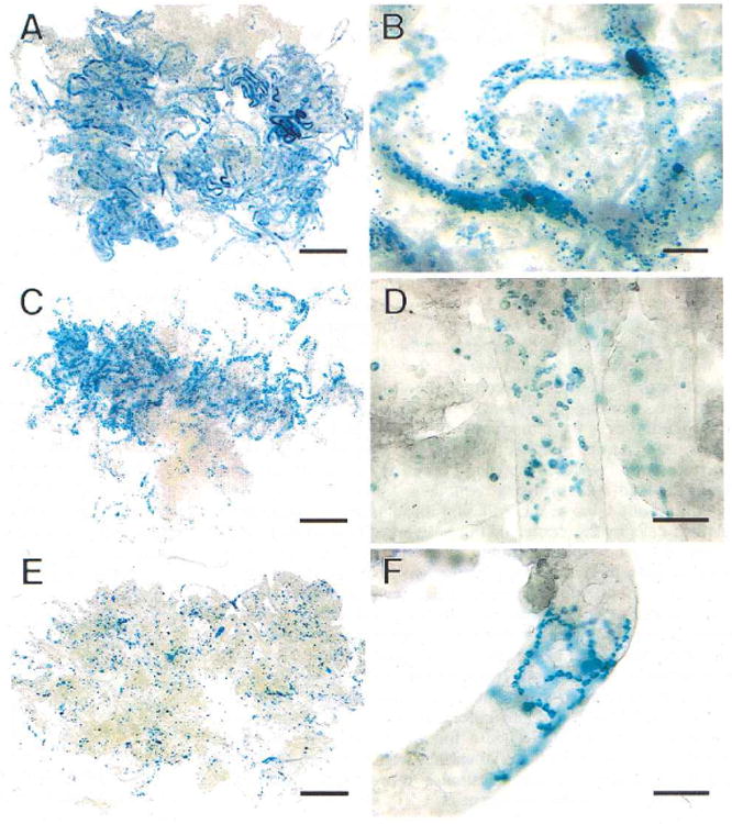
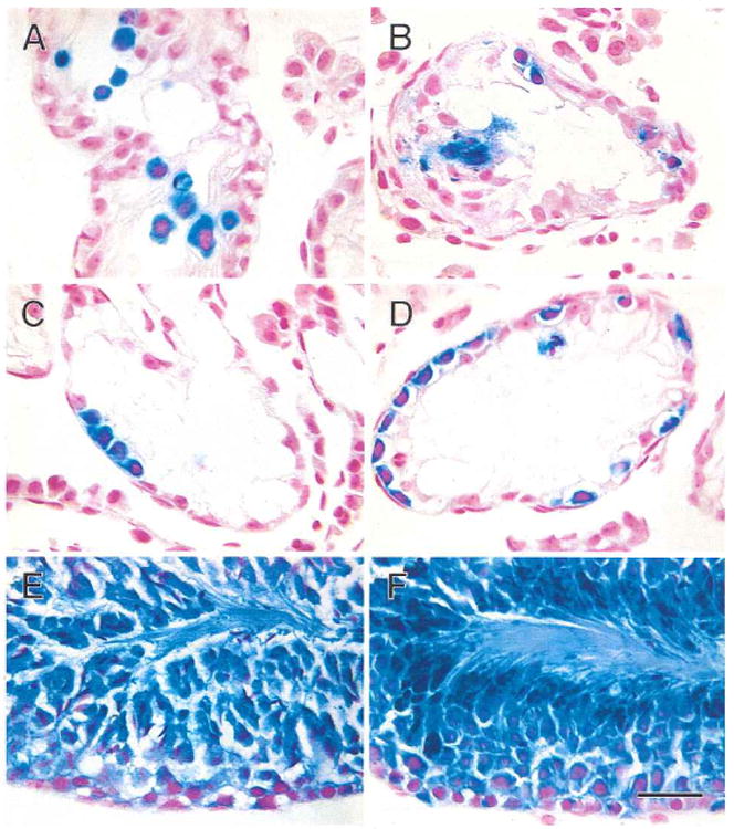
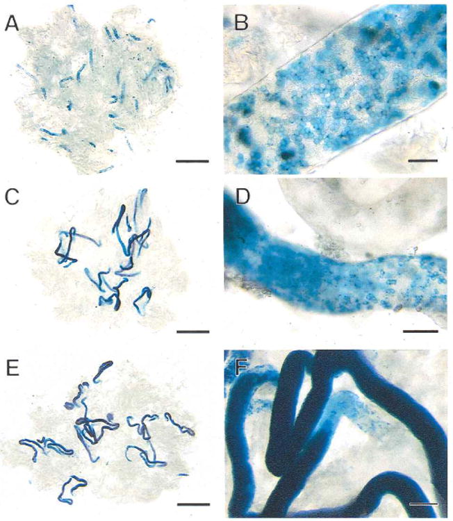
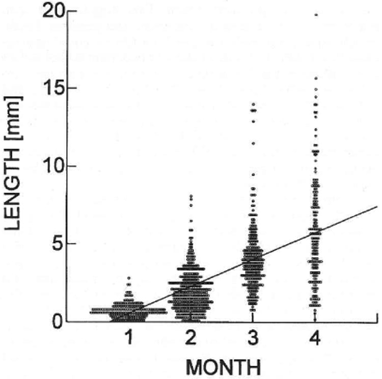
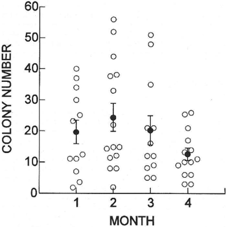
Recently a system was developed in which transplanted donor spermatogonial stem cells establish complete spermatogenesis in the testes of an infertile recipient. To obtain insight into stem cell activity and the behavior of donor germ cells, the pattern and kinetics of mouse spermatogonial colonization in recipient seminiferous tubules were analyzed during the 4 mo following transplantation. The colonization process can be divided into three continuous phases. First, during the initial week, transplanted cells were randomly distributed throughout the tubules, and a small number reached the basement membrane. Second, from 1 wk to 1 mo, donor cells on the basement membrane divided and formed a monolayer network. Third, beginning at about 1 mo and continuing throughout the observation period, cells in the center of the network differentiated extensively and established a colony of spermatogenesis, which expanded laterally by repeating phase two and then three. An average of 19 donor cell-derived colonies developed from 10(6) cells transplanted to the seminiferous tubules of a recipient testis; the number of colonized sites did not change between 1 and 4 mo. However, the length of the colonies increased from 0.73 to 5.78 mm between 1 and 4 mo. These experiments establish the feasibility of studying in a systematic and quantitative manner the pattern and kinetics of the colonization process. Using spermatogonial transplantation as a functional assay, it should be possible to assess the effects of various treatments on stem cells and on recipient seminiferous tubules to provide unique insight into the process of spermatogenesis.


Figures

Fig. 1
Fig. 1
Colonization of recipient mouse seminiferous tubules by transgenic donor testis cells (stained blue) at intervals up to 2 wk post-transplantation. A) One day after transplantation, blue donor cells are widely dispersed in recipient tubules. B) Cells are primarily single or in small groups at one day after transplantation. C) One week after transplantation, blue cells are still spread throughout tubules, but small clusters of cells can be seen. D) These small clusters of cells at 1 wk represent the beginning of chains of spermatogonia. E) Two weeks after transplantation, small groups of cells can be seen at low magnification. F) These groups of cells at 2 wk represent chains of cells rapidly spreading and branching on the basement membrane. Stain, X-gal. Scale bars are 2 mm in A, C, E; 200 μm in B; and 100 μm in D and F.
Fig. 2
Fig. 2
Transgenic donor cells (stained blue) in recipient mouse seminiferous tubules following transplantation. A) One day after transplantation, blue donor cells are widely scattered in the central area of the tubule. B) One week after transplantation, some donor cells have reached the basement membrane and are dividing. C) Two weeks after transplantation, blue donor cells on the basement membrane of the tubule are forming chains of spermatogonia. D) One month after transplantation, many blue cells are found on the basement membrane in the areas of colonization. E) Two months after transplantation, spermatogenesis has been established in the central areas of colonies. F) Three months after transplantation, spermatogenesis is well organized in the dark blue areas of the colonies. Stain, X-gal and nuclear fast red. Scale bar = 30 μm.
Fig. 3
Fig. 3
Colonization of recipient mouse seminiferous tubules by donor cells from the first to third month after transplantation. A) One month after transplantation, extensive blue areas are present in the tubules representing areas of germ cell colonization. B) These blue areas of colonization at 1 mo are composed of cellular chains and a network of cells on the basement membrane of the tubule. C) Two months after transplantation, the blue areas are dark in the center, indicating multiple layers of germ cells and development of spermatogenesis. D) The ends of the colonies at 2 mo consist of a network of cells on the basement membrane comparable to early stages of colonization. E) Three months after transplantation, the structure of the colonies is similar to that found at 2 mo, but each colony is longer. F) The intense blue color in the center of each colony at 3 mo represents complete spermatogenesis. The network of cells at the end of the colony, where colony extension is still continuing, is pale blue. Stain, X-gal. Scale bars are 2 mm in A, C, E; 50 μm in B; 100 μm in D; and 200 μm in F.
Fig. 4
Fig. 4
Length of donor cell-derived colonies of spermatogenesis in recipient seminiferous tubules. Each circle represents the measurement of an individual colony determined at the time indicated following cell transplantation. The average colony lengths in millimeters were 0.73 ± 0.03 (237), 2.05 ± 0.07 (367), 4.13 ± 0.14 (243), and 5.78 ± 0.27 (174) at 1, 2, 3, and 4 mo, respectively. Values are mean ± SEM (number of colonies measured). The increase in length at each month was significant (p < 0.001), and the regression line has the equation y = 1.73x − 1.188.
Fig. 5
Fig. 5
Number of donor cell-derived colonies of spermatogenesis in recipient testes. Open circles represent the number of colonies in a single testis determined at the time indicated after cell transplantation. The average numbers of colonies (filled circles) were 19.8 ± 3.9 (12), 24.5 ± 4.5 (15), 20.3 ± 4.6 (12), and 12.4 ± 2.0 (14) at 1, 2, 3, and 4 mo, respectively. Values are mean ± SEM (number of testes examined). There was no significant difference in the number of colonies present in recipient testes during the 4 mo of observation (ANOVA).

Similar articles

Cited by

References

    1. Potten CS. Cell lineages. In: McGee JO'D, Issacson PG, Wright NA, editors. Oxford Textbook of Pathology. Vol. 1. Oxford: Oxford University Press; 1992. pp. 43–52.
    1. Huckins C. The spcrmatogonial stem cell population in adult rats. I. Their morphology, proliferation, and maturation. Anat Rec. 1971;169:533–558. - PubMed
    1. Clermont Y. Kinetics of spermatogenesis in mammals: seminiferous epithelium cycle and spermatogonial renewal. Physiol Rev. 1972;52:198–236. - PubMed
    1. Russell LD, Ettlin RA, Sinha Hikim AP, Clcgg ED. Histological and Histopathological Evaluation of the Testis. Clearwater, FL: Cache River Press; 1990. pp. 1–40.
    1. Dym M. The male reproductive system. In: Weiss L, editor. Histology: Cell and Tissue Biology. 5th. New York: Elsevier Science Publishing Co. Inc.; 1983. pp. 1000–1053.

Publication types