Comparative Study
. 1999 May;9(5):437-48.

Genomic structure and comparative analysis of nine Fugu genes: conservation of synteny with human chromosome Xp22.2-p22.1

Affiliations
Comparative Study

Genomic structure and comparative analysis of nine Fugu genes: conservation of synteny with human chromosome Xp22.2-p22.1

B Brunner et al. Genome Res. 1999 May.

Abstract

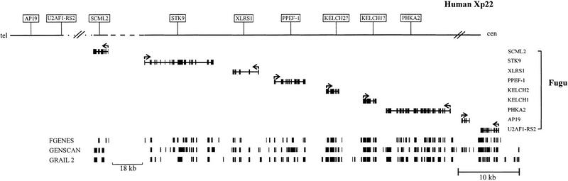
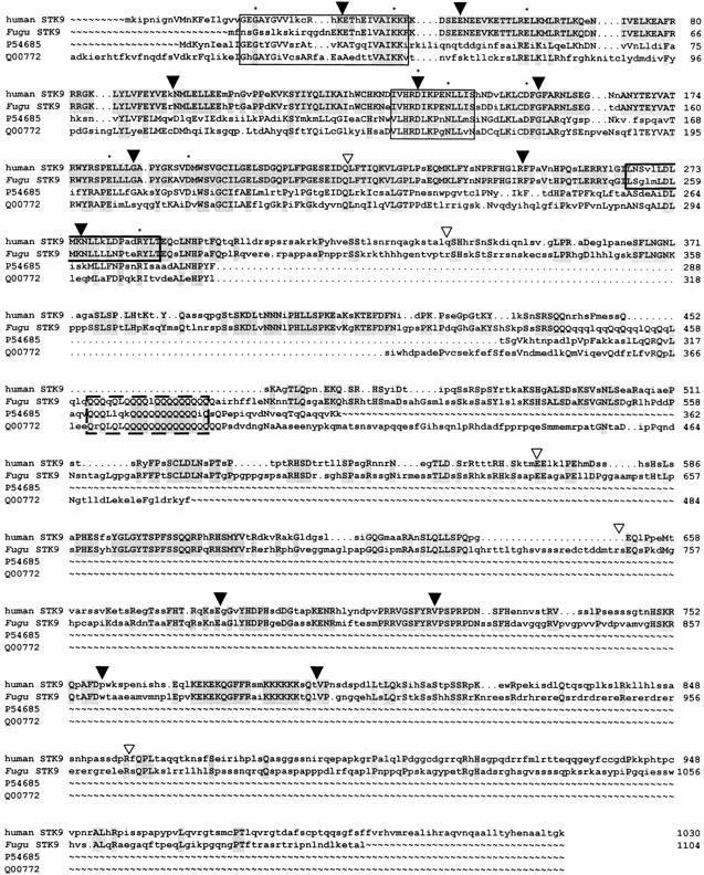
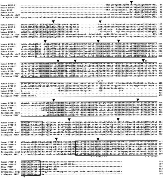
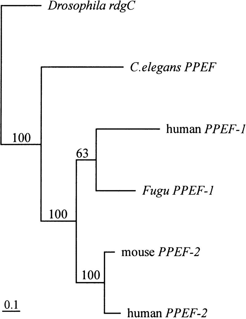
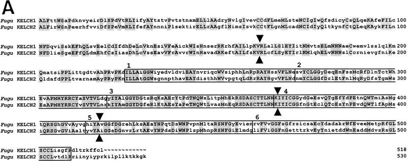
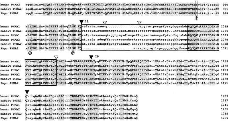
The pufferfish Fugu rubripes has a compact 400-Mb genome that is approximately 7.5 times smaller than the human genome but contains a similar number of genes. Focusing on the distal short arm of the human X chromosome, we have studied the evolutionary conservation of gene orders in Fugu and man. Sequencing of 68 kb of Fugu genomic DNA identified nine genes in the following order: (SCML2)-STK9, XLRS1, PPEF-1, KELCH2, KELCH1, PHKA2, AP19, and U2AF1-RS2. Apart from an evolutionary inversion separating AP19 and U2AF1-RS2 from PHKA2, gene orders are identical in Fugu and man, and all nine human homologs map to the Xp22 band. All Fugu genes were found to be smaller than their human counterparts, but gene structures were mostly identical. These data suggest that genomic sequencing in Fugu is a powerful and economical strategy to predict gene orders in the human genome and to elucidate the structure of human genes.


Figures

Figure 1
Figure 1
Scale representation of the Fugu genomic region encompassing 80 kb compared with the equivalent genomic region of human Xp22 (not drawn to scale). In total, 68 kb of Fugu genomic DNA have been sequenced, of which 62 kb are contiguous. (Broken horizontal line) The region not completely sequenced in both Fugu and humans, which, in Fugu, spans ∼18 kb. (Dotted line) A distance of ∼1 Mb in human DNA. (Cross-lines) The borders of conserved gene order. The complete coding region of each Fugu gene is represented by aligned solid boxes and the transcriptional direction is indicated by the arrow above each gene. The results of the exon prediction programs FGENES, GENSCAN, and GRAIL 2 are shown (bottom), with each predicted exon shown by a solid box.
Figure 2
Figure 2
Multiple alignment of the human, Fugu, Dictyostelium (P54685), and yeast (Q00772) STK9 proteins. The alignment was performed with the GCG programs PILEUP and LINEUP. Amino acids identical in at least two proteins are shown in uppercase letters, shading indicates residues identical in Fugu and human and in all proteins, respectively. (▾) Intron/exon boundaries identical in Fugu and human STK9; (▿) splice sites only present in Fugu. The protein kinase ATP-binding region is presented by the first box. The second box indicates the predicted serine-threonine protein kinase active site. The predicted leucine zipper is boxed in bold and the polyglutamine stretch in broken lines. (Asterisks) Amino acid residues invariant in 60 members of the eukaryotic protein kinase superfamily (Hanks and Hunter 1995).
Figure 3
Figure 3
Alignment of Fugu and C. elegans PPEF, Drosophila RdgC, mouse PPEF-2, and human PPEF-1 and PPEF-2 proteins. The alignment was performed with the GCG programs PILEUP and PRETTY. Amino acids at least identical in two aligned sequences are shown in uppercase letters. Residues identical in Fugu and human PPEF-1 and in all proteins, respectively, are shaded. (▾) Identical intron/exon boundaries of Fugu PPEF-1 and human PPEF-1; (▿) Position of the putative additional splice site in Fugu. Fugu exons 10–11 are numbered. The catalytic domain and the EF hand motif of the PPEF proteins are boxed. The two positions of the Ca2+ chelating side chains are labeled X–Z.
Figure 4
Figure 4
Phylogenetic tree of the PPEF subfamily. The unrooted tree was generated from aligned nucleotide sequences by the DNAML program of the PHYLIP software package. Regions of ambiguous alignment were excluded from the analysis. Scale bar, 0.1 expected substitutions per nucleotide site. Bootstrap values were generated from 100 bootstrap replicates of the alignment and are shown. Fugu PPEF-1 and human PPEF-1 cluster in the same branch; human and mouse PPEF-2 cluster in a second branch.
Figure 5
Figure 5
(A) Alignment of Fugu KELCH1 and KELCH2 proteins. The alignment was performed with the GCG programs PILEUP and PRETTY. Residues identical at each position are shown in uppercase letters and shaded. Intron/exon boundaries are shown by closed arrowheads on either site of the alignment. Each repeated segment is boxed and the beginning of each repeat is indicated by its respective number. (B) Alignment of the six repeated segments of Fugu KELCH1 and KELCH2 proteins. The alignments were performed with the GCG programs PILEUP and PRETTY. Amino acid residues at least identical in three repeats are shaded and shown in the consensus sequence. Dashes were introduced at less conserved positions.
Figure 5
Figure 5
(A) Alignment of Fugu KELCH1 and KELCH2 proteins. The alignment was performed with the GCG programs PILEUP and PRETTY. Residues identical at each position are shown in uppercase letters and shaded. Intron/exon boundaries are shown by closed arrowheads on either site of the alignment. Each repeated segment is boxed and the beginning of each repeat is indicated by its respective number. (B) Alignment of the six repeated segments of Fugu KELCH1 and KELCH2 proteins. The alignments were performed with the GCG programs PILEUP and PRETTY. Amino acid residues at least identical in three repeats are shaded and shown in the consensus sequence. Dashes were introduced at less conserved positions.
Figure 6
Figure 6
Alignment of Fugu, rabbit, and human PHKA2 and mouse, rabbit, and human PHKA1 proteins. The alignment was performed with the GCG programs PILEUP and PRETTY. Residues identical at each position are shown in uppercase letters and are shaded. (▾) Intron/exon boundaries of Fugu PHKA2. Fugu exons 18–29 are numbered. (▿) Known positions of splice sites in the human PHKA2 gene. The PHKA muscle and liver isoforms have two subunit-specific domains (boxed) with comparatively low amino acid sequence similarities of <50%. These domains encompass amino acids 612–788 and amino acids 1027–1055 in Fugu with a negatively charged cluster spanning amino acids 620–645. The hypothetical 5′ and 3′ calmodulin binding sites of all Phk α subunits are highly conserved (boxed in bold) and encompass amino acids 828–855 and amino acids 1056–1087 in Fugu. Three autophosphorylation sites, serine 987, serine 1001, and serine 1022, labeled by an P underneath the alignment and their vicinity are conserved.
Figure 6
Figure 6
Alignment of Fugu, rabbit, and human PHKA2 and mouse, rabbit, and human PHKA1 proteins. The alignment was performed with the GCG programs PILEUP and PRETTY. Residues identical at each position are shown in uppercase letters and are shaded. (▾) Intron/exon boundaries of Fugu PHKA2. Fugu exons 18–29 are numbered. (▿) Known positions of splice sites in the human PHKA2 gene. The PHKA muscle and liver isoforms have two subunit-specific domains (boxed) with comparatively low amino acid sequence similarities of <50%. These domains encompass amino acids 612–788 and amino acids 1027–1055 in Fugu with a negatively charged cluster spanning amino acids 620–645. The hypothetical 5′ and 3′ calmodulin binding sites of all Phk α subunits are highly conserved (boxed in bold) and encompass amino acids 828–855 and amino acids 1056–1087 in Fugu. Three autophosphorylation sites, serine 987, serine 1001, and serine 1022, labeled by an P underneath the alignment and their vicinity are conserved.

Similar articles

Cited by

References

    1. Aparicio S, Brenner S. How good a model is the Fugu genome. Nature. 1997;387:140. - PubMed
    1. Armes N, Gilley J, Fried M. The comparative genomic structure and sequence of the surfeit gene homologs in the puffer fish Fugu rubripes and their association with CpG-rich islands. Genome Res. 1997;7:138–152. - PubMed
    1. Baxendale S, Abdulla S, Elgar G, Buck D, Berks M, Micklem G, Durbin R, Bates G, Brenner S, Beck S. Comparative sequence analysis of the human and pufferfish Huntington’s disease genes. Nat Genet. 1995;10:67–76. - PubMed
    1. Birney E, Thompson JD, Gibson TJ. Pairwise and searchwise—Finding the optimal alignment in a simultaneous comparison of a protein profile against all DNA translation frames. Nucleic Acids Res. 1996;24:2730–2739. - PMC - PubMed
    1. Bork P, Doolittle RF. Drosophila kelch motif is derived from a common enzyme fold. J Mol Biol. 1994;236:1277–1282. - PubMed

Publication types

Associated data

LinkOut - more resources