Comparative Study
. 1999 Jun;19(6):3940-50.
doi: 10.1128/MCB.19.6.3940.

Dimeric RFX proteins contribute to the activity and lineage specificity of the interleukin-5 receptor alpha promoter through activation and repression domains

Affiliations
Comparative Study

Dimeric RFX proteins contribute to the activity and lineage specificity of the interleukin-5 receptor alpha promoter through activation and repression domains

A Iwama et al. Mol Cell Biol. 1999 Jun.

Abstract

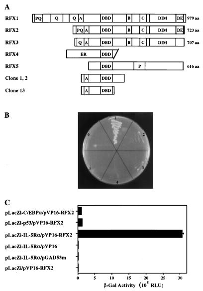
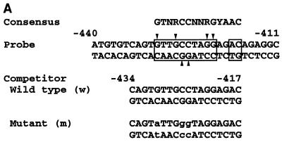
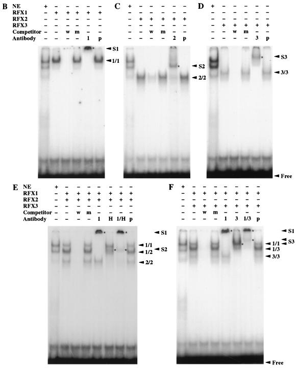
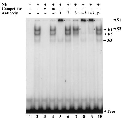
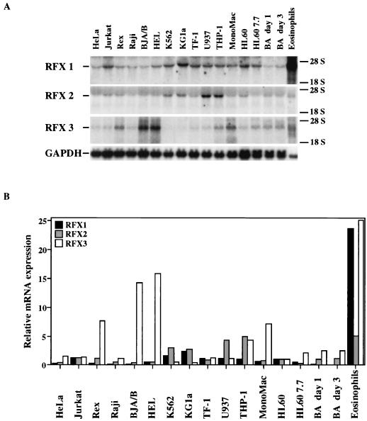
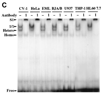
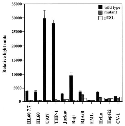
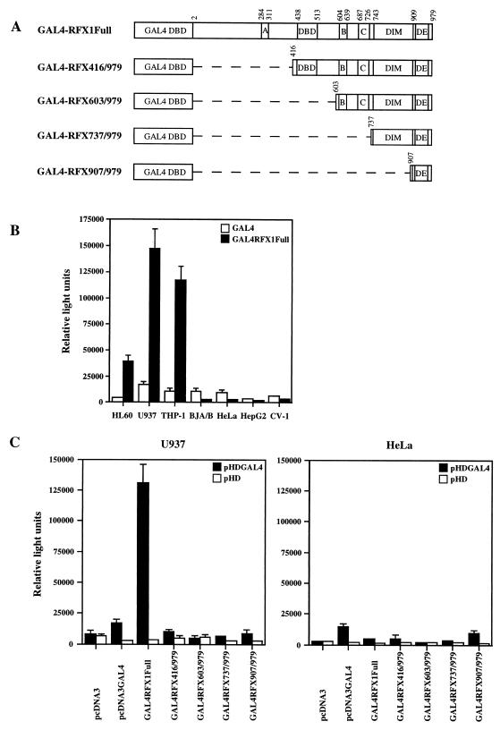
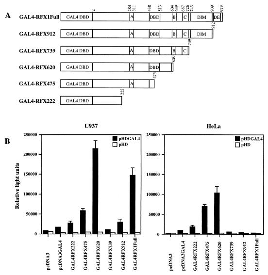
Interleukin-5 (IL-5) plays a central role in the differentiation, proliferation, and functional activation of eosinophils. The specific action of IL-5 on eosinophils and hematopoietically related basophils is regulated by the restricted expression of IL-5 receptor alpha (IL-5Ralpha), a subunit of high-affinity IL-5R, on these cells. We have previously identified an enhancer-like cis element in the IL-5Ralpha promoter that is important for both full promoter function and lineage-specific activity. Here, we demonstrate by yeast one-hybrid screening that RFX2 protein specifically binds to this cis element. RFX2 belongs to the RFX DNA-binding protein family, the biological role of which remains obscure. Using an electrophoretic mobility shift assay, we further show that RFX1, RFX2, and RFX3 homodimers and heterodimers specifically bind to the cis element of the IL-5Ralpha promoter. The mRNA expression of RFX1, RFX2, and RFX3 was detected ubiquitously, but in transient-transfection assays, multimerized RFX binding sites in front of a basal promoter efficiently functioned in a tissue- and lineage-specific manner. To further investigate RFX functions on transcription, full-length and deletion mutants of RFX1 were targeted to DNA through fusion to the GAL4 DNA binding domain. Tissue- and lineage-specific transcriptional activation with the full-length RFX1 fusion plasmid on a reporter controlled by GAL4 binding sites was observed. Distinct activation and repression domains within the RFX1 protein were further mapped. Our findings suggest that RFX proteins are transcription factors that contribute to the activity and lineage specificity of the IL-5Ralpha promoter by directly binding to a target cis element and cooperating with other tissue- and lineage-specific cofactors.


Figures

FIG. 1
FIG. 1
Binding of RFX2 to the cis element of the IL-5Rα promoter in yeast. (A) Schematic representation of RFX1 to -5 (7, 28) and three RFX2 clones isolated from yeast one-hybrid screening. The DNA binding domain (DBD), dimerization domain (DIM), conserved regions A, B, and C, and estrogen receptor (ER) regions rich in proline (P), glutamine (Q), or acidic amino acids (DE) are indicated (7, 28). (B) Reporter yeast strains grown on Sabouraud dextrose plates without histidine and with 5 mM 3-amino-1,2,4-triazole at 30°C for 3 days. The reporter strain carrying pHISi-1-IL-5Rα was transformed with pVP16-RFX2 clone 1 (section 1), pVP16 (section 4), and pGAD53m (section 5). At the same time, negative-control reporter strains carrying pHISi-1-C/EBPα (section 2), pHISi-1-p53 (section 3), and pHISi-1 (section 6) were transformed with pVP16-RFX2 clone 1. (C) Binding of RFX2 to the cis element of the IL-5Rα promoter detected by liquid β-galactosidase assay. Bars depict β-galactosidase (β-Gal) reporter gene activity in yeast extracts from the reporter strains. The reporter strain carrying pLacZi-IL-5Rα was transformed with pVP16-RFX2 clone 1, pVP16, and pGAD53m. Negative-control reporter strains carrying pLacZi-C/EBPα, pLacZi-p53, and pLacZi were also transformed with pVP16-RFX2 clone 1. The results are shown as the means ± the standard deviations for three transformants.
FIG. 2
FIG. 2
Binding of in vitro-synthesized RFX1, RFX2, and RFX3 to the cis element of the IL-5Rα promoter in EMSA. (A) The probe used in the gel shift assay is the cis element of the IL-5Rα promoter, bp −440 to −411 (37), containing a centrally located RFX binding site (8). The boxes enclose nucleotides that match the consensus sequence. R, Y, and N represent a purine, a pyrimidine, and any nucleotide, respectively. The mutant competitor was designed to contain mutations (in lowercase) of three of the six G residues on which DNA methylation interfered with nuclear factor binding in our previous study (arrows) (38). (B through F) RFX1 (B), RFX2 (C), and RFX3 (D) were individually synthesized in vitro, and RFX1 was cosynthesized with HA-tagged RFX-2 (E) or RFX3 (F); RFX-DNA complexes were analyzed by EMSA. For comparison, RFX-DNA complexes formed with AML14 nuclear extracts (NE) were loaded in the first lanes. The legends above the autoradiographs indicate the source of the in vitro translated protein, competitor, and antibody used. Antibodies 1, 2, 3, H, and p denote antibodies against RFX1, RFX2, RFX3, and HA and preimmune serum, respectively. Positions of free DNA probe (Free), RFX1 homodimers (1/1), RFX2 homodimers (2/2), RFX3 homodimers (3/3), RFX1-RFX2 heterodimers (1/2), RFX1-RFX3 heterodimers (1/3), all supershifted complexes (*), and complexes supershifted by the RFX1 antiserum (S1), the RFX3 antiserum (S3), or the RFX2 antiserum and anti-HA antibody (S2) are indicated.
FIG. 2
FIG. 2
Binding of in vitro-synthesized RFX1, RFX2, and RFX3 to the cis element of the IL-5Rα promoter in EMSA. (A) The probe used in the gel shift assay is the cis element of the IL-5Rα promoter, bp −440 to −411 (37), containing a centrally located RFX binding site (8). The boxes enclose nucleotides that match the consensus sequence. R, Y, and N represent a purine, a pyrimidine, and any nucleotide, respectively. The mutant competitor was designed to contain mutations (in lowercase) of three of the six G residues on which DNA methylation interfered with nuclear factor binding in our previous study (arrows) (38). (B through F) RFX1 (B), RFX2 (C), and RFX3 (D) were individually synthesized in vitro, and RFX1 was cosynthesized with HA-tagged RFX-2 (E) or RFX3 (F); RFX-DNA complexes were analyzed by EMSA. For comparison, RFX-DNA complexes formed with AML14 nuclear extracts (NE) were loaded in the first lanes. The legends above the autoradiographs indicate the source of the in vitro translated protein, competitor, and antibody used. Antibodies 1, 2, 3, H, and p denote antibodies against RFX1, RFX2, RFX3, and HA and preimmune serum, respectively. Positions of free DNA probe (Free), RFX1 homodimers (1/1), RFX2 homodimers (2/2), RFX3 homodimers (3/3), RFX1-RFX2 heterodimers (1/2), RFX1-RFX3 heterodimers (1/3), all supershifted complexes (*), and complexes supershifted by the RFX1 antiserum (S1), the RFX3 antiserum (S3), or the RFX2 antiserum and anti-HA antibody (S2) are indicated.
FIG. 3
FIG. 3
Binding of native RFX protein complexes in a myeloid nuclear extract to the cis element of the IL-5Rα promoter in EMSA. RFX-DNA complexes were formed by using an AML14 nuclear extract and were detected by EMSA. The probe and competitors were the same as for Fig. 2. The legends above the autoradiograph indicate the source of competitor and antibody used. Abbreviations are defined in the legend to Fig. 2.
FIG. 4
FIG. 4
Expression of RFX1, RFX2, and RFX3 in human cells. (A) Northern blot analysis of the mRNA of RFX1, RFX2, and RFX3 in human hematopoietic cells. Total RNAs from human cell lines (15 μg) and primary eosinophils (2 μg) were loaded and probed with cDNA fragments specific for human RFX1, RFX2, RFX3, or GAPDH. (B) Relative expression levels of RFX1, RFX2, and RFX3 in human cell lines and primary eosinophils. The levels of RFX mRNAs in each cell line were normalized to the levels of GAPDH mRNA and then measured relative to the levels in HL-60 cells. Evaluation of mRNA levels are consistent for each gene but cannot be compared among genes. (C) Binding of native RFX complexes in nuclear extracts from various human cell lines to the cis element of the IL-5Rα promoter in EMSA. RFX-DNA complexes were formed in crude nuclear extracts and were detected by EMSA. The probe and competitors were the same as for Fig. 2. Positions of RFX1 homodimers (1/1), homodimers of RFX2 or RFX3 (Homo), heterodimers of RFX1 and RFX2 or RFX3 (Hetero), and complexes supershifted by RFX1 antiserum (S1) are indicated. Free, free DNA probe.
FIG. 4
FIG. 4
Expression of RFX1, RFX2, and RFX3 in human cells. (A) Northern blot analysis of the mRNA of RFX1, RFX2, and RFX3 in human hematopoietic cells. Total RNAs from human cell lines (15 μg) and primary eosinophils (2 μg) were loaded and probed with cDNA fragments specific for human RFX1, RFX2, RFX3, or GAPDH. (B) Relative expression levels of RFX1, RFX2, and RFX3 in human cell lines and primary eosinophils. The levels of RFX mRNAs in each cell line were normalized to the levels of GAPDH mRNA and then measured relative to the levels in HL-60 cells. Evaluation of mRNA levels are consistent for each gene but cannot be compared among genes. (C) Binding of native RFX complexes in nuclear extracts from various human cell lines to the cis element of the IL-5Rα promoter in EMSA. RFX-DNA complexes were formed in crude nuclear extracts and were detected by EMSA. The probe and competitors were the same as for Fig. 2. Positions of RFX1 homodimers (1/1), homodimers of RFX2 or RFX3 (Homo), heterodimers of RFX1 and RFX2 or RFX3 (Hetero), and complexes supershifted by RFX1 antiserum (S1) are indicated. Free, free DNA probe.
FIG. 5
FIG. 5
Tissue- and lineage-specific enhancer activity of multimerized RFX binding sites. Trimerized RFX binding sites from the IL-5Rα promoter (bp −434 to −417), containing either wild-type or mutant sequences, were subcloned into the pT81-luc vectors in front of basal thymidine kinase promoters. The reporter plasmids and the parent pT81-luc were transiently transfected into various hematopoietic and nonhematopoietic cell lines. The minimum promoter activity of pT81-luc was approximately 300 RLU, while the background luciferase activity was below 100 RLU in all cells analyzed.
FIG. 6
FIG. 6
Tissue- and lineage-specific transactivation by GAL4-RFX1 fusion proteins. (A) Schematic representation of RFX1 and its 5′ deletions fused to the GAL4 DNA binding domain (DBD). The numbers correspond to amino acid residues of each domain and the deletion sites (dashed lines). DE, acidic amino acids; DIM, dimerization domain. (B) A reporter plasmid containing multimerized GAL4 binding sites (pHDGAL4 luciferase) or its parent pHD luciferase plasmid was transiently cotransfected with either pcDNA3 containing the GAL4 DNA binding domain only (GAL4) or full-length RFX1 fused to the GAL4 DNA binding domain (GAL4-RFX1Full) into various cell lines. The lowest luciferase activity was obtained in HepG2 (2,360 RLU), while the background luciferase activity was below 100 RLU in all cells analyzed. (C) Mapping of RFX1 regions that activate transcription. A series of 5′ deletion mutants of RFX1 fused to the GAL4 DNA binding domain were cotransfected with the reporter plasmid into U937 and HeLa cells. The minimum promoter activity of pHD luciferase plasmid was 6,053 RLU in U937 cells and 1,556 RLU in HeLa cells, while the background luciferase activity was 42 RLU in U937 cells and 85 RLU in HeLa cells.
FIG. 7
FIG. 7
Localization of the activation and inhibitory domains of RFX1. (A) Schematic representation of RFX1 and its 3′ deletions fused to the GAL4 DNA binding domain (DBD). Numbers correspond to amino acid residues of each domain; DIM, dimerization domain; DE, acidic amino acids. (B) A series of 3′ deletion mutants of RFX1 fused to the GAL4 DNA binding domain were cotransfected with the reporter plasmid into U937 and HeLa cells. The minimum promoter activity of pHD luciferase plasmid was 6,053 RLU in U937 cells and 1,556 RLU in HeLa cells, while the background luciferase activity was 42 RLU in U937 cells and 85 RLU in HeLa cells.

Similar articles

Cited by

References

    1. Ayer D E, Laherty C D, Lawrence Q A, Armstrong A P, Eisenman R N. Mad proteins contain a dominant transcription repression domain. Mol Cell Biol. 1996;16:5772–5781. - PMC - PubMed
    1. Baltus B, van Dijk T B, Caldenhoven E, Zanders E, Raaijmakers J A M, Lammers J-W J, Koenderman L, de Groot R P. An AP-1 site in the promoter of the human IL-5Rα gene is necessary for promoter activity in eosinophilic HL60 cells. FEBS Lett. 1998;434:251–254. - PubMed
    1. Chen H M, Zhang P, Voso M T, Hohaus S, Gonzalez D A, Christopher K G, Zhang D-E, Tenen D G. Neutrophils and monocytes express high levels of PU.1 (Spi-1) but not Spi-B. Blood. 1995;85:2918–2928. - PubMed
    1. Chirgwin J M, Przybyla A E, MacDonald R J, Rutter W J. Isolation of biologically active ribonucleic acid from sources rich in ribonuclease. Biochemistry. 1979;18:5294–5299. - PubMed
    1. Clausen B E, Waldburger J-M, Schwenk F, Barras E, Mach B, Rajewsky K, Förster I, Reith W. Residual MHC class II expression on mature dendritic cells and activated B cells in RFX5-deficient mice. Immunity. 1998;8:143–155. - PubMed

Publication types

MeSH terms

LinkOut - more resources