. 1999 Jun;19(6):4056-64.
doi: 10.1128/MCB.19.6.4056.

Regulation of AUF1 expression via conserved alternatively spliced elements in the 3' untranslated region

Affiliations

Regulation of AUF1 expression via conserved alternatively spliced elements in the 3' untranslated region

G M Wilson et al. Mol Cell Biol. 1999 Jun.

Abstract

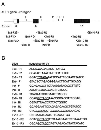
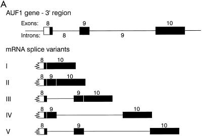
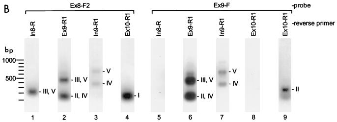
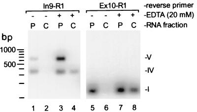
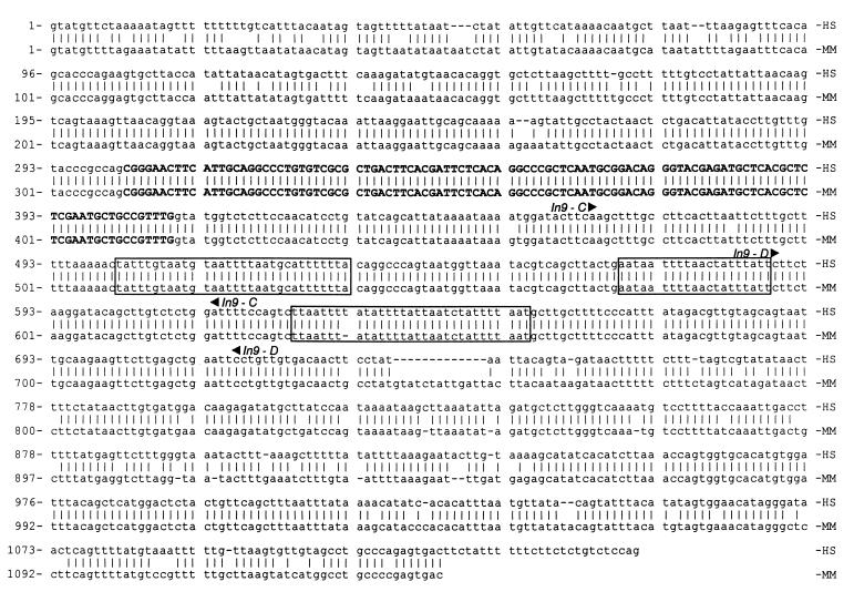
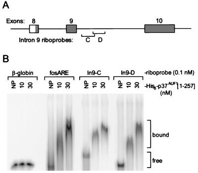
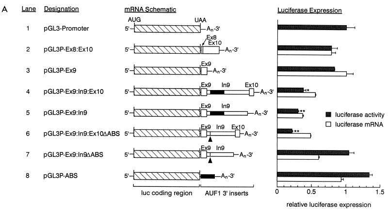
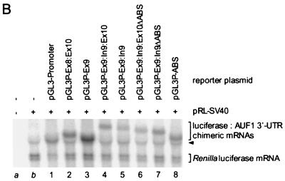
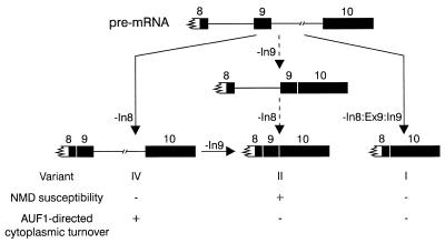
The A+U-rich RNA-binding factor AUF1 exhibits characteristics of a trans-acting factor contributing to the rapid turnover of many cellular mRNAs. Structural mapping of the AUF1 gene and its transcribed mRNA has revealed alternative splicing events within the 3' untranslated region (3'-UTR). In K562 erythroleukemia cells, we have identified four alternatively spliced AUF1 3'-UTR variants, including a population of AUF1 mRNA containing a highly conserved 107-nucleotide (nt) 3'-UTR exon (exon 9) and the adjacent downstream intron (intron 9). Functional analyses using luciferase-AUF1 3'-UTR chimeric transcripts demonstrated that the presence of either a spliceable or an unspliceable intron 9 in the 3'-UTR repressed luciferase expression in cis, indicating that intron 9 sequences may down-regulate gene expression by two distinct mechanisms. In the case of the unspliceable intron, repression of luciferase expression likely involved two AUF1-binding sequences, since luciferase expression was increased by deletion of these sites. However, inclusion of the spliceable intron in the luciferase 3'-UTR down-regulated expression independent of the AUF1-binding sequences. This is likely due to nonsense-mediated mRNA decay (NMD) owing to the generation of exon-exon junctions more than 50 nt downstream of the luciferase termination codon. AUF1 mRNA splice variants generated by selective excision of intron 9 are thus also likely to be subject to NMD since intron 9 is always positioned >137 nt downstream of the stop codon. The distribution of alternatively spliced AUF1 transcripts in K562 cells is consistent with this model of regulated AUF1 expression.


Figures

FIG. 1
FIG. 1
Oligonucleotides used in this study. (A) Schematic of the 3′ end of the AUF1 gene. Untranslated exon sequences are shaded; restriction endonuclease cleavage sites used in subsequent plasmid constructions (H, HindIII; E, EcoRI) are shown. Below, the location and orientation of each oligonucleotide is noted. (B) Sequence of each oligonucleotide. Incorporated restriction sites are underlined.
FIG. 2
FIG. 2
Splicing variants of the AUF1 3′-UTR. (A) Potential 3′-UTR splice variants based on exon-intron organization of the AUF1 gene. (B) RT-PCR was performed from K562 cell total RNA as described in Materials and Methods. Oligonucleotide Ex8-F1 was used as the forward primer for each reaction, with reverse primers shown. Southern blots of RT-PCR products were probed with 32P-labeled oligonucleotides specific for exon 8 (Ex8-F2) or exon 9 (Ex9-F). A longer exposure of lane 8 yielded the signal depicted in lane 9. Potential AUF1 3′-UTR splice variants encoding hybridized fragments are indicated to the right of each band.
FIG. 2
FIG. 2
Splicing variants of the AUF1 3′-UTR. (A) Potential 3′-UTR splice variants based on exon-intron organization of the AUF1 gene. (B) RT-PCR was performed from K562 cell total RNA as described in Materials and Methods. Oligonucleotide Ex8-F1 was used as the forward primer for each reaction, with reverse primers shown. Southern blots of RT-PCR products were probed with 32P-labeled oligonucleotides specific for exon 8 (Ex8-F2) or exon 9 (Ex9-F). A longer exposure of lane 8 yielded the signal depicted in lane 9. Potential AUF1 3′-UTR splice variants encoding hybridized fragments are indicated to the right of each band.
FIG. 3
FIG. 3
Detection of AUF1 3′-UTR splicing variants by RPA. (A) 32P-labeled riboprobe A was generated as described in Materials and Methods. The position of the riboprobe is shown along with the predicted sizes of RNA fragments protected by hybridization with each AUF1 3′-UTR splice variant (left). Riboprobe A (5 fmol) was used to program RPA reactions (right) containing 20 μg of total RNA from K562 cells (whole cell) or an equal mass of yeast tRNA. In experiments with subcellular fractionated RNA, 20 μg of cytoplasmic RNA was assayed pairwise with equal cellular equivalents of nuclear RNA (≈15 μg). A lane containing undigested probe (0.1 fmol) was also included to verify probe excess and to ensure that sample digests were complete. (B) Similar analyses performed with 32P-labeled riboprobe B to discriminate AUF1 mRNAs containing fully spliced 3′-UTRs (variant I) from those containing inserts in this region. An estimate of the relative levels of variant I versus variants II to V was calculated by PhosphorImager analysis of the 140- and 100-nt bands, respectively, and normalization to the number of uridylate residues protected in each fragment (see text).
FIG. 4
FIG. 4
Detection of AUF1 3′-UTR splice variants in polysomes. Fragments of AUF1 3′-UTR splice variants were amplified by RT-PCR from polysomal (P) or postpolysomal cytosolic (C) RNA fractions prepared with or without EDTA (20 mM) from K562 cell cytoplasm as described in Materials and Methods. Amplification reactions were programmed with equal cellular equivalents of RNA from each fraction. Oligonucleotide Ex8-F1 was used as the forward primer for each reaction, with reverse primers shown. Products from one-fifth of each reaction were fractionated by agarose gel electrophoresis and Southern blotted. Amplified fragments were detected by probing with 32P-labeled oligonucleotide Ex8-F2. The locations of fragments corresponding to selected 3′-UTR splice variants are indicated.
FIG. 5
FIG. 5
Sequence identity between human and murine AUF1 3′-UTR inserts. A fragment of the human AUF1 gene spanning intron 8, exon 9, and intron 9 was amplified from cosmid 10A (44) by using oligonucleotide primers Ex8-F2 and Ex10-R1 to generate plasmid pG7(+)In8-10 as described in Materials and Methods. Sequence was obtained on both strands of this insert by automated sequencing and showed only minor variations from an archived sequence (16) (GenBank accession no. AF026126). This sequence (HS [Homo sapiens]) was then compared by using NALIGN (PC/GENE; Intelligenetics) to the murine (MM [Mus musculus]) 3′-UTR insert (24), with sequence modifications based on murine EST submissions (25, 26) identified by BLAST homology search (3). Identical nucleotides are indicated with vertical lines. Exon 9 sequences are in boldface. Conserved intron 9 sequences similar to AREs are boxed. The 5′ and 3′ limits of riboprobes In9-C and In9-D (Fig. 6) are indicated by arrowheads above the corresponding human sequence.
FIG. 6
FIG. 6
Association of recombinant AUF1 with A+U-rich sequences in intron 9. (A) Schematic showing the relative positions of intron 9 sense riboprobes In9-C and In9-D. Riboprobe In9-C spans 151 nt from bases 464 to 614 (Fig. 5, human sequence). Riboprobe In9-D is 130 nt in length and corresponds to the sequence from bases 588 to 717 (Fig. 5, human sequence). (B) Riboprobe binding to recombinant AUF1 was monitored by gel mobility shift assay either without added AUF1 (NP) or in the presence of 10 or 30 nM His6-p37AUF1[1-257] as described in Materials and Methods. Identical reactions using fos ARE and β-globin riboprobes as positive and negative controls, respectively, were assembled. The positions of free and bound riboprobes are indicated.
FIG. 7
FIG. 7
Identification of cis-acting regulatory elements in AUF1 3′-UTR splice variants. (A) A series of luciferase–AUF1 3′-UTR chimeric constructs was assembled as described in Materials and Methods. A schematic of each chimeric mRNA is shown along with a lane number for text reference (left). Exon and intron sequences are labeled, and the ABSs in intron 9 are denoted by black boxes. In lanes 6 and 7, the locations of the deleted ABSs are indicated with arrowheads. Transfection into HeLa cells and analyses of luciferase activities and mRNA levels were performed for each plasmid as described in Materials and Methods. Normalized firefly luciferase activity from each construct (shaded bars) is presented relative to the expression from pGL3-Promoter (lane 1) and represents the mean + standard deviation of triplicate transfections. (∗, P < 0.01; ∗∗, P < 0.005 relative to pGL3-Promoter). Levels of cytoplasmic firefly luciferase and luciferase–AUF1 3′-UTR chimeric mRNAs normalized to Renilla luciferase mRNA (open bars) represent the mean + spread of duplicate samples. Since firefly luciferase mRNA (lane 1) comigrated with an extended transcript generated from the pRL-SV40 expression cassette, accurate quantitation of this mRNA was not possible. Accordingly, all mRNA levels are expressed relative to the mRNA encoded by pGL3P-Ex8:Ex10. (B) Representative blot of cytoplasmic RNA from transiently transfected HeLa cells performed as described in Materials and Methods. RNA was purified from the cytoplasm of mock-transfected cells (lane a), cells transfected with the control Renilla vector pRL-SV40 alone (lane b), or cells cotransfected with pRL-SV40 and a luciferase–AUF1 3′-UTR chimera (lanes 1 to 8). Positions of luciferase–AUF1 3′-UTR chimeric transcripts and the Renilla luciferase mRNA are bracketed; the extended transcript generated from pRL-SV40 is indicated by the arrowhead.
FIG. 7
FIG. 7
Identification of cis-acting regulatory elements in AUF1 3′-UTR splice variants. (A) A series of luciferase–AUF1 3′-UTR chimeric constructs was assembled as described in Materials and Methods. A schematic of each chimeric mRNA is shown along with a lane number for text reference (left). Exon and intron sequences are labeled, and the ABSs in intron 9 are denoted by black boxes. In lanes 6 and 7, the locations of the deleted ABSs are indicated with arrowheads. Transfection into HeLa cells and analyses of luciferase activities and mRNA levels were performed for each plasmid as described in Materials and Methods. Normalized firefly luciferase activity from each construct (shaded bars) is presented relative to the expression from pGL3-Promoter (lane 1) and represents the mean + standard deviation of triplicate transfections. (∗, P < 0.01; ∗∗, P < 0.005 relative to pGL3-Promoter). Levels of cytoplasmic firefly luciferase and luciferase–AUF1 3′-UTR chimeric mRNAs normalized to Renilla luciferase mRNA (open bars) represent the mean + spread of duplicate samples. Since firefly luciferase mRNA (lane 1) comigrated with an extended transcript generated from the pRL-SV40 expression cassette, accurate quantitation of this mRNA was not possible. Accordingly, all mRNA levels are expressed relative to the mRNA encoded by pGL3P-Ex8:Ex10. (B) Representative blot of cytoplasmic RNA from transiently transfected HeLa cells performed as described in Materials and Methods. RNA was purified from the cytoplasm of mock-transfected cells (lane a), cells transfected with the control Renilla vector pRL-SV40 alone (lane b), or cells cotransfected with pRL-SV40 and a luciferase–AUF1 3′-UTR chimera (lanes 1 to 8). Positions of luciferase–AUF1 3′-UTR chimeric transcripts and the Renilla luciferase mRNA are bracketed; the extended transcript generated from pRL-SV40 is indicated by the arrowhead.
FIG. 8
FIG. 8
Putative modulation of AUF1 3′-UTR splice variants through regulated splicing or RNA turnover rates. A schematic of the 3′ end of the AUF1 pre-mRNA is shown (top) with the splicing pathways necessary to generate each possible splicing variant. Exon sequences downstream of the translation termination codon in exon 8 are shaded. Potential regulatory events modulating the levels of splice variants I, II, and IV are indicated and further described in the text. The arrows flanking the intron 9-excised mRNA intermediate are dashed (right) because this splice variant (III) was not observed in K562 cells.

Similar articles

Cited by

References

    1. Aharon T, Schneider R J. Selective destabilization of short-lived mRNAs with the granulocyte-macrophage colony-stimulating factor AU-rich 3′ noncoding region is mediated by a cotranslational mechanism. Mol Cell Biol. 1993;13:1971–1980. - PMC - PubMed
    1. Alfonso-Pizzaro A, Carlson D P, Ross J. Subcellular localization of RNAs in transfected cells: role of sequences at the 5′ terminus. Nucleic Acids Res. 1984;12:8363–8380. - PMC - PubMed
    1. Altschul S F, Madden T L, Schäffer A A, Zhang J, Zhang Z, Miller W, Lipman D J. Gapped BLAST and PSI-BLAST: a new generation of protein database search programs. Nucleic Acids Res. 1997;25:3389–3402. - PMC - PubMed
    1. Atwater J A, Wisdom R, Verma I M. Regulated mRNA stability. Annu Rev Genet. 1990;24:519–541. - PubMed
    1. Brewer G. An A+U-rich element RNA-binding factor regulates c-myc mRNA stability in vitro. Mol Cell Biol. 1991;11:2460–2466. - PMC - PubMed

Publication types

MeSH terms

LinkOut - more resources