. 1999 Jun;19(6):4065-78.
doi: 10.1128/MCB.19.6.4065.

Ku antigen-DNA conformation determines the activation of DNA-dependent protein kinase and DNA sequence-directed repression of mouse mammary tumor virus transcription

Affiliations

Ku antigen-DNA conformation determines the activation of DNA-dependent protein kinase and DNA sequence-directed repression of mouse mammary tumor virus transcription

W Giffin et al. Mol Cell Biol. 1999 Jun.

Abstract

Mouse mammary tumor virus (MMTV) transcription is repressed by DNA-dependent protein kinase (DNA-PK) through a DNA sequence element, NRE1, in the viral long terminal repeat that is a sequence-specific DNA binding site for the Ku antigen subunit of the kinase. While Ku is an essential component of the active kinase, how the catalytic subunit of DNA-PK (DNA-PKcs) is regulated through its association with Ku is only beginning to be understood. We report that activation of DNA-PKcs and the repression of MMTV transcription from NRE1 are dependent upon Ku conformation, the manipulation of DNA structure by Ku, and the contact of Ku80 with DNA. Truncation of one copy of the overlapping direct repeat that comprises NRE1 abrogated the repression of MMTV transcription by Ku-DNA-PKcs. Remarkably, the truncated element was recognized by Ku-DNA-PKcs with affinity similar to that of the full-length element but was unable to promote the activation of DNA-PKcs. Analysis of Ku-DNA-PKcs interactions with DNA ends, double- and single-stranded forms of NRE1, and the truncated NRE1 element revealed striking differences in Ku conformation that differentially affected the recruitment of DNA-PKcs and the activation of kinase activity.


Figures
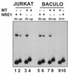
FIG. 1
FIG. 1
Differential binding of Ku to double- and single-stranded DNAs upon truncation of NRE1. EMSA on a 4% polyacrylamide gel of Ku binding to full-length and truncated NRE1 elements through incubation of Jurkat nuclear extract (lanes 1 to 4) or purified recombinant Ku expressed from baculovirus (lanes 5 to 10) with 4 to 5 pmol of double-stranded (ds) (lanes 1, 3, 5, and 7), upper-strand (up) (lanes 2, 4, 6, and 8), and lower-strand (lo) (lanes 9 and 10) 23-mer NRE1 and 17-mer MT oligonucleotides as indicated above the gel. Binding was performed in the presence of 2 μg of highly sheared calf thymus DNA.
FIG. 2
FIG. 2
MT is a direct, sequence-specific, double-stranded DNA binding site for Ku. EMSA on a 4% polyacrylamide gel with recombinant Ku was performed with 4 to 5 pmol of covalently closed circular DNA microcircles comprised of a 223-bp DNA microcircle from pBluescript (lanes 1 to 3 and 13 to 15), a 246-bp microcircle containing the same pBluescript fragment with a 23-bp NRE1 insert (lanes 4 to 6 and 16 to 18), a 240-bp microcircle containing the pBluescript fragment with a 17-bp MT insert (lanes 7 to 9 and 19 to 21), and a 240-bp microcircle containing the pBluescript fragment with a 17-mer MT oligonucleotide containing the substitution GAGATAGA for the GAGAAAGA MT core sequence (MTmt) (lanes 10 to 12 and 22 to 24) in the presence of 2 μg of highly sheared calf thymus DNA. Lanes 1 to 12 show the results obtained with Jurkat crude nuclear extract, and lanes 13 to 24 show results obtained with recombinant Ku. Ku binding was verified by inclusion of a Ku antibody in lanes 3, 6, 9, 12, 15, 18, 21, and 24. Inclusion of a nonspecific antibody had no effect on complex migration (28).
FIG. 3
FIG. 3
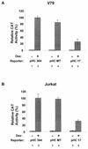
Mutation of NRE1 to MT abrogates the repression of MMTV transcription by Ku. (A) Transient transfection analysis of glucocorticoid-induced (2 × 10⁻⁷ M dex) transcription from the MMTV LTR in V79 Chinese hamster ovary fibroblasts transfected with MMTV CAT reporter genes pHC364, which is truncated prior to NRE1 (MMTV sequences −364 to +125; lanes 1 and 2); pHCMT (−421 to +125; lanes 3 and 4), in which one copy of the GAGAAAGA repeat in NRE1 had been mutated; and pHC17 (MMTV sequences −421 to +125; lanes 5 and 6), containing wild-type NRE1. (B) Similar transient transfection analysis in Jurkat T cells. Results are expressed as percentages of the maximal activity of the pHC364 construct treated with dex. In both panels A and B, CAT values were corrected against a constitutive RSV–β-gal reporter construct to control for transfection. Results are the averages (± standard errors of the means) of three to five individual experiments performed in duplicate.
FIG. 4
FIG. 4
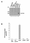
MT and single-stranded NRE1 are unable to activate DNA-PK in vitro. (A) Phosphorylation of GST-GR substrate by DNA-PK in the presence of covalently closed circular was determined for MMTV LTR plasmids pHC364 (lane 4), pHCMT (lane 5), and pHC17 (lane 6). Results of control reactions performed with pHC17 in the absence of DNA-PK and GST-GR are displayed in lanes 1 and 3, while phosphorylation of GST-GR in the absence of added DNA is shown in lane 2. Samples were resolved on 12% polyacrylamide gels. The molecular size markers are showed to the left, while the position of GST-GR is highlighted to the right. (B) p53 peptide phosphorylation by DNA-PK in the presence of 58-mer oligonucleotides encoding a nonspecific double-stranded DNA sequence (dsNS), the upper, single strand of the MMTV LTR centered over NRE1 (upNRE), or the same sequence in which mutations have been introduced into one copy of the NRE1 repeat (upMT). The results are expressed as counts per minute of 32P incorporated, and the error bars represent the standard errors of the means for three independent experiments performed in duplicate.
FIG. 5
FIG. 5
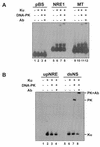
Binding of DNA-PK to NRE1 and MT. (A) EMSA on a 4% polyacrylamide gel of the binding of recombinant Ku (lanes 2, 6, and 10) or purified DNA-PK (lanes 3, 4, 7, 8, 11, and 12) to 32P-labeled, covalently closed microcircles containing no insert (lanes 1 to 4), the wild-type NRE1 element (lanes 5 to 8), or the MT element (lanes 9 to 12) in the presence of 2 μg of highly sheared calf thymus DNA. The incubations in lanes 4, 8, and 12 included a DNA-PK antibody. (B) EMSA of recombinant Ku (lanes 2 and 6) or purified DNA-PK (lanes 3, 4, 7, and 8) binding to single-stranded 58-mer oligonucleotides encoding upNRE1 in the presence of 2 μg of sheared, denatured calf thymus DNA (lanes 1 to 4) or dsNS (lanes 5 to 8) in the presence of 100 ng of highly sheared calf thymus DNA. A DNA-PK antibody (Ab) was added to the incubations in lanes 4 and 8. The positions of the Ku–DNA-PK complexes and the shift in migration by the DNA-PK antibody to just below the well are indicated to the right.
FIG. 6
FIG. 6
Decreased translocation of Ku from MT. EMSA on 4% polyacrylamide gel of recombinant Ku binding to closed 240- and 246-bp microcircles containing the wild-type NRE1 element (lanes 1 to 5) or the MT element (lanes 6 to 10) in the presence of 2 μg of highly sheared calf thymus DNA. The microcircles were coincubated with Ku, 10 mM MgCl2, and Ku antibody (Ab) as summarized above the autoradiograph.
FIG. 7
FIG. 7
Mg2+/ATP-dependent contact of Ku80 with DNA is specific for NRE1. A 23-mer NRE1 nucleotide and a 17-mer MT oligonucleotide were 32P-labeled on the upper, purine-rich strands, while a 39-mer nonspecific oligonucleotide was labeled on both strands. Following the incubation of recombinant Ku with the purine-rich upNRE1 (lane 1), dsNRE1 (lanes 2 to 4), or dsMT (lanes 5 to 7) or the dsNS oligonucleotide (lanes 8 to 10) under standard binding conditions in the presence of 10 mM MgCl2 and/or 10 mM ATP as indicated above the autoradiograph, the samples were irradiated with UV. Following irradiation, the samples were electrophoresed through an SDS–12% polyacrylamide gel to resolve the cross-linked products.
FIG. 8
FIG. 8
Ku induces an NRE1-dependent structural transition in the MMTV LTR immediately upstream of NRE1. (A) Upper LTR strand. (B) Lower LTR strand. Covalently closed circular relaxed pHC17 and pHC17MT plasmids were incubated with Jurkat nuclear extract (lanes 2 to 6 and 15 to 20) or purified recombinant Ku (lanes 8 to 12 and 22 to 27). Following the equilibration of binding, samples were treated with Mg2+ and KMnO4 as indicated. KMnO4-modified bases were identified following piperidine treatment by linear PCR from oligonucleotides extended downstream from the plasmid backbone (upper strand, panel A), or upstream from the MMTV LTR at position −289, and visualized following electrophoresis on 8% (8 M urea) polyacrylamide gels. The short arrows beside each gel highlight the positions of the modified T’s in the LTR sequences as determined by comparison to KMnO4-generated T sequencing tracks generated with single-stranded DNAs (lanes 1, 7, 13, 14, 21, and 28). The long arrows parallel to the sequences highlight the positions of the GAGAAGA repeat units that occur twice in NRE1 (left) but only once in the MT substitution. (C) Summary of Mg2+-dependent, KMnO4-mediated thymidine modifications in the MMTV LTR. The overlapping direct repeat of NRE1 is highlighted by the overlapping arrows parallel to the DNA sequence, while the positions of the modification are indicated by the perpendicular arrows. The sizes of the arrows are approximately proportional to the intensity of cleavage.
FIG. 9
FIG. 9
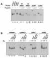
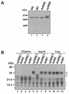
EMSA of Ku conformation and protease sensitivity when bound to different DNA forms. (A) EMSA on 10% polyacrylamide gel of Ku binding to 23-mer dsNRE1, 17-bp dsMT oligonucleotides, a 39-mer dsNS oligonucleotide, or upNRE1 oligonucleotide on a nondenaturing 10% polyacrylamide gel with or without a 5-min incubation with 2 or 5 μg of trypsin following the equilibration of binding. dsNRE1 and dsMT binding were performed in the presence of 2 μg of highly sheared calf thymus DNA, DNA end binding was performed in the presence of 100 ng of the same DNA, and binding to upNRE1 was performed in the presence of 2 μg of heat-denatured calf thymus DNA. (B) EMSA results obtained as described for panel A, except that protease digestion was performed with 2 μg of trypsin (Tryp), 1 μg of chymotrypsin (Chymo), or 2 μg of Asp-N as indicated, prior to electrophoresis. (C) Competition of Ku from DNA following trypsin digestion. Following the incubation of Ku with the DNAs described for panel A, trypsin was added to the incubations as indicated for 5 min. EMSA was performed following a further 5-min incubation in the presence or absence of a 100-fold excess of unlabeled 23-mer dsNRE1 DNA.
FIG. 9
FIG. 9
EMSA of Ku conformation and protease sensitivity when bound to different DNA forms. (A) EMSA on 10% polyacrylamide gel of Ku binding to 23-mer dsNRE1, 17-bp dsMT oligonucleotides, a 39-mer dsNS oligonucleotide, or upNRE1 oligonucleotide on a nondenaturing 10% polyacrylamide gel with or without a 5-min incubation with 2 or 5 μg of trypsin following the equilibration of binding. dsNRE1 and dsMT binding were performed in the presence of 2 μg of highly sheared calf thymus DNA, DNA end binding was performed in the presence of 100 ng of the same DNA, and binding to upNRE1 was performed in the presence of 2 μg of heat-denatured calf thymus DNA. (B) EMSA results obtained as described for panel A, except that protease digestion was performed with 2 μg of trypsin (Tryp), 1 μg of chymotrypsin (Chymo), or 2 μg of Asp-N as indicated, prior to electrophoresis. (C) Competition of Ku from DNA following trypsin digestion. Following the incubation of Ku with the DNAs described for panel A, trypsin was added to the incubations as indicated for 5 min. EMSA was performed following a further 5-min incubation in the presence or absence of a 100-fold excess of unlabeled 23-mer dsNRE1 DNA.
FIG. 10
FIG. 10
In-gel cross-linking of protease-digested Ku-DNA complexes reveals a difference in Ku binding to dsNRE1 and DNA ends. SDS-PAGE analysis of Ku cross-linked to dsNRE1, upNRE1, dsMT, and a 39-mer dsNS oligonucleotide following EMSA of protease-treated Ku-DNA complexes. Ku binding to the dsNS oligonucleotide was performed in the presence of 100 ng of calf thymus DNA, while binding to dsNRE1, dsMT, and upNRE1 was performed in the presence of 2 μg of double-stranded or denatured calf thymus DNA. Protease digestion was performed with chymotrypsin (Chymo) (lanes 1 to 4), Asp-N (lanes 5 to 8), and trypsin (Tryp) (lanes 9 to 12). Following EMSA, the wet gel was irradiated with UV. Polyacrylamide slices containing Ku-DNA complexes were excised from the gel, washed and heated in SDS sample buffer, and then electrophoresed through an SDS–20% polyacrylamide gel and subjected to autoradiography. The asterisks highlight two bands that migrated within the salt front on the gel and which had mobilities identical to those of the 32P-labeled free oligonucleotides cut from the EMSA gel and electrophoresed in parallel with the Ku-DNA complexes (28). Complexes specific for individual DNAs and/or protease treatments are highlighted by the numeric labels to the right of the autoradiograph.
FIG. 11
FIG. 11
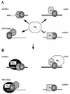
Schematic summary of the association of Ku and DNA-PKcs with the DNAs employed in this study. (A) In solution, DNA-PKcs occurs in an inactive conformation (PKI) that is attracted to Ku bound to dsNRE1 (upper left), dsMT (upper right), and double-stranded DNA ends (lower left). It does not appear to interact with Ku bound to the single, upper strand of NRE1 (ssNRE1) (lower right). The association of Ku with each of these DNAs is reflected by differences in Ku conformation, DNA conformation, and Ku-DNA contacts. In this pictogram, differences in conformation of the Ku heterodimer on DNA, as reflected by differences in protease sensitivity in EMSA and crosslinking experiments, are illustrated through differences in the shape of the Ku subunits. The Mg2+-dependent structural transition upstream of NRE1 is indicated by the dotted lines in the absence of definitive information on the exact structure of the DNA. The relative positioning of the Ku subunits with respect to the DNAs has been assigned arbitrarily. The DNA bound by Ku at the end is shown in grey to illustrate that it is not known whether a transition in DNA end structure is also important for the activation of DNA-PKcs from DNA ends. (B) The association of DNA-PKcs with Ku and DNA at NRE1 and DNA ends induces an allosteric change in the kinase that activates catalytic activity (PKA). By contrast, when associated with Ku on the MT element, DNA-PKcs remains in an inactive conformation. The positioning of DNA-PKcs over the structural transition upstream of NRE1 reflects our anticipation that this change in DNA structure may be in some way directly important for activation of the kinase at NRE1. The positioning of DNA-PKcs at the double-stranded DNA end, with Ku70 moved internally on the DNA, reflects the findings and model of Hammarsten and Chu for the assembly of Ku on DNA ends (38), with the refinement that Ku80 may not directly contact the DNA.

Similar articles

Cited by

References

    1. Anderson C W, Lees-Miller S P. The nuclear serine/threonine protein kinase DNA-PK. Crit Rev Eukaryot Gene Expr. 1992;2:283–314. - PubMed
    1. Archer T K, Lefebvre P, Wolford R G, Hager G L. Transcription factor loading on the MMTV promoter: a bimodal mechanism for promoter activation. Science. 1992;255:1573–1576. - PubMed
    1. Ball J K, Diggelmann H, Dekaban G A, Grossi G F, Semmler R, Waight P A, Fletcher R F. Alterations in the U3 region of the long terminal repeat of an infectious thymotropic type B retrovirus. J Virol. 1988;62:2985–2993. - PMC - PubMed
    1. Barlev N A, Poltoratsky V, Owen-Hughes T, Ying C, Liu L, Workman J L, Berger S L. Repression of GCN5 histone acetyltransferase activity via bromodomain-mediated binding and phosphorylation by the Ku–DNA-dependent protein kinase complex. Mol Cell Biol. 1998;18:1349–1358. - PMC - PubMed
    1. Beato M. Chromatin structure and the regulation of gene expression: remodeling at the MMTV promoter. J Mol Med. 1996;74:711–724. - PubMed

Publication types

MeSH terms