. 1999 Jun;19(6):4121-33.
doi: 10.1128/MCB.19.6.4121.

Transcriptional repressor ERF is a Ras/mitogen-activated protein kinase target that regulates cellular proliferation

Affiliations

Transcriptional repressor ERF is a Ras/mitogen-activated protein kinase target that regulates cellular proliferation

L Le Gallic et al. Mol Cell Biol. 1999 Jun.

Abstract

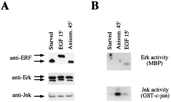
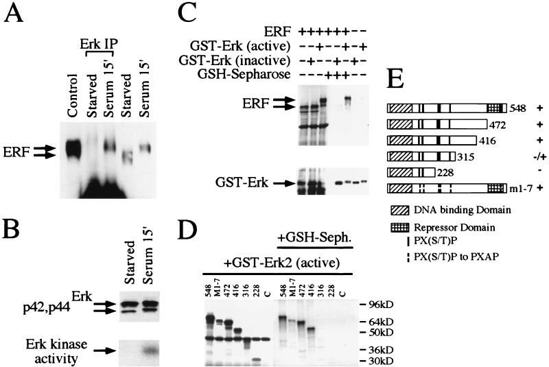
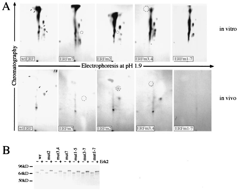
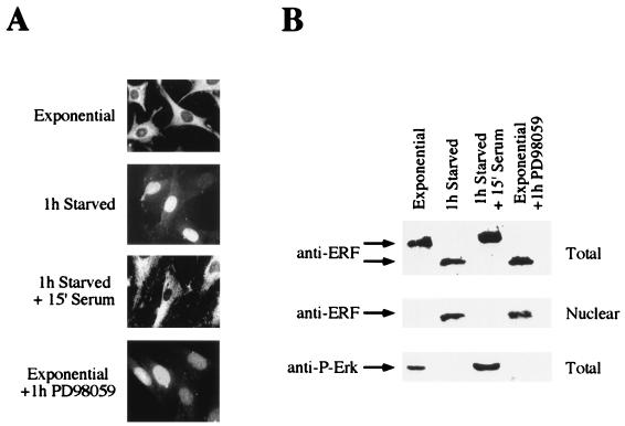
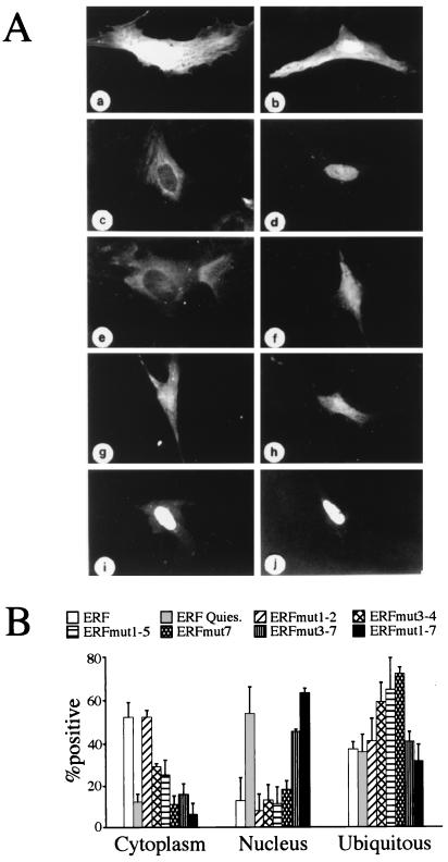
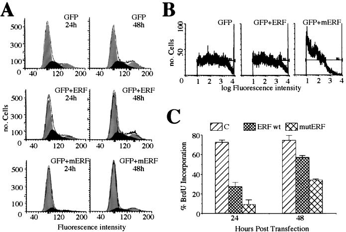
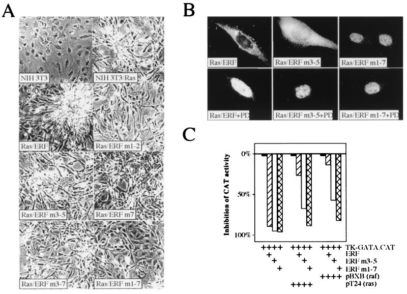
A limited number of transcription factors have been suggested to be regulated directly by Erks within the Ras/mitogen-activated protein kinase signaling pathway. In this paper we demonstrate that ERF, a ubiquitously expressed transcriptional repressor that belongs to the Ets family, is physically associated with and phosphorylated in vitro and in vivo by Erks. This phosphorylation determines the ERF subcellular localization. Upon mitogenic stimulation, ERF is immediately phosphorylated and exported to the cytoplasm. The export is blocked by specific Erk inhibitors and is abolished when residues undergoing phosphorylation are mutated to alanine. Upon growth factor deprivation, ERF is rapidly dephosphorylated and transported back into the nucleus. Phosphorylation-defective ERF mutations suppress Ras-induced tumorigenicity and arrest the cells at the G0/G1 phase of the cell cycle. Our findings strongly suggest that ERF may be important in the control of cellular proliferation during the G0/G1 transition and that it may be one of the effectors in the mammalian Ras signaling pathway.


Figures

FIG. 1
FIG. 1
ERF is phosphorylated in response to mitogens but not in response to stress. (A) Ref-1 cells expressing the ERF gene were arrested by serum starvation for 20 h and then induced for the indicated times with either 100 ng of epidermal growth factor (EGF) per ml or 25 ng of anisomycin (Anisom.) per ml. Thirty micrograms of cellular extract was analyzed by SDS gel electrophoresis, and proteins were detected by immunoblotting with the indicated antibodies. (B) Three hundred micrograms of the same cellular extracts was immunoprecipitated with anti-Erk or anti-JNK antibodies, as indicated. The kinase activity was detected by in-gel kinase assay with, as substrates, myelin basic protein (MBP) for Erks and the amino terminus of c-Jun for JNKs.
FIG. 2
FIG. 2
Physical interaction of ERF and Erk in vivo and in vitro. (A) Ref-1 cells were grown in the absence of serum for 20 h (starved) and then were induced for 15 min with 20% fetal calf serum (serum). Five hundred micrograms of cellular extracts from Ref-1 cells expressing ERF was immunoprecipitated with an anti-Erk rabbit polyclonal antibody under nondenaturing conditions (Erk IP) and analyzed by SDS gel electrophoresis. Twenty micrograms of cellular extracts before the addition of the anti-Erk antibody and of extracts from cells growing in complete medium (control) was also analyzed on the same gel. The presence of the ERF protein was detected by immunoblotting with the S17S anti-ERF specific antibody and is indicated by arrows. (B) Erk presence and activity in the same extracts were detected by immunoblotting (upper panel) and in-gel kinase assay (lower panel) as described for Fig. 1. (C) In vitro-translated ERF was mixed with active or inactive GST-Erk2 (Upstate Biotechnology Inc.), and the complexes were precipitated with GSH-Sepharose and analyzed by SDS gel electrophoresis. The presence or absence of ERF was detected by autoradiography (upper panel), and that of GST-Erk2 was detected by immunoblotting with an anti-Erk specific antibody (lower panel). (D) The presence or absence of truncated ERF proteins in complex with active GST-Erk2 was detected as described for panel C. The numbers indicate the carboxy-terminal amino acid of each deletion. M1-7 is a full-length ERF with all seven putative MAPK sites mutated to alanine. (E) Diagrammatic representation of the deletions and mutation used for panel D. The Ets DNA-binding domain, the previously identified repression domain, and the putative MAPK phosphorylation sites are indicated. Plus and minus indicate the interaction of each protein with active GST-Erk2 kinase.
FIG. 3
FIG. 3
Identification of MAPK phosphorylation sites. (A) Upper panels, the indicated in vitro-produced proteins were purified by immunoprecipitation and labeled with [γ-32P]ATP and recombinant Erk2 kinase. Lower panels, for the in vivo labeling, plasmids encoding the indicated proteins were transfected into NIH 3T3 cells. Twenty hours after transfection, the cells were serum deprived, labeled for 4 h with 32P, and stimulated for 10 min with serum, and the ERF proteins were immunoprecipitated with the S17S antibody. The labeled proteins were isolated after SDS gel electrophoresis and digested with trypsin, and the tryptic peptides were analyzed by chromatography and electrophoresis. The arrows in the wt ERF panels indicate the phosphopeptides eliminated or modified by the subsequent mutations. The circles indicate the positions of the eliminated or modified peptides. (B) The indicated ERF proteins were synthesized in vitro and labeled with [35S]methionine. Half of each sample (lanes +) was phosphorylated with active GST-Erk2 and ATP. The effect of the mutations on ERF mobility was determined after SDS gel electrophoresis.
FIG. 4
FIG. 4
Phosphorylation-dependent subcellular localization of ERF. (A) Ref-1 cells growing under the indicated conditions were fixed and stained with the S17S anti-ERF specific antibody and visualized by fluorescence microscopy (magnification, ×70). (B) Total and nuclear protein extracts from the same cells were analyzed by immunoblotting with the same anti-ERF specific antibody. Activated Erks were detected with an antibody directed against their phosphorylated form.
FIG. 5
FIG. 5
Site-specific phosphorylation determines ERF localization. (A) Ref-1 cells were transfected with a GFP-expressing plasmid (a and b) or with plasmids expressing GFP-ERF fusion proteins (c to j). The localization of the proteins was determined by the GFP fluorescence in exponentially growing cells (a, c, and e to j) or 1 h after serum withdrawal (b and d). (a and b) GFP; (c and d) GFP-ERF; (e) GFP-ERFmut1-2; (f) GFP-ERFmut3-4; (g) GFP-ERFmut7; (h) GFP-ERFmut1-5; (i) GFP-ERFmut3-7; (j) GFP-ERFmut1-7. (B) The localization of hybrid proteins expressed from the same plasmids as for panel A was scored in at least three different experiments. The values are the averages from at least 100 positive cells. Cells with proteins localized exclusively in the nucleus or in the cytoplasm were scored in the respective category. Ubiquitous distribution includes cells with detectable fluorescence in both compartments even when the distribution was not even.
FIG. 6
FIG. 6
Cell cycle arrest by ERF. (A) NIH 3T3 cells were cotransfected with a GFP-expressing plasmid and an empty vector plasmid, a plasmid expressing wt ERF (ERF), or a plasmid expressing the ERFm1-7 mutant (mERF). At 24 and 48 h after transfection, the cells were harvested and the DNA content of GFP-positive cells was determined by flow cytometry from the fluorescence of Hoechst 33342 dye. The data were analyzed with the ModFit LT 2.0 program. The black area represents S phase cells. (B) The protein expression level in the transfected cells was determined by the intensity of the GFP autofluorescence. (C) Ref-1 cells were cotransfected with a CD4-expressing plasmid and an empty vector plasmid (C), a plasmid expressing wt ERF (ERF), or a plasmid expressing the ERFm1-7 mutant (mutERF). Cells were scored for CD4 expression and BrdU incorporation by immunofluorescence. The values are the averages for at least 300 CD4+ cells from three independent experiments. The bars indicate statistical error.
FIG. 7
FIG. 7
ERF mutants can suppress the ras-transformed phenotype. (A) NIH 3T3 cells transformed with pT24 Ha-ras were transfected with wt or phosphorylation-deficient ERF mutants. Transformed cells were selected with G418 to establish colonies and cell lines. Cells from established cell lines were grown in the presence of 1% serum for 10 days to determine effects on cell morphology and photographed with a 10× phase-contrast lens. The numbers indicate the mutation(s) for each plasmid: 1, T148A; 2, S161A; 3, S246A; 4, S251A; 5, T271A; 6, T357A, and 7, T526A. (B) The subcellular localization of ERF mutants in ras-transformed NIH 3T3 cells was determined by immunofluorescence in exponentially growing cells in the presence or absence of the specific MEK1 inhibitor PD98059 (PD). (C) One microgram of the pTK-GATA.CAT reporter plasmid was cotransfected into HeLa cells with 0.5 μg of the indicated ERF plasmid and 1 μg of the indicated Ha-ras and c-raf-1 plasmids. The bars indicate inhibition of chloramphenicol acetyltransferase (CAT) activity relative to that in the samples in the absence of exogenous ERF.

References

    1. Albanese C, Johnson J, Watanabe G, Eklund N, Vu D, Arnold A, Pestell R G. Transforming p21ras mutants and c-Ets-2 activate the cyclin D1 promoter through distinguishable regions. J Biol Chem. 1995;270:23589–23597. - PubMed
    1. Brunner D, Ducker K, Oellers N, Hafen E, Scholz H, Klambt C. The ETS domain protein pointed-P2 is a target of MAP kinase in the sevenless signal transduction pathway. Nature. 1994;370:386–389. - PubMed
    1. Chow C W, Rincon M, Cavanagh J, Dickens M, Davis R J. Nuclear accumulation of NFAT4 opposed by the JNK signal transduction pathway. Science. 1997;278:1638–1641. - PubMed
    1. Corbett A H, Silver P A. Nucleocytoplasmic transport of macromolecules. Microbiol Mol Biol Rev. 1997;61:193–211. - PMC - PubMed
    1. Coso O A, Chiariello M, Yu J C, Teramoto H, Crespo P, Xu N, Miki T, Gutkind J S. The small GTP-binding proteins Rac1 and Cdc42 regulate the activity of the JNK/SAPK signaling pathway. Cell. 1995;81:1137–1146. - PubMed

Publication types

MeSH terms