. 1999 Jun;19(6):4167-81.
doi: 10.1128/MCB.19.6.4167.

GCD14p, a repressor of GCN4 translation, cooperates with Gcd10p and Lhp1p in the maturation of initiator methionyl-tRNA in Saccharomyces cerevisiae

Affiliations

GCD14p, a repressor of GCN4 translation, cooperates with Gcd10p and Lhp1p in the maturation of initiator methionyl-tRNA in Saccharomyces cerevisiae

O Calvo et al. Mol Cell Biol. 1999 Jun.

Abstract

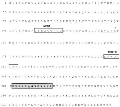
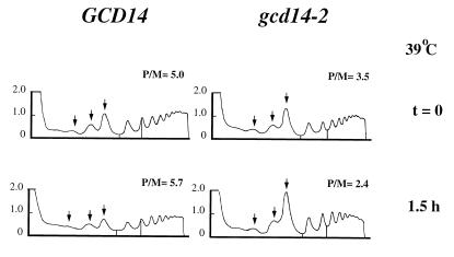
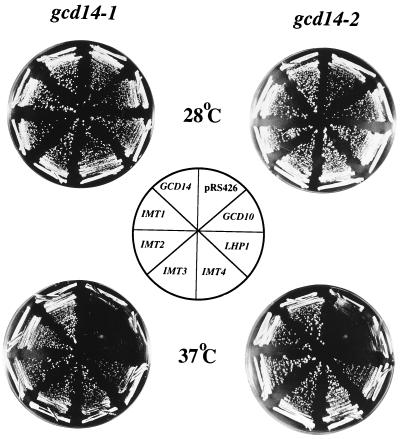
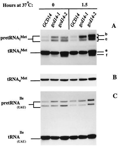
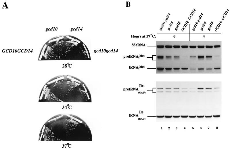
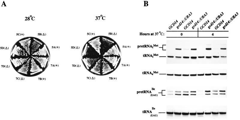
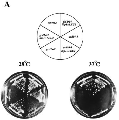
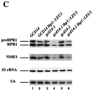
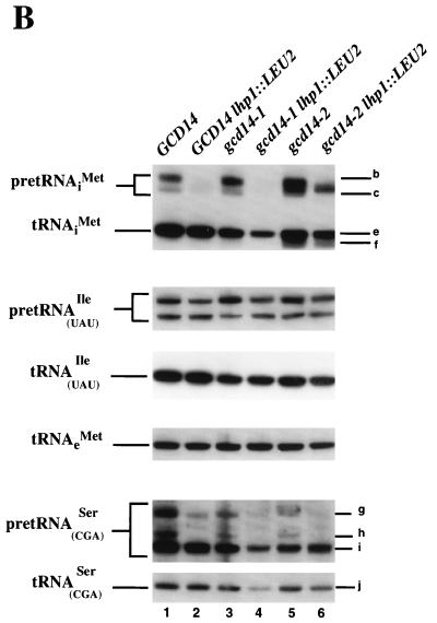
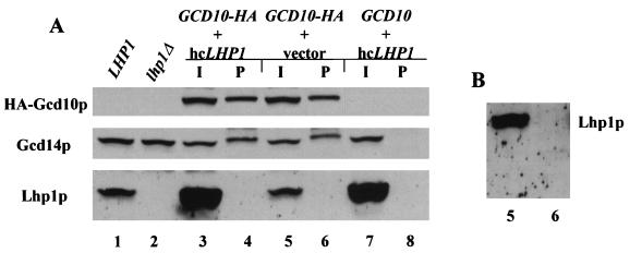
Gcd10p and Gcd14p were first identified genetically as repressors of GCN4 mRNA translation in Saccharomyces cerevisiae. Recent findings indicate that Gcd10p and Gcd14p reside in a nuclear complex required for the presence of 1-methyladenosine in tRNAs. Here we show that Gcd14p is an essential protein with predicted binding motifs for S-adenosylmethionine, consistent with a direct function in tRNA methylation. Two different gcd14 mutants exhibit defects in cell growth and accumulate high levels of initiator methionyl-tRNA (tRNAiMet) precursors containing 5' and 3' extensions, suggesting a defect in processing of the primary transcript. Dosage suppressors of gcd10 mutations, encoding tRNAiMet (hcIMT1 to hcIMT4; hc indicates that the gene is carried on a high-copy-number plasmid) or a homologue of human La protein implicated in tRNA 3'-end formation (hcLHP1), also suppressed gcd14 mutations. In fact, the lethality of a GCD14 deletion was suppressed by hcIMT4, indicating that the essential function of Gcd14p is required for biogenesis of tRNAiMet. A mutation in GCD10 or deletion of LHP1 exacerbated the defects in cell growth and expression of mature tRNAiMet in gcd14 mutants, consistent with functional interactions between Gcd14p, Gcd10p, and Lhp1p in vivo. Surprisingly, the amounts of NME1 and RPR1, the RNA components of RNases P and MRP, were substantially lower in gcd14 lhp1::LEU2 double mutants than in the corresponding single mutants, whereas 5S rRNA was present at wild-type levels. Our findings suggest that Gcd14p and Lhp1p cooperate in the maturation of a subset of RNA polymerase III transcripts.


Figures

FIG. 1
FIG. 1
Analysis of the GCD14 coding region. A partial restriction map of the GCD14 region is shown in the center, indicating the positions of the two ORFs, GCD14 and SPT10 (44), identified in the plasmid isolated from the genomic library bearing GCD14 (pRC5). The direction of transcription is indicated by the arrows. The top line depicts the genomic DNA insert in pRC5, and below that are depicted the fragments present in subclones constructed from pRC5 in low-copy-number vector pRS316 (pRC55 and pRC56). The ability of each subclone to complement the phenotypes of gcd14-1 and gcd14-2 mutants is shown on the right. Below the map of the GCD14 region are shown enlargements of the 2.6-kb SpeI-ClaI region in plasmid pRC56 and of the 3.2-kb SpeI-ClaI region in pRC560 bearing the gcd14::URA3 allele, in which a 1.1-kb URA3 fragment replaces ∼730 bp of the GCD14 coding sequence. Restriction enzyme sites: B, BamHI; Bg, BglII; C, ClaI; H, HindIII; Hp, HpaI; S, Sau3A; X, XbaI; Sp, SpeI.
FIG. 2
FIG. 2
Deduced amino acid sequence of Gcd14p. The predicted amino acid sequence of S. cerevisiae Gcd14p is given in single-letter code. Two regions of sequence similarity observed in several S-AdoMet-dependent methyltransferases are boxed and indicated as motif I and motif II (35). The conserved G residue at position 5 of motif I is G118 in Gcd14p. A glutamate residue commonly located 17 to 19 residues C terminal to motif I is found 17 residues C terminal to motif I in Gcd14p, at position 139 (*), and a cluster of hydrophobic residues, hhXh (D/E), at positions 135 to 138 (underlined, LFSF) precedes the E element. The central invariant aspartate in motif II is conserved at position 209 in Gcd14p. Motifs I and II are separated by 83 residues in Gcd14p. A putative nuclear localization signal (7) (shaded box) is located between residues K282 R294 in the Gcd14p sequence.
FIG. 3
FIG. 3
Polysome profiles of isogenic gcd14-2 and GCD14 strains. Isogenic strains Hm296g and Hm296G were cultured overnight in yeast extract-peptone-dextrose at 23°C and used to inoculate two flasks containing 300 ml of yeast extract-peptone-dextrose to an optical density at 600 nm of ∼0.1. Cultures were incubated at room temperature in a rotary shaker at 250 rpm. At an optical density at 600 nm of ∼4.0, cells were collected and resuspended in the appropriate volume of yeast extract-peptone-dextrose prewarmed to 39°C and grown to an optical density at 600 nm of ∼2.0. From both flasks, 150 ml was collected immediately for the 39°C, t = 0 samples, and 150 ml of fresh prewarmed yeast extract-peptone-dextrose was added back to each flask. Samples of 150 ml were collected from both flasks after 1.5 h and processed for polysome analysis by velocity sedimentation of whole-cell extracts on sucrose gradients (22). Gradients were fractionated while being scanned at 254 nm, and the resulting absorbance profiles are shown, with the tops of the gradients on the left. The positions of 40S, 60S, and 80S ribosomal species are indicated by arrows. For each strain, the top two gradients correspond to samples incubated at 39°C for 0 h (t = 0) and the two bottom gradients contain samples obtained after 1.5 h of incubation at 39°C. Areas delimited inside the profiles correspond to the fractions of the total absorbance profiles represented by 2-mer and 3-mer polysomes. In the gcd14-2 strain, the 2-mers and 3-mers represented 26% of the polysome mass at the permissive temperature but comprised 32% of the polysomes after 1.5 h at 39°C. In contrast, 2-mers and 3-mers represented 24 and 25% of the polysome mass at 23°C and after 1.5 h at 39°C, respectively, in the wild-type strain Hm296G. The P/M ratios were calculated at t = 0 and t = 1.5 h for three independent experiments, and the following mean values and standard errors (in parentheses) were obtained: at t = 0, P/M = 4.2 (0.83) for GCD14 and 2.8 (0.44) for gcd14-2; at t = 1.5 h, P/M = 5.2 (0.35) for GCD14 and 3.5 (0.72) for gcd14-2.
FIG. 4
FIG. 4
Phenotypes of gcd14 dosage suppressors. Eight independent transformants of strains Hm295 (gcd14-1) and Hm296 (gcd14-2) carrying empty vector pRS426 (gcd14), a low-copy-number plasmid bearing GCD14 (pRC56), or high-copy-number plasmids bearing IMT1 (p2632), IMT2 (p2633), IMT3 (p2634), IMT4 (p2635), GCD10 (pE107), or LHP1 (p2636) were streaked for single colonies on minimally supplemented SD plates and incubated at 28°C (top plates) or 37°C (bottom plates) for 2 days. The locations of the transformants on plates are indicated inside the central schematic.
FIG. 5
FIG. 5
gcd14 mutants are defective in processing the primary precursors of initiator tRNAMet and tRNAUAUIle. Northern blot analysis of total RNA (10 μg) isolated from strains H117 (GCD14), H160 (gcd14-1), and H168 (gcd14-2) grown in yeast extract-peptone-dextrose medium at 28°C to mid-exponential phase (0 h at 37°C) and shifted to 37°C for 1.5 h is shown. The blot was probed with a radiolabeled oligonucleotide that specifically hybridized to tRNAiMet (A) and then was stripped and reprobed with radiolabeled oligonucleotides specific for tRNAeMet (B) or tRNAUAUIle (C) (see Materials and Methods). The positions of pre-tRNAiMet species, mature tRNAiMet, tRNAeMet, and primary (upper band) and 5′- and 3′-end-processed intron-containing (lower band) pre-tRNAUAUIle and mature tRNAUAUIle are indicated on the left. Indicated on the right are the positions of pre-tRNAiMet containing 5′ and 3′ extensions encoded by IMT2 and IMT3 (b), pre-tRNAiMet containing 5′ and 3′ extensions encoded by IMT1 and IMT4 (c), mature tRNAiMet (e), and aberrantly processed tRNAiMet species (f) (see text for details).
FIG. 6
FIG. 6
Exacerbation of gcd14-2 phenotypes by a gcd10-505 mutation. (A) The double mutant Hm423 (gcd10-505 gcd14-2) and isogenic GCD10 gcd14-2 (Hm420), gcd10-505 GCD14 (Hm421), and GCD10 GCD14 (Hm422) strains were streaked for single colonies on minimally supplemented SD plates and incubated at 28, 34, or 37°C for 2 days. The genotypes of the transformants are indicated adjacent to the appropriate sectors of the 28°C plate. (B) Northern blot analysis of total RNA (10 μg) isolated from the same strains analyzed in panel A, conducted as described for Fig. 5 except that cells were grown in minimally supplemented SD at 28°C (t = 0) (lanes 1 to 4) and then shifted to 37°C for 4 h (lanes 5 to 8). The blot was probed for 5S rRNA, tRNAiMet, and tRNAUAUIle as described for Fig. 5.
FIG. 7
FIG. 7
GCD14 is not essential in the presence of hcIMT4. (A) Ascospore clones from two four-spored tetrads (designated 5 and 7) were obtained from a heterozygous GCD14/gcd14::URA3 diploid (YNG1) containing high-copy-number plasmid p1775 (hcIMT4) (20). The four strains from each tetrad (5A to 5D and 7A to 7D), all containing hcIMT4, were streaked for single colonies on minimally supplemented SD medium and incubated at 28 or 37°C for 3 days. The position of each spore clone and its GCD14 genotype is indicated adjacent to the appropriate plate sector. +, wild-type GCD14; Δ, gcd14::URA3. (B) Northern blot analysis of total RNA (10 μg) isolated from the spore clones of tetrad 5 (strains 5A to 5D) bearing hcIMT4 (described for panel A). The strains were grown to mid-logarithmic phase in minimally supplemented SD medium at 28°C (t = 0 at 37°C) and shifted to 37°C for 4 h. The blot was probed for tRNAiMet, tRNAeMet, or tRNAUAUIle as described for Fig. 5.
FIG. 8
FIG. 8
Synthetic interactions of gcd14 and lhp1::LEU2 mutations. (A) Strains YNG174 (GCD14), YNG175 (gcd14-1), and YNG176 (gcd14-2) and the isogenic lhp1::LEU2-containing derivatives Hm406 (GCD14 lhp1::LEU2), Hm407 (gcd14-1 lhp1::LEU2), and Hm408 (gcd14-2 lhp1::LEU2) were streaked for single colonies on minimally supplemented SD plates and incubated at 28 or 37°C for 2 days. The locations of the strains are indicated by their relevant genotypes in the schematic at the top. (B) Northern blot analysis of total RNA (20 μg) isolated from the strains described for panel A and separated by electrophoresis in a 6% polyacrylamide–8.3 M urea gel. Strains were grown in minimally supplemented SD at 28°C. The blot was probed for tRNAiMet, tRNAUAUIle, tRNAeMet, and tRNACGASer by using the appropriate oligonucleotides (see Materials and Methods). Indicated on the right of the top panel are the positions of various precursor and processed forms of tRNAiMet, as described in the legend to Fig. 5. The positions of precursor and mature tRNACGASer species are indicated on the right of the bottom panel: a primary precursor containing 5′ and 3′ extensions (g), a processing intermediate containing only the 3′ extension (h), a 5′- and 3′-end-processed intron-containing precursor (i), and mature tRNACGASer (j). (C) The Northern blot in panel B was probed for RPR1 RNA, NME1 RNA, 5S rRNA, and U6 RNA by using the appropriate oligonucleotides (see Materials and Methods). The positions of precursor and mature RPR1 species are indicated on the left.
FIG. 8
FIG. 8
Synthetic interactions of gcd14 and lhp1::LEU2 mutations. (A) Strains YNG174 (GCD14), YNG175 (gcd14-1), and YNG176 (gcd14-2) and the isogenic lhp1::LEU2-containing derivatives Hm406 (GCD14 lhp1::LEU2), Hm407 (gcd14-1 lhp1::LEU2), and Hm408 (gcd14-2 lhp1::LEU2) were streaked for single colonies on minimally supplemented SD plates and incubated at 28 or 37°C for 2 days. The locations of the strains are indicated by their relevant genotypes in the schematic at the top. (B) Northern blot analysis of total RNA (20 μg) isolated from the strains described for panel A and separated by electrophoresis in a 6% polyacrylamide–8.3 M urea gel. Strains were grown in minimally supplemented SD at 28°C. The blot was probed for tRNAiMet, tRNAUAUIle, tRNAeMet, and tRNACGASer by using the appropriate oligonucleotides (see Materials and Methods). Indicated on the right of the top panel are the positions of various precursor and processed forms of tRNAiMet, as described in the legend to Fig. 5. The positions of precursor and mature tRNACGASer species are indicated on the right of the bottom panel: a primary precursor containing 5′ and 3′ extensions (g), a processing intermediate containing only the 3′ extension (h), a 5′- and 3′-end-processed intron-containing precursor (i), and mature tRNACGASer (j). (C) The Northern blot in panel B was probed for RPR1 RNA, NME1 RNA, 5S rRNA, and U6 RNA by using the appropriate oligonucleotides (see Materials and Methods). The positions of precursor and mature RPR1 species are indicated on the left.
FIG. 9
FIG. 9
Gcd14p, but not Lhp1p, is tightly associated with Gcd10p in cell extracts. (A) Whole-cell extracts were prepared as described previously (47) from strains H1515 (LHP1) and YJA113 (lhp1) and from transformants of YJA142 (GCD10HA) or YJA143 (GCD10) bearing high-copy-number plasmid p2626 containing LHP1 or empty vector YEp24. Aliquots containing 200 μg of total cell protein were mixed with 2 to 4 μg of anti-HA monoclonal antibody HA.11 (Babco), which was prebound to protein A-Sepharose for 2 h on ice, and the mixture was incubated for 1 h at 4°C. After three washes with lysis buffer (20 mM Tris-HCl [pH 7.4], 100 mM KCl, 1.0 mM Mg acetate, 0.1% [vol/vol] Triton X-100) containing one complete protease inhibitor tablet (Boehringer Mannheim) per 25 ml, immunoprecipitates were collected by centrifugation and proteins were eluted by boiling in Laemmli buffer (37). The total proteins immunoprecipitated from 200 μg (pellet [P]; lanes 4, 6, and 8) or 50 μg of the starting whole-cell extracts (input [I]; lanes 3, 5, and 7) were separated by SDS-polyacrylamide gel electrophoresis (36) and transferred to a nitrocellulose membrane (Millipore) in 25 mM Tris–192 mM glycine–0.1% SDS containing 20% (vol/vol) methanol. Lanes 1 and 2 contain 50 μg of the starting whole-cell extracts from strains H1515 and YJA113, respectively, which were included to establish the identity of Lhp1p. The membrane was blocked overnight at 4°C in BLOTTO (5% [wt/vol] nonfat dry milk, 10 mM Tris-HCl [pH 7.4], 150 mM NaCl, 0.05% [vol/vol] Tween 20). A single immunoblot was probed with 3 μg of rabbit anti-HA antibody HA.11 (Babco) per ml, stripped, reprobed with a 1:1,000 dilution of anti-Gcd14p antibodies (see Materials and Methods), and reprobed with a 1:1,000 dilution of anti-Lhp1p antibodies (60). Immune complexes were detected with horseradish peroxidase-conjugated sheep antimouse (Amersham) or donkey antirabbit (Amersham) secondary antibodies and an enhanced chemiluminescence kit (ECL; Amersham). (B) A longer exposure of lanes 5 and 6 of the blot in panel A probed with anti-Lhp1p antibodies, included to detect small amounts of Lhp1p in the immunoprecipitates.

Similar articles

Cited by

References

    1. Alani E, Cao L, Kleckner N. A method for gene disruption that allows repeated use of URA3 selection in the construction of multiply disrupted yeast strains. Genetics. 1987;116:541–545. - PMC - PubMed
    1. Altschul S F, Boguski M S, Gish W, Wootton J C. Issues in searching molecular sequence databases. Nat Genet. 1994;6:119–129. - PubMed
    1. Anderson J, Phan L, Cuesta R, Carlson B A, Pak M, Asano K, Björk G R, Tamame M, Hinnebusch A G. The essential Gcd10p-Gcd14p nuclear complex is required for 1-methyladenosine modification and maturation of initiator methionyl-tRNA. Genes Dev. 1998;12:3650–3662. - PMC - PubMed
    1. Anderson, J., and A. G. Hinnebusch. Unpublished observations.
    1. Asano K, Phan L, Anderson J, Hinnebusch A G. Complex formation by all five homologues of mammalian translation initiation factor 3 subunits from yeast Saccharomyces cerevisiae. J Biol Chem. 1998;273:18573–18585. - PubMed

Publication types

MeSH terms

Associated data