. 1999 Jun;19(6):4219-30.
doi: 10.1128/MCB.19.6.4219.

Platelet-derived growth factor-stimulated expression of the MCP-1 immediate-early gene involves an inhibitory multiprotein complex

Affiliations

Platelet-derived growth factor-stimulated expression of the MCP-1 immediate-early gene involves an inhibitory multiprotein complex

P Sridhar et al. Mol Cell Biol. 1999 Jun.

Abstract

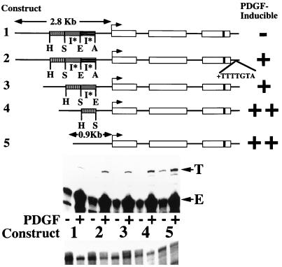
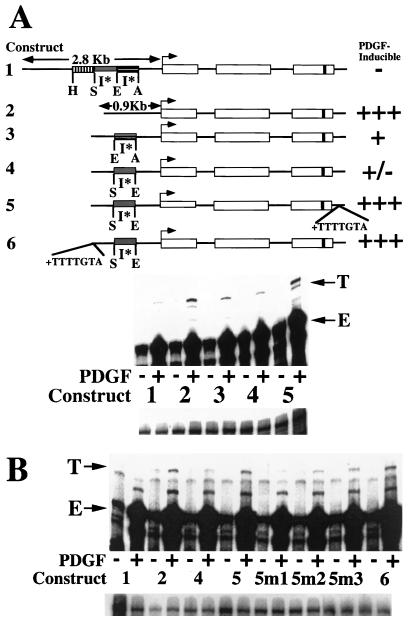
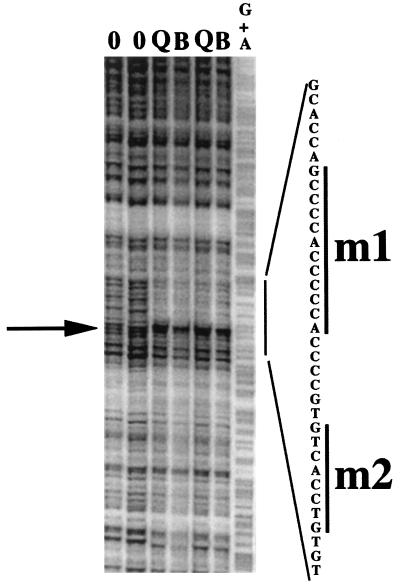
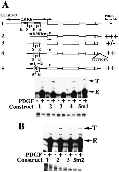
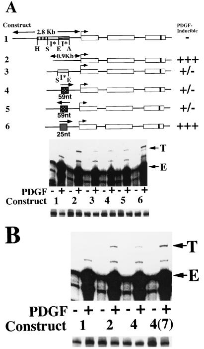
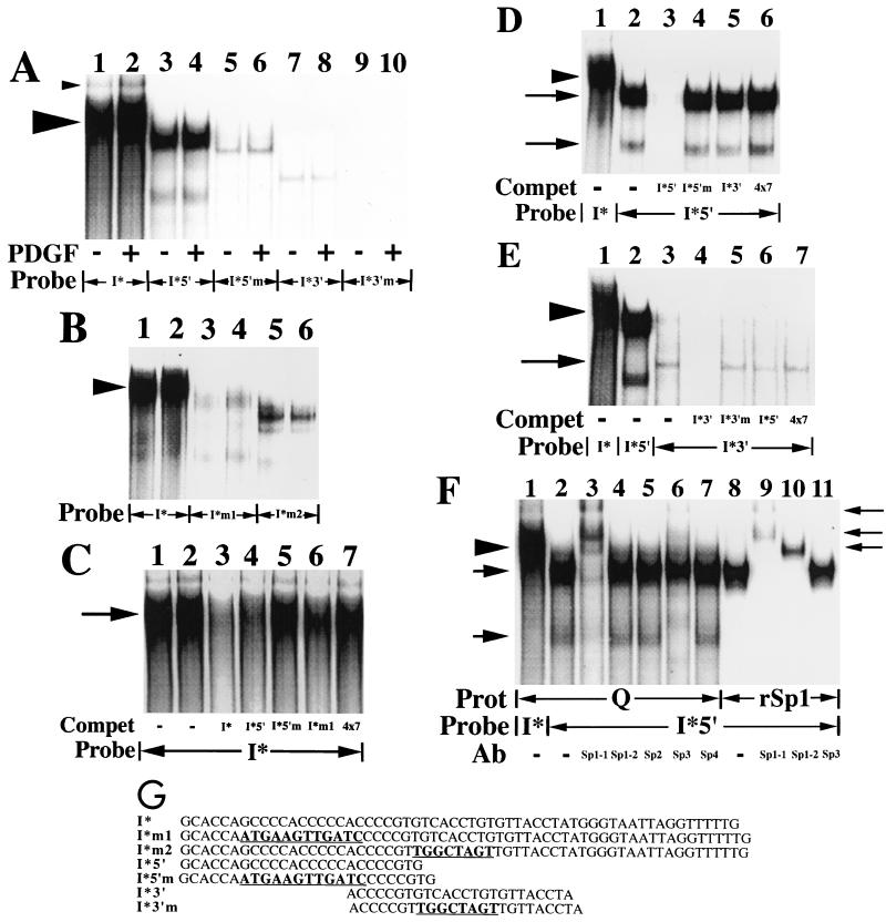
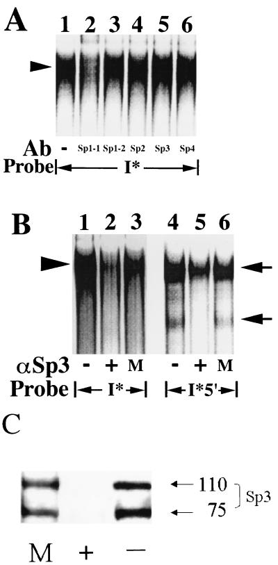
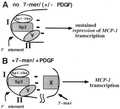
We have demonstrated previously that the seven-nucleotide (nt) motif TTTTGTA (the heptamer) that is present within the proximal 3' untranslated sequences of numerous immediate-early genes is essential for platelet-derived growth factor (PDGF)-stimulated induction of the MCP-1 immediate-early gene. On this basis, the heptamer was suggested to be a conserved regulatory element involved in immediate-early gene expression, although its mechanism of action was unknown. Herein, we demonstrate that the heptamer functions to remove an inhibition of PDGF induction of MCP-1 maintained by two independently acting inhibitory elements present in the MCP-1 5' flanking sequences (designated I* elements). PDGF treatment relieves the I*-mediated inhibition of MCP-1 expression only if the heptamer is also present. One inhibitory element is contained within a 59-nt portion of MCP-1 5' flanking sequences and functions in an orientation-independent and heptamer-regulated manner. Significantly, proteins binding to two DNA sequences contribute to the formation of a single multiprotein complex on the 59-nt I* element. The I*-binding complex contains Sp3, an Sp1-like protein, and a novel DNA-binding protein. Moreover, the complex does not form on two 59-nt sequences containing mutations that reverse the inhibition of PDGF induction maintained by the wild-type I* element. We propose to call the multiprotein I*-binding complex a repressosome and suggest that it acts to repress PDGF-stimulated transcription of MCP-1 in the absence of the heptamer TTTTGTA.


Figures

FIG. 1
FIG. 1
Deletion of 5′ flanking sequences results in heptamerless PDGF-inducible MCP-1 reporter genes. At the top is a schematic of the structures of five tagged MCP-1 reporter constructs. Open rectangles represent exons, and introns are represented by the dark lines between the exons. The lengths of 5′ flanking MCP-1 sequences contained within constructs are given in kilobases. The start of transcription is shown by the bent arrow. The 33-bp tag is represented by the dark band in the third exon. Constructs 1, 3, 4, and 5 contain 104 bp of 3′ untranslated sequences (that include the polyadenylation signal but not the heptamer). Only construct 2 contains the heptamer within its 3′ untranslated sequences. The relative positions of AocI (A), EcoRI (E), SpeI (S), and HincII (H) sites, located in the MCP-1 5′ flanking sequences and defining the endpoints of three MCP-1 5′ sequence fragments, are shown. The overall AocI-HincII 5′ fragment includes 1,936 bp. Construct 2 is derived from construct 1 by readdition of the heptamer TTTTGTA to the 3′ untranslated sequences of construct 1. Construct 3 is derived from construct 1 by deletion of the 707-bp EcoRI-AocI fragment. Construct 4 is derived from construct 3 by deletion of the 695-bp SpeI-EcoRI fragment. Construct 5 is derived from construct 4 by deletion of the 534-bp HincII-SpeI fragment. The PDGF-regulated 5′ I* elements are indicated. The PDGF inducibilities of the five constructs in transfection experiments are summarized on the right. In the middle are RNase protection assays of 40 μg of total cellular RNA that was prepared from NIH 3T3 fibroblasts transiently transfected with 5 μg of the constructs shown, allowed to become quiescent, and then not exposed (−) or exposed (+) to the B-B isoform of PDGF (30 ng/ml) for 3 h. The numbers refer to the tagged MCP-1 constructs diagrammed at the top. The positions of the 305- and 241-nt protected fragments corresponding to expression of the transfected and tagged (T) and endogenous (E) MCP-1 genes, respectively, are shown. The experiment was performed four times with similar results. The PDGF inductions obtained with constructs 4 and 5 were 2.6 to 4.0 and 3.0 to 8.1 times greater, respectively, than those obtained with construct 1 in these transfections. The PDGF induction increases observed with constructs 2, 3, 4, and 5, compared to construct 1, are all significant (P < 0.05 by the Wilcoxon two-sample test). At the bottom are RNase protection assays of 15 μg of total cellular RNA taken from the transfections shown above and analyzed with an alpha-globin riboprobe.
FIG. 2
FIG. 2
PDGF induction of MCP-1 involves interactions between the heptamer TTTTGTA and a 5′ I* element. (A) At the top is a schematic of the structures of six tagged MCP-1 reporter constructs. Constructs 1 through 4 and 6 contain 104 bp of 3′ untranslated sequences (that include the polyadenylation signal but not the heptamer). Only construct 5 contains the heptamer within its 3′ untranslated sequences. The details of the MCP-1 schematics are otherwise as described in the legend to Fig. 1. Constructs 1 and 2 in this figure are identical to constructs 1 and 5, respectively, in Fig. 1. Construct 3 is derived from construct 2 by readdition of the 707-bp EcoRI-AocI fragment. Construct 4 is derived from construct 2 by readdition of the 695-bp SpeI-EcoRI fragment. Construct 5 is derived from construct 4 by readdition of the heptamer to the proximal 3′ untranslated sequences. Construct 6 is derived from construct 4 by readdition of the heptamer to the distal 5′ flanking sequences. The PDGF-regulated 5′ I* elements are indicated. The PDGF inducibilities of the six constructs in transfection experiments are summarized on the right. At the middle are RNase protection assays of 40 μg of total cellular RNA that was prepared from NIH 3T3 fibroblasts transiently transfected with 5 μg of the constructs shown, allowed to become quiescent, and then not exposed (−) or exposed (+) to the B-B isoform of PDGF (30 ng/ml) for 3 h. The numbers refer to the tagged MCP-1 constructs diagrammed at the top. The 305- and 241-nt protected fragments corresponding to expression of the transfected and tagged (T) or endogenous (E) MCP-1 genes, respectively, are indicated. The experiment was performed four times with similar results. The PDGF inductions obtained with constructs 3 and 4 were 10 to 64 and 10 to 15%, respectively, of those obtained with construct 2 in these transfections. The PDGF inductions obtained with construct 5 were 2.1 to 9.7 times greater than those obtained with construct 4 in these transfections. The PDGF induction decreases observed with constructs 3 and 4, compared to construct 2, are both statistically significant (P < 0.05 by the Wilcoxon two-sample test). The PDGF induction increases observed with construct 5, compared to construct 4, are significant (P < 0.05 by the Wilcoxon two-sample test). At the bottom are RNase protection assays of 15 μg of total cellular RNA taken from the transfections shown above and analyzed with an alpha-globin riboprobe. (B) At the top are RNase protection assays of 40 μg of total cellular RNA that was prepared from NIH 3T3 fibroblasts transiently transfected with 4 μg of the constructs shown, allowed to become quiescent, and then not exposed (−) or exposed (+) to the B-B isoform of PDGF (30 ng/ml) for 3 h. The numbers refer to the tagged MCP-1 constructs described at the top, except for constructs 5m1, 5m2, and 5m3. The latter constructs are derived from construct 4 by addition of the mutant heptamers TTTTATG, GGGGGTA, and TTTTGGA, respectively, to the 3′ untranslated sequences. Mutations of the wild-type heptamer sequence are underlined. The 305- and 241-nt protected fragments corresponding to expression of the transfected and tagged (T) and endogenous (E) MCP-1 genes, respectively, are indicated. The experiment was performed four times with similar results. The decreased PDGF inductions obtained with constructs 5m1, 5m2, and 5m3 varied from 6 to 15%, 8 to 25%, and 50 to 60% of those obtained with construct 5, respectively, in these transfections. The PDGF induction differences obtained with constructs 5m1, 5m2, and 5m3, compared to construct 5, are all statistically significant (P < 0.05 by the Wilcoxon two-sample test). The PDGF induction increases observed with constructs 5 and 6, compared to construct 4, are statistically significant (P < 0.05 by the Wilcoxon two-sample test). At the bottom are RNase protection assays of 15 μg of total cellular RNA taken from the transfections shown above and analyzed with an alpha-globin riboprobe.
FIG. 3
FIG. 3
A single DNase I-protected sequence is detected within the 695-bp SpeI-EcoRI inhibitory fragment. A top-strand-labeled probe corresponding to the 695-bp inhibitory element-containing SpeI-EcoRI MCP-1 5′ fragment was used in DNase I footprinting assays. Nuclear protein extracts for these assays were prepared from two groups of quiescent fibroblasts (lanes Q) and fibroblasts treated with 30 ng of the B-B isoform of PDGF per ml for 2 h (lanes B). Lanes 0 contained no protein, and lane G+A contained the purine sequence of the top-strand-labeled probe. The nucleotide sequence of the overall protected region is given on the right. The arrow highlights a DNase I-hypersensitive site separating the 5′ and 3′ protected subregions. The 12-nt (m1) and 8-nt (m2) sequences mutated in this study are highlighted by the bars on the right.
FIG. 4
FIG. 4
Necessity of 12-nt (5′) and 8-nt (3′) footprinted sequences for inhibition of MCP-1 induction. (A) At the top is a schematic of the structures of five tagged MCP-1 reporter constructs. Constructs 1 through 3 and 5 contain 104 bp of 3′ untranslated sequences (that include the polyadenylation signal but not the heptamer). Only construct 4 contains the heptamer within its 3′ untranslated sequences. The details of the MCP-1 schematics are otherwise as described in the legend to Fig. 1. Construct 2 is derived from construct 1 by removal of the 1,936-bp AocI-HincII 5′ fragment. Construct 3 is derived from construct 2 by readdition of the 695-bp SpeI-EcoRI fragment. Construct 4 is derived from construct 3 by addition of the heptamer TTTTGTA to the 3′ untranslated sequences. Constructs 5m1 and m2 are derived from construct 3 by site-directed mutation of nonoverlapping 12-nt (5′ subregion) and 8-nt (3′ subregion) portions, respectively, of the footprinted region shown in Fig. 3 (i.e., mutating the 12-base sequence GCCCCACCCCCA to ATGAAGTTGATC and the 8-base sequence GTCACCTG to TGGCTAGT within the otherwise unaltered 695-bp SpeI-EcoRI fragment). The PDGF-regulated 5′ inhibitory I* elements are indicated. The PDGF inducibilities of the constructs in transfection experiments are summarized on the right. At the middle are RNase protection assays of 40 μg of total cellular RNA that was prepared from NIH 3T3 fibroblasts transiently transfected with 5 μg of the constructs shown, allowed to become quiescent, and then not exposed (−) or exposed (+) to the B-B isoform of PDGF (30 ng/ml) for 3 h. The numbers refer to the tagged MCP-1 constructs described at the top. The 305- and 241-nt protected fragments corresponding to expression of the transfected and tagged (T) and endogenous (E) MCP-1 genes, respectively, are indicated. The experiment was performed four times with similar results. The PDGF inductions obtained with construct 5m1 were 1.6 to 5.2 times greater than those obtained with construct 3 in these transfections. The PDGF induction increases observed with construct 5m1, compared to construct 3, are statistically significant (P < 0.05 by the Wilcoxon two-sample test). At the bottom are RNase protection assays of 15 μg of total cellular RNA taken from the transfections shown above and analyzed with an alpha-globin riboprobe. (B) At the top are RNase protection assays of 40 μg of total cellular RNA prepared from NIH 3T3 fibroblasts transiently transfected with 4 μg of the constructs shown, allowed to become quiescent, and then not exposed (−) or exposed (+) to the B-B isoform of PDGF (30 ng/ml) for 3 h. The numbers refer to the tagged MCP-1 constructs described at the top. The 305- and 241-nt protected fragments corresponding to expression of the transfected and tagged (T) and endogenous (E) MCP-1 genes, respectively, are indicated. The experiment was performed four times with similar results. The PDGF inductions obtained with construct 5m2 were 1.8 to 2.7 times greater than those obtained with construct 3 in these transfections. The PDGF induction increases observed with construct 5m2, compared to construct 3, are statistically significant (P < 0.05 by the Wilcoxon two-sample test). At the bottom are RNase protection assays of 15 μg of total cellular RNA taken from the transfections shown above and analyzed with an alpha-globin riboprobe.
FIG. 5
FIG. 5
A 59-nt sequence is sufficient for inhibition of PDGF induction of MCP-1. (A) At the top is a schematic of the structures of six tagged MCP-1 reporter constructs. All of the constructs contain 104 bp of 3′ untranslated sequences (that include the polyadenylation signal but not the heptamer). The details of the MCP-1 schematics are otherwise as described in the legend to Fig. 1. Construct 2 is derived from construct 1 by removal of the 1,936-bp AocI-HincII 5′ fragment. Construct 3 is derived from construct 2 by readdition of the 695-bp SpeI-EcoRI fragment. Constructs 4 and 5 are derived from construct 2 by readdition of a 59-nt subfragment of the 695-bp SpeI-EcoRI fragment in the in vivo (construct 4) and reverse (construct 5) orientations. The 59-nt subfragment includes the complete footprinted region shown in Fig. 3 and a short stretch of additional MCP-1 sequences immediately 3′ to the footprinted region. Construct 6 is derived from construct 2 by readdition of a 25-nt oligonucleotide (I*5′) containing the 5′ footprinted subregion shown in Fig. 3. The 25-nt sequence is entirely contained within the 59-nt sequences. The PDGF-regulated 5′ I* elements are indicated. The PDGF inducibilities of the six constructs in transfection experiments are summarized on the right. Below the schematic are results from RNase protection assays of 40 μg of total cellular RNA that was prepared from NIH 3T3 fibroblasts transiently transfected with 4 μg of the constructs shown, allowed to become quiescent, and then not exposed (−) or exposed (+) to the B-B isoform of PDGF (30 ng/ml) for 3 h. The numbers refer to the tagged MCP-1 constructs described at the top. The 305- and 241-nt protected fragments corresponding to expression of the transfected and tagged (T) and endogenous (E) MCP-1 genes, respectively, are indicated. The experiment was performed three times with similar results. The PDGF inductions obtained with constructs 3, 4, and 5 varied from 3.5 to 24% of those obtained with construct 2 in these transfections. Differences among the PDGF inductions observed with constructs 3, 4, and 5 are not statistically significant (P = 0.49 by the Kruskal-Wallis test). The PDGF induction decreases observed with constructs 3, 4, and 5, compared to construct 2, are all statistically significant (P < 0.05 by the Wilcoxon two- sample test). At the bottom are RNase protection assays of 15 μg of total cellular RNA taken from the transfections shown above and analyzed with an alpha-globin riboprobe. (B) At the top are RNase protection assays of 40 μg of total cellular RNA that was prepared from NIH 3T3 fibroblasts transiently transfected with 4 μg of the constructs shown, allowed to become quiescent, and then not exposed (−) or exposed (+) to the B-B isoform of PDGF (30 ng/ml) for 3 h. The numbers refer to the tagged MCP-1 constructs described at the top, except for construct 4(7), which is derived from construct 4 by addition of the heptamer TTTTGTA to the 3′ untranslated sequences. The 305- and 241-nt protected fragments corresponding to expression of the transfected and tagged (T) and endogenous (E) MCP-1 genes, respectively, are indicated. The experiment was performed four times with similar results. The PDGF inductions obtained with construct 4(7) were 2.5 to 4.2 times greater than those obtained with construct 4 in these transfections. The PDGF induction increases observed with construct 4(7), compared to construct 4, are statistically significant (P < 0.05 by the Wilcoxon two-sample test). At the bottom are RNase protection assays of 15 μg of total cellular RNA taken from the transfections shown above and analyzed with an alpha-globin riboprobe.
FIG. 6
FIG. 6
A slowly migrating protein complex binds to the 59-nt I* element-containing sequence. (A) Nuclear extracts (20 μg) prepared from quiescent fibroblasts (−) or fibroblasts treated with the B-B isoform of PDGF (30 ng/ml) for 2 h (+) were used in mobility shift assays along with the radiolabeled double-stranded oligonucleotide probes shown. The large arrowhead highlights the predominant complex that binds to the 59-nt fragment (I*) probe. The small arrowhead highlights an additional protein complex, of unclear significance, that also binds to the 59-nt fragment probe. (B) Nuclear extracts (15 μg) prepared from quiescent fibroblasts were used in mobility shift assays along with the radiolabeled double-stranded oligonucleotide probes shown. The arrowhead highlights the predominant complex that binds to the I* probe. (C) Nuclear extracts (15 μg) prepared from quiescent fibroblasts were used in mobility shift assays along with the radiolabeled double-stranded oligonucleotide probe shown. Unlabeled double-stranded oligonucleotides (300-fold excesses) were used as competitors where indicated. The 4×7 competitor is a double-stranded oligonucleotide containing four copies of the heptamer TTTTGTA. The arrow highlights the predominant complex that binds to the I* probe. Compet, competitor. (D) Nuclear extracts (15 μg) prepared from quiescent fibroblasts were used in mobility shift assays along with the radiolabeled double-stranded oligonucleotide probes shown. Unlabeled double-stranded oligonucleotides (300-fold excesses) were used as competitors where indicated. The arrowhead highlights the predominant complex that binds to the 59-nt fragment (I*) probe. The upper and lower arrows show the positions of two complexes that bind specifically to the I*5′ probe. (E) Nuclear extracts (20 μg) prepared from quiescent fibroblasts were used in mobility shift assays along with the radiolabeled double-stranded oligonucleotide probes shown. Unlabeled double-stranded oligonucleotides (200-fold excesses) were used as competitors where indicated. The arrowhead highlights the predominant complex that binds to the 59-nt fragment (I*) probe. The arrow shows the position of a complex that binds specifically to the I*3′ probe. (F) Nuclear extracts (15 μg) prepared from quiescent fibroblasts (Q) and recombinant Sp1 (rSp1, 1fpu) were used in mobility shift assays along with the radiolabeled double-stranded oligonucleotide probes shown. Antibodies specific for individual members of the Sp1 family of transcription factors (5 μg) were added where indicated. The Sp1-1 and Sp1-2 antibodies bind to distinct, nonoverlapping portions of the Sp1 transcription factor (amino acids 436 to 454 and 520 to 538, respectively). Prot, protein. The arrowhead on the left highlights the predominant complex that binds specifically to the 59-nt fragment (I*) probe. The arrows on the left show the positions of the two complexes that bind specifically to the 5′ subregion (I*5′) probe. The arrows on the right show the positions of complexes supershifted by the anti-Sp1 and anti-Sp3 antibodies (Ab). (G) The sequences of seven oligonucleotides and the relative position of each oligonucleotide within the overall 59-nt footprinted (I*) region are shown. These oligonucleotides were used as double-stranded probes or competitors in the six mobility shift assays whose results are shown here and correspond to the 25-nt (5′ subregion), 24-nt (3′ subregion), and 59-nt (full-length) footprinted regions shown in Fig. 3 (designated I*5′, I*3′, and I*, respectively). Mutant 25- and 24-nt probes, containing the 12- and 8-nt mutations described in the legend to Fig. 4 are designated I*5′m and I*3′m, respectively. Mutant I* probes containing the same 12- and 8-nt mutations are designated I*m1 and I*m2, respectively. The mutated sequences are boldfaced and underlined for each oligonucleotide. Free probe is not shown in these mobility shift assays. No complexes were observed with any of the probes alone in the absence of extract (data not shown).
FIG. 7
FIG. 7
The I* element-binding complex contains the Sp3 transcription factor and an Sp1-like protein. (A) Nuclear extracts (12.5 μg) prepared from quiescent fibroblasts were used in mobility shift assays along with the radiolabeled double-stranded oligonucleotide probe shown. The sequence of the probe is given in Fig. 6G. Antibodies (Ab) specific for individual members of the Sp1 family of transcription factors (2 μg) were added where indicated. The Sp1-1 and Sp1-2 antibodies are as described in the legend to Fig. 6F. The arrowhead on the left highlights the predominant complex that binds specifically to the I* probe. Free probe is not shown. No complexes were observed with the probe alone in the absence of extract (data not shown). (B) Nuclear extracts (12.5 μg) prepared from quiescent fibroblasts were used in mobility shift assays along with the radiolabeled double-stranded oligonucleotide probes shown. The sequences of the probes are given in Fig. 6G. Extracts were immunodepleted initially with anti-Sp3 antibodies (+), mock immunodepleted with rabbit immunoglobulin G (M), or not treated (−). The arrowhead on the left highlights the predominant complex that binds specifically to the I* probe. The arrows on the right highlight the positions of the two complexes that bind specifically to the 5′ subregion (I*5′) probe. Free probe is not shown. No complexes were observed with the probes alone in the absence of extract (data not shown). (C) Immunoblot of an aliquot (30 μg) of the extracts used for panel B with the anti-Sp3 antibody. The designations +, M, and − are as described for panel B. Molecular sizes are given in kilodaltons.
FIG. 8
FIG. 8
Repressosome model of inhibition of PDGF induction of MCP-1. (A) A multiprotein complex (repressosome) binds to the I* element. At least three proteins form the repressosome and bind to two distinct sites on the I* element, designated I and II and corresponding to the I*5′ and I*3′ sequences, respectively. The proteins include the Sp3 transcription factor, an Sp1-like protein, and an apparently novel regulatory protein that binds to site II (represented by oval Y). The net effect is the creation on the I* element of a repressor surface that represses MCP-1 transcription in the absence of the heptamer TTTTGTA (denoted by the multiple minus signs). PDGF stimulation does not result in MCP-1 expression in the absence of the heptamer due to the unopposed action of the repressosome. (B) After PDGF stimulation and in the presence of the heptamer, the effect of the repressor surface is relieved or neutralized and MCP-1 transcription proceeds. An assumption inherent in this model is that the heptamer-binding protein(s) (represented by rectangle X) is altered after PDGF stimulation, thereby allowing it to interact with the repressor surface formed on the I* element. Although Sp3 and the Sp1-like protein are drawn as interacting, this does not have to obtain. This model does not exclude the possibility that additional regulatory or architectural proteins are involved in the formation of the repressosome.

Similar articles

Cited by

References

    1. Almendral J M, Sommer D, Macdonald-Bravo H, Burckhardt J, Perera J, Bravo R. Complexity of the early genetic response to growth factors in mouse fibroblasts. Mol Cell Biol. 1988;8:2140–2148. - PMC - PubMed
    1. Ayer D E, Eisenman R N. A switch from Myc:Max to Mad:Max heterocomplexes accompanies monocyte/macrophage differentiation. Genes Dev. 1993;7:2110–2119. - PubMed
    1. Berkowitz L A, Riabowol K T, Gilman M Z. Multiple sequence elements of a single functional class are required for cyclic AMP responsiveness of the mouse c-fos promoter. Mol Cell Biol. 1989;9:4272–4281. - PMC - PubMed
    1. Bigger C B, Melnikova I N, Gardner P D. Sp1 and Sp3 regulate expression of the neuronal nicotinic acetylcholine receptor beta-4 subunit gene. J Biol Chem. 1997;272:25976–25982. - PubMed
    1. Boring L, Gosling J, Cleary M, Charo I F. Decreased lesion formation in CCR2 −/− mice reveals a role for chemokines in the initiation of atherosclerosis. Nature. 1998;394:894–897. - PubMed

Publication types

MeSH terms