. 1999 Jun;19(6):4262-9.
doi: 10.1128/MCB.19.6.4262.

Basis for the checkpoint signal specificity that regulates Chk1 and Cds1 protein kinases

Affiliations

Basis for the checkpoint signal specificity that regulates Chk1 and Cds1 protein kinases

J M Brondello et al. Mol Cell Biol. 1999 Jun.

Abstract

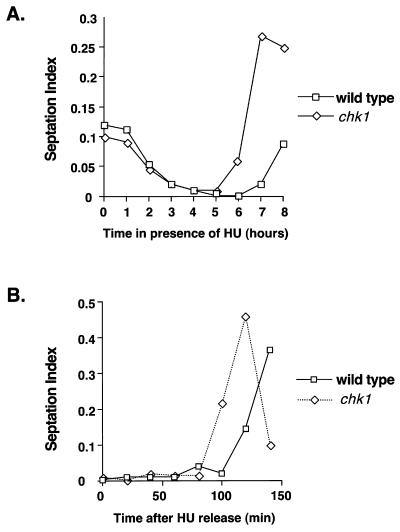
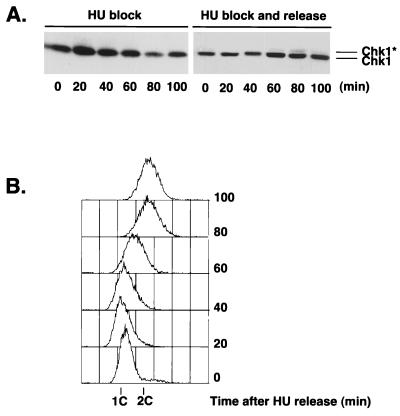
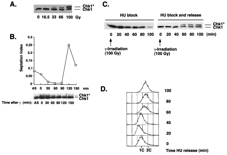
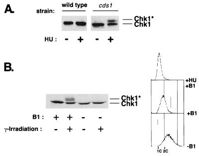
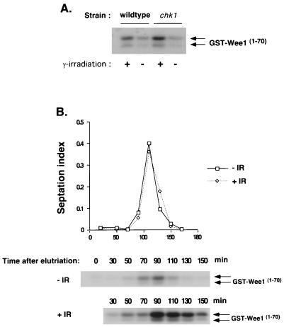
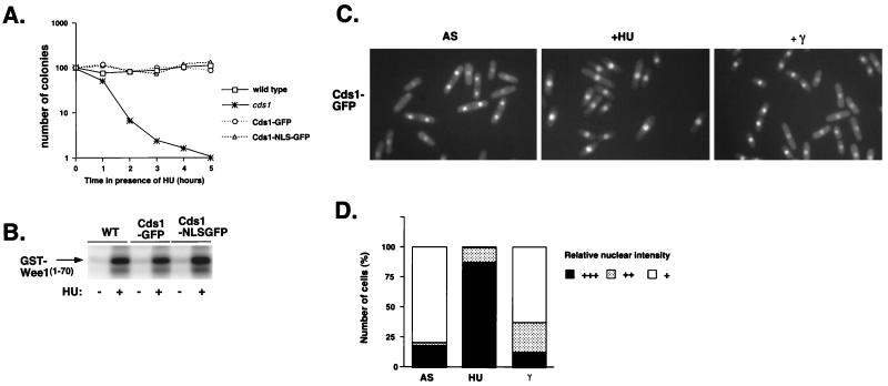
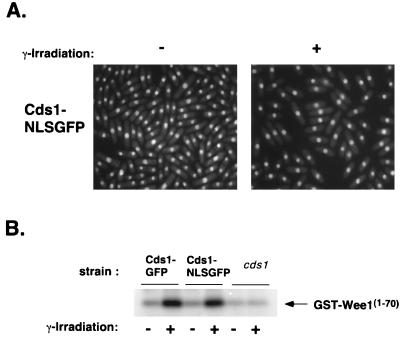
Six checkpoint Rad proteins (Rad1, Rad3, Rad9, Rad17, Rad26, and Hus1) are needed to regulate checkpoint protein kinases Chk1 and Cds1 in fission yeast. Chk1 is required to prevent mitosis when DNA is damaged by ionizing radiation (IR), whereas either kinase is sufficient to prevent mitosis when DNA replication is inhibited by hydroxyurea (HU). Checkpoint Rad proteins are required for IR-induced phosphorylation of Chk1 and HU-induced activation of Cds1. IR activates Cds1 only during the DNA synthesis (S) phase, whereas HU induces Chk1 phosphorylation only in cds1 mutants. Here, we investigate the basis of the checkpoint signal specificity of Chk1 phosphorylation and Cds1 activation. We show that IR fails to induce Chk1 phosphorylation in HU-arrested cells. Release from the HU arrest following IR causes substantial Chk1 phosphorylation. These and other data indicate that Cds1 prevents Chk1 phosphorylation in HU-arrested cells, which suggests that Cds1 actively suppresses a repair process that leads to Chk1 phosphorylation. Cds1 becomes more highly concentrated in the nucleus only during the S phase of the cell cycle. This finding correlates with S-phase specificity of IR-induced activation of Cds1. However, constitutive nuclear localization of Cds1 does not enhance IR-induced activation of Cds1. This result suggests that Cds1 activation requires DNA structures or protein activities that are present only during S phase. These findings help to explain how Chk1 and Cds1 respond to different checkpoint signals.


Figures

FIG. 1
FIG. 1
Chk1 delays mitosis during recovery from an HU-induced arrest. (A) Wild-type or chk1 cells were treated with HU, and the septation index was monitored at hourly intervals. The chk1 strain underwent division before the wild-type strain. (B) Wild-type or chk1 strains were treated with HU for 4 h. HU was then removed by washing cells in YES media, and the septation index was monitored every 20 min. Division was advanced in the chk1 cells relative to that in the wild type.
FIG. 2
FIG. 2
Chk1 is phosphorylated as cells recover from an HU-induced arrest. Cells were treated with HU for 4 h at 30°C. HU was removed from half of the culture by washing in YES medium. The other half of the culture was left in the presence of HU. Cells were harvested every 20 min. (A) Samples were processed for immunoblot analysis of Chk1. (B) Samples were processed for FACS analysis to determine DNA content after HU release. ∗, phosphorylated form of Chk1.
FIG. 3
FIG. 3
IR fails to cause Chk1 phosphorylation in cells arrested with HU. (A) An asynchronous culture of wild-type cells was exposed to γ-irradiation (0 to 100 Gy). Samples were processed immediately for immunoblot analysis of Chk1. (B) Wild-type cells were exposed to a 100-Gy dose of γ-irradiation. Samples were processed for immunoblot analysis of Chk1 and measurement of septation index during a 120-min time course. (C) Cells were treated with HU for 3.5 h at 30°C, followed by exposure to a 100-Gy dose of γ-irradiation. HU was removed from half of the culture by washing in YES medium. The other half of the culture was left in the presence of HU. Cells were harvested every 20 min. (D) Samples from the experiment described in the legend for panel B were processed for FACS analysis to determine the DNA content after HU release. ∗, phosphorylated form of Chk1.
FIG. 4
FIG. 4
Overproduction of Cds1 prevents phosphorylation of Chk1. (A) HU induces Chk1 phosphorylation in a cds1 background. Wild-type or cds1 cells were HU treated for 3.5 h. Samples were processed for immunoblot analysis of Chk1. (B) GST-Cds1 overexpression in G2 prevents phosphorylation of Chk1 that is induced by DNA damage. Cells that expressed GST-Cds1 under the control of the thiamine-repressible nmt1 promoter were grown in minimal media containing thiamine (+B1; nmt1-repressing conditions) or lacking thiamine (−B1; nmt1-inducing conditions) for 20 h. Cells were γ irradiated or mock irradiated. Samples were processed for FACS analysis to determine DNA content (upper panel) or for immunoblot analysis of Chk1 (lower panel). ∗, phosphorylated form of Chk1.
FIG. 5
FIG. 5
Chk1 does not prevent Cds1 activation in G2 phase. (A) Asynchronous cultures of wild-type or chk1 cells were irradiated with 100 Gy. Samples were harvested to measure Cds1 kinase activity by using GST-Wee11–152 as a substrate. (B) A chk1 strain was synchronized in G2 by elutriation. Cells were γ irradiated (+IR) or mock irradiated (−IR). Samples were harvested to measure septation index and Cds1 kinase activity by using GST-Wee11–152 as a substrate. The band shown corresponds to the GST-Wee11–70 degradation product as previously described (7). DNA damage incurred during the G2 phase activated Cds1 only after cells had completed mitosis and entered S phase. Arrows indicate two different forms of GST-Wee11–70.
FIG. 6
FIG. 6
Cds1 accumulates in the nucleus during S phase. (A) Addition of GFP or NLS-GFP to the C terminus of Cds1 does not compromise its function. The HU sensitivity of wild-type (WT), Cds1-GFP, Cds1-NLS-GFP, and cds1 cells was determined by monitoring the colony formation during the time course of HU exposure. (B) Modified forms of Cds1 behave the same as the WT in the Cds1 kinase assay following treatment or mock treatment with HU. (C) Cds1-GFP localization was determined by fluorescence microscopy in an asynchronous population (left panel), after 4 h of HU treatment (middle panel), or after a 100-Gy dose of irradiation (right panel). Cds1 is nuclear in septated cells and attached daughters. During HU-induced arrest, Cds1 is strongly nuclear. In contrast, after irradiation, Cds1 is not accumulated in cells arrested at G2. (D) Percentages of cells with relative nuclear staining intensities for Cds1 in an asynchronous population (AS), HU-arrested cells, or 100-Gy-irradiated cells (γ).
FIG. 7
FIG. 7
Cds1-NLS-GFP that is constitutively present in the nuclei of G2 cells is not activated by irradiation. (A) A strain expressing Cds1-NLS-GFP was grown in minimal media at 30°C. Cds1-NLS-GFP localization was determined by fluorescence microscopy in an asynchronous population (−γ) or after 100 Gy of irradiation (+ γ). Cds1 remains nuclear at all stages of the cell cycle, even during a DNA-damage-induced arrest at G2. (B) Activities of Cds1-GFP and Cds1-NLS-GFP after irradiation were measured with GST-Wee11–70 as a substrate. Cds1-NLS-GFP and Cds1-GFP are both weakly activated by irradiation, while a strain deleted for Cds1 has no detectable activity.

References

    1. Aligue R, Wu L, Russell P. Regulation of Schizosaccharomyces pombe Wee1 tyrosine kinase. J Biol Chem. 1997;272:13320–13325. - PubMed
    1. al-Khodairy F, Carr A M. DNA repair mutants defining G2 checkpoint pathways in Schizosaccharomyces pombe. EMBO J. 1992;11:1343–1350. - PMC - PubMed
    1. al-Khodairy F, Fotou E, Sheldrick K S, Griffiths D J, Lehmann A R, Carr A M. Identification and characterization of new elements involved in checkpoint and feedback controls in fission yeast. Mol Biol Cell. 1994;5:147–160. - PMC - PubMed
    1. Basi G, Schmid E, Maundrell K. TATA box mutations in the Schizosaccharomyces pombe nmt1 promoter affect transcription efficiency but not the transcription start point or thiamine repressibility. Gene. 1993;123:131–136. - PubMed
    1. Bentley N J, Holtzman D A, Flaggs G, Keegan K S, DeMaggio A, Ford J C, Hoekstra M, Carr A M. The Schizosaccharomyces pombe rad3 checkpoint gene. EMBO J. 1996;15:6641–6651. - PMC - PubMed

Publication types