. 1999 Jun;19(6):4311-23.
doi: 10.1128/MCB.19.6.4311.

Evidence that tristetraprolin binds to AU-rich elements and promotes the deadenylation and destabilization of tumor necrosis factor alpha mRNA

Affiliations

Evidence that tristetraprolin binds to AU-rich elements and promotes the deadenylation and destabilization of tumor necrosis factor alpha mRNA

W S Lai et al. Mol Cell Biol. 1999 Jun.

Abstract

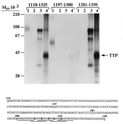
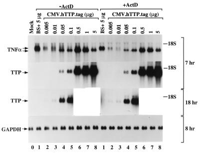
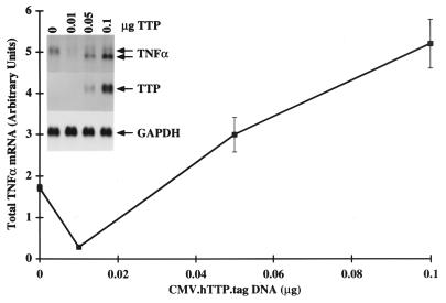
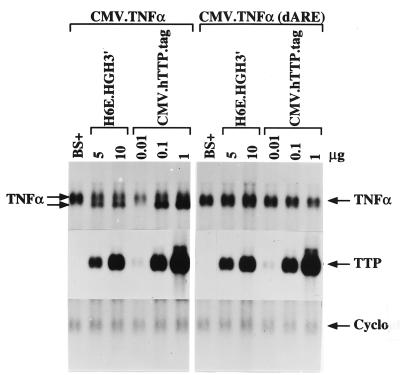
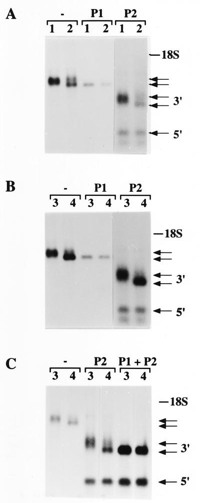
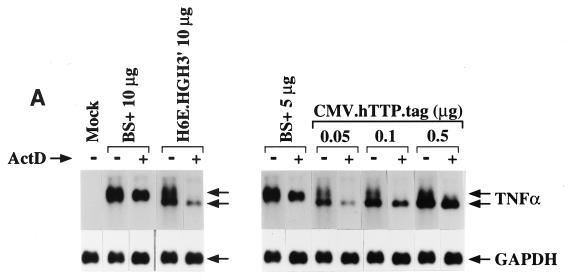
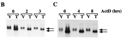
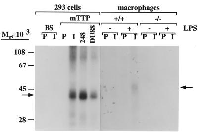
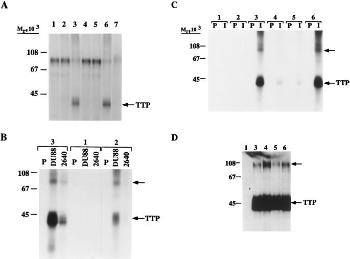
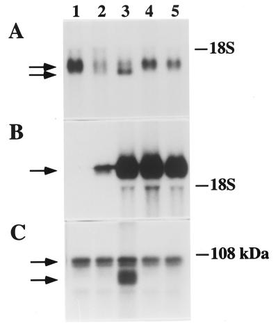
Mice deficient in tristetraprolin (TTP), the prototype of a family of CCCH zinc finger proteins, develop an inflammatory syndrome mediated by excess tumor necrosis factor alpha (TNF-alpha). Macrophages derived from these mice oversecrete TNF-alpha, by a mechanism that involves stabilization of TNF-alpha mRNA, and TTP can bind directly to the AU-rich element (ARE) in TNF-alpha mRNA (E. Carballo, W. S. Lai, and P. J. Blackshear, Science 281:1001-1005, 1998). We show here that TTP binding to the TNF-alpha ARE is dependent upon the integrity of both zinc fingers, since mutation of a single cysteine residue in either zinc finger to arginine severely attenuated the binding of TTP to the TNF-alpha ARE. In intact cells, TTP at low expression levels promoted a decrease in size of the TNF-alpha mRNA as well as a decrease in its amount; at higher expression levels, the shift to a smaller TNF-alpha mRNA size persisted, while the accumulation of this smaller species increased. RNase H experiments indicated that the shift to a smaller size was due to TTP-promoted deadenylation of TNF-alpha mRNA. This CCCH protein is likely to be important in the deadenylation and degradation of TNF-alpha mRNA and perhaps other ARE-containing mRNAs, both in normal physiology and in certain pathological conditions.


Figures

FIG. 1
FIG. 1
UV cross-linking of hTTP to TNF-α mRNA ARE probes. Cytosolic extracts were prepared from 293 cells transfected with 5 μg of either CMV.hTTP.tag or vector alone as described in Materials and Methods. Extract (20 μg of protein) was incubated with the indicated 32P-labeled TNF-α RNA probes (2 × 10⁶ cpm). The numbers at the top of each set refer to the base numbers in the mTNF-α mRNA, as shown at the bottom of the figure. Probe 1110–1325 contained approximately 35% U residues, probe 1197–1300 contained 40% U residues, and probe 1281–1350 contained 62% U residues. Heparin and yeast tRNA were then added to decrease nonspecific binding. After UV cross-linking of the probes to cellular proteins, RNases T1 and A were added to digest probe not cross-linked to protein. The RNase-resistant RNA-protein complexes were resolved by SDS–10% PAGE followed by autoradiography. Lanes 1, probe alone (5,000 cpm); lanes 2, probe (2 × 10⁶ cpm) treated with RNases T1 and A; lanes 3, extract (20 μg of protein) from 293 cells transfected with vector alone (5 μg of DNA); lanes 4, extract (20 μg of protein) from 293 cells transfected with CMV.hTTP.tag (5 μg). The position of TTP cross-linked to 32P-labeled RNA is indicated by the arrow. The positions of protein molecular weight standards are indicated on the left. Shown at the bottom is a portion of the mTNF-α mRNA 3′ UTR (GenBank accession no. X02611), from which the probes were derived. The five AU-rich nanomers are underlined. The five flanking A’s within the ARE that were mutated to form a nonbinding probe are indicated in boldface.
FIG. 2
FIG. 2
Effect of TTP on TNF-α mRNA stability. CMV.mTNF-α was cotransfected into 293 cells with either TTP expression constructs or vector alone. After addition of actinomycin D to a final concentration of 10 μg/ml (+ActD) or buffer alone (−ActD) for 4 h, total cellular RNA was harvested. Each lane was loaded with 10 μg of total RNA. Electrophoresis and Northern blot hybridization were performed as described in Materials and Methods. Lane 0, RNA from mock-transfected 293 cells. Lanes 1 to 8, RNA from 293 cells cotransfected with CMV.mTNF-α (1 μg) and vector or CMV.hTTP.tag as follows: lanes 1, vector alone (BS+; 5 μg/plate); lanes 2 to 8, CMV.hTTP.tag (0.005, 0.01, 0.05, 0.1, 0.5, 1, and 5 μg/plate, respectively). Vector was also added in lanes 2 to 7 to make the total amount of cotransfected plasmids 5 μg/plate. The Northern blots were probed with either a 32P-labeled mTNF-α cDNA or a 32P-labeled mTTP cDNA. The two arrows indicate the two species of TNF-α mRNA discussed in the text. The position of transfected-cell-expressed TTP mRNA is indicated by an arrow. Film exposure was 7 h for the filters hybridized with mTNF-α and TTP cDNAs, as indicated; portions of the same blot probed with the TTP cDNA were also exposed to film for 18 h to show the expression of TTP mRNA in 293 cells transfected with low concentrations of CMV.hTTP.tag. The positions of the 18S rRNA are indicated. The blot hybridized with the mTNF-α cDNA was stripped and reprobed with a GAPDH cDNA probe; the filter was then exposed to film for 8 h and is shown at the bottom to demonstrate equivalent loading.
FIG. 3
FIG. 3
Effect of low amounts of transfected CMV.hTTP.tag on the accumulation of TNF-α mRNA. 293 cells were cotransfected with CMV.mTNF-α and pXGH5 (1 μg each per plate) and either vector alone (5 μg/plate) or CMV.hTTP.tag (0.01, 0.05, and 0.1 μg/plate). Vector was also added to make the total amount of cotransfected plasmids 5 μg/plate. One day after replacement of the transfection medium, the medium was collected from each plate for the assay of released HGH. Total cellular RNA was then harvested. Each lane of the gel was loaded with 10 μg of total RNA. Electrophoresis and Northern blot hybridization were performed as described in Materials and Methods. The Northern blots were probed with either an mTNF-α cDNA probe or an mTTP cDNA probe and exposed to film. The blot that hybridized with the mTNF-α cDNA was stripped and reprobed with a GAPDH cDNA probe. The film showing the expressed mTNF-α mRNA was scanned with a laser scanning densitometer, and the results were normalized to HGH expression as well as to the PhosphorImager value for GAPDH mRNA. The graph shows the average results (± standard errors) from four such experiments; the inset shows the Northern blots from a representative experiment. The two species of TNF-α mRNA discussed in the text are indicated with two arrows; the positions of TTP mRNA and GAPDH mRNA are also indicated by arrows.
FIG. 4
FIG. 4
Effect of TTP expression on the expression of TNF-α constructs containing or lacking the ARE. CMV.mTNF-α (1 μg/plate) or CMV.mTNF-α (dARE) (1 μg/plate) was cotransfected into 293 cells with either vector alone (BS+, 5 μg/plate), H6E.HGH3′ (5 or 10 μg/plate), or CMV.hTTP.tag (0.01, 0.1, or 1 μg/plate). Vector was also added to make the total amount of cotransfected plasmids 5 μg/plate. Preparation of total cellular RNA, electrophoresis, and Northern blot analysis were performed as described in Materials and Methods. Each lane was loaded with 10 μg of total RNA. The Northern blots were probed with an mTNF-α cDNA, together with probes for cyclophilin (Cyclo) or mTTP as indicated. On the left, the arrows indicate the two species of mTNF-α mRNA formed in the presence of TTP; on the right, the arrow indicates the single band of mTNF-α mRNA expressed from the plasmid lacking the ARE. TTP mRNA expressed from the cotransfected plasmid and the endogenous cyclophilin mRNA are also indicated by arrows.
FIG. 5
FIG. 5
Evidence that the smaller species of TNF-α mRNA formed in the presence of TTP is a deadenylated intermediate. 293 cells were cotransfected with CMV.mTNF-α (1 μg/plate) and either vector alone or TTP expression constructs as follows: lanes 1, vector alone (10 μg/plate); lanes 2, H6E.HGH3′ (10 μg/plate); lanes 3, vector alone (5 μg/plate); lanes 4, CMV.hTTP.tag (5 μg/plate). As indicated, the RNA samples were treated with 0.8 U of RNase H or not treated (−) as described in Materials and Methods. In panels A and B, 5 μg of RNA was loaded into each lane; in panel C, 3 μg of RNA was loaded into each lane when no RNase H (−) was used. P1, oligonucleotide poly(dT)12–18 (0.5 μg) was added to 10 μg of 293 cell RNA. P2, an oligonucleotide (0.6 μg) complementary to bases 506 to 528 of the TNF-α mRNA was added to 10 μg of 293 cell RNA. P1 + P2, both oligonucleotides were added to 15 μg of 293 cell RNA (C). The Northern blots were probed with an mTNF-α cDNA. The position of the 18S rRNA is indicated. The two pairs of arrows in each panel indicate TNF-α mRNA species that contained (top arrow of each pair) or did not contain (bottom arrow of each pair) their poly(A) tails. The top pair of arrows in each panel points to full-length and deadenylated TNF-α mRNA; the bottom pair of arrows in each panel (3′) points to the 810-bp 3′ fragment of TNF-α mRNA and its deadenylated form in lanes P2-2 and P2-4. The single arrow at the bottom (5′) indicates the ∼400-bp 5′ fragment of TNF-α mRNA, which is the same size in control and in TTP-expressing cells.
FIG. 6
FIG. 6
Effect of TTP on the formation of the two species of TNF-α mRNA in the presence and absence of actinomycin D. CMV.mTNF-α (1 μg/plate) was cotransfected into 293 cells with either TTP expression constructs, as indicated, or vector alone (BS+). After addition of buffer alone (−) or actinomycin D (ActD) to a final concentration of 10 μg/ml (+), total cellular RNA was harvested. Each lane was loaded with 10 μg of total RNA. Electrophoresis and Northern blot hybridization were performed as described in Materials and Methods. (A) (Left) TNF-α and GAPDH mRNA from mock-transfected 293 cells or from cells cotransfected with CMV.mTNF-α and vector alone (10 μg/plate) or CMV.mTNF-α with TTP expression construct H6E.HGH3′ (10 μg/plate). (Right) TNF-α and GAPDH mRNA from 293 cells cotransfected with CMV.mTNF-α and vector alone (5 μg/plate) or CMV.mTNF-α with CMV.hTTP.tag (0.05, 0.1, and 0.5 μg/plate). Vector was also added to make the total amount of cotransfected plasmids 5 μg/plate. Actinomycin D (+) was added for 4 h as indicated. The two arrows labeled TNF-α indicate the two species of TNF-α mRNA formed in the presence of TTP. (B) H6E.HGH3′ (T) (10 μg/plate) or an equivalent amount of vector (V) was used in the cotransfection, and actinomycin D (10 μg/ml) was added for 0, 2, and 3 h as indicated. (C) CMV.hTTP.tag (T) (0.1 μg/plate) or an equivalent amount of vector (V) was used, and actinomycin D (10 μg/ml) was added for 0, 4, and 8 h as indicated. The two arrows indicate the two forms of TNF-α mRNA in panels B and C.
FIG. 6
FIG. 6
Effect of TTP on the formation of the two species of TNF-α mRNA in the presence and absence of actinomycin D. CMV.mTNF-α (1 μg/plate) was cotransfected into 293 cells with either TTP expression constructs, as indicated, or vector alone (BS+). After addition of buffer alone (−) or actinomycin D (ActD) to a final concentration of 10 μg/ml (+), total cellular RNA was harvested. Each lane was loaded with 10 μg of total RNA. Electrophoresis and Northern blot hybridization were performed as described in Materials and Methods. (A) (Left) TNF-α and GAPDH mRNA from mock-transfected 293 cells or from cells cotransfected with CMV.mTNF-α and vector alone (10 μg/plate) or CMV.mTNF-α with TTP expression construct H6E.HGH3′ (10 μg/plate). (Right) TNF-α and GAPDH mRNA from 293 cells cotransfected with CMV.mTNF-α and vector alone (5 μg/plate) or CMV.mTNF-α with CMV.hTTP.tag (0.05, 0.1, and 0.5 μg/plate). Vector was also added to make the total amount of cotransfected plasmids 5 μg/plate. Actinomycin D (+) was added for 4 h as indicated. The two arrows labeled TNF-α indicate the two species of TNF-α mRNA formed in the presence of TTP. (B) H6E.HGH3′ (T) (10 μg/plate) or an equivalent amount of vector (V) was used in the cotransfection, and actinomycin D (10 μg/ml) was added for 0, 2, and 3 h as indicated. (C) CMV.hTTP.tag (T) (0.1 μg/plate) or an equivalent amount of vector (V) was used, and actinomycin D (10 μg/ml) was added for 0, 4, and 8 h as indicated. The two arrows indicate the two forms of TNF-α mRNA in panels B and C.
FIG. 7
FIG. 7
UV cross-linking and immunoprecipitation of TTP-RNA complexes from 293 cells or TTP+/+ or TTP−/− macrophages. Cytosolic extracts from 293 cells transfected either with CMV.mTTP (5 μg) or with vector (BS) alone (5 μg) were prepared as described in Materials and Methods. Cytosolic extracts from TTP+/+ or TTP−/− macrophages untreated (−) or treated with (+) 1 μg of LPS per ml for 4 h were prepared as described elsewhere (7). Incubation of extracts (each sample contained 20 μg of protein from 293 cells or 40 μg of macrophage protein) with the 32P-labeled TNF-α probe (1281–1350), UV cross-linking, and RNase digestion were performed as described in Materials and Methods. The samples were then precleared with preimmune serum and divided into two portions, which were then incubated with preimmune serum (P), a polyclonal antibody to an mTTP–glutathione S-transferase fusion protein (I), a polyclonal antibody raised against an amino-terminal peptide of mTTP (248), or a polyclonal antibody to an hTTP–glutathione S-transferase fusion protein (DU88). The immunoprecipitated complexes were resolved by SDS-PAGE (10% polyacrylamide gel) and autoradiography. Film exposure was 8 h for the 293 cell gel and 6 days for the macrophage gel. The positions of radiolabeled transfected-cell-expressed TTP (293 cells) and endogenous macrophage TTP are indicated by the arrows. The positions of protein molecular weight standards are indicated.
FIG. 8
FIG. 8
UV cross-linking assays of TTP–mTNF-α–ARE complexes. Cytosolic extracts of 293 cells transfected with either vector alone or constructs expressing hTTP were prepared as described in Materials and Methods. (A) Lanes 1, extracts from 293 cells transfected with 5 μg of vector plasmid; lanes 2, extracts from 293 cells transfected with 10 μg of plasmid H6E.HGH3′; lanes 3 to 6, extracts from 293 cells transfected with 5 μg of wild-type CMV.hTTP.tag (lane 3), zinc finger mutant C124R (lane 4), zinc finger mutant C147R (lane 5), or phosphorylation site mutant S228A (lane 6); lane 7, TNF-α probe (1197–1350) alone (5,000 cpm). UV cross-linking and RNase digestion were performed as described in Materials and Methods. The RNA-protein complexes were resolved by SDS-PAGE (10% polyacrylamide gel) followed by autoradiography. The bands at ∼42,000 Mr in lanes 3 and 6 (arrow) represent radiolabeled TTP. Note the endogenous cellular protein of ∼80,000 in Mr that is cross-linked to the TNF-α ARE in the absence of expressed TTP; this cross-linking was markedly decreased in the presence of TTP (lanes 3 and 6). (B) UV cross-linked and RNase-digested samples as described for panel A (lanes 1 to 3) were precleared with preimmune serum and divided into three portions that were incubated with preimmune serum (P), a polyclonal antibody to an hTTP–glutathione S-transferase fusion protein (DU88), or a polyclonal antibody to an mTTP–glutathione S-transferase fusion protein (2640). The immunoprecipitated complexes were resolved by SDS-PAGE (10% polyacrylamide gel) and autoradiography. The higher-molecular-weight immunoprecipitated complex is indicated by the upper arrow, and the position of TTP is indicated by the lower one. (C) UV cross-linked and RNase-digested samples as described for panel A (lanes 1 to 6) were precleared with preimmune serum and divided into two portions that were incubated with preimmune serum (P) or a polyclonal anti-epitope tag antibody (I). The immunoprecipitated complexes were resolved by SDS-PAGE (10% polyacrylamide gel) and autoradiography. A higher-molecular-weight immunoprecipitated complex is indicated by the upper arrow, and the position of TTP is indicated by the lower one. (D) Five micrograms of protein extract prepared from 293 cells as described above (lanes 1 and 3 to 6) was analyzed by Western blotting with a polyclonal antibody to the epitope tag of the fusion protein hTTG.tag. The positions of molecular weight standards (in thousands) are indicated to the left of each gel. The position of TTP is indicated by the lower arrow; the position of the ∼100,000-Mr complex that contains TTP is indicated by the upper arrow.
FIG. 9
FIG. 9
Electrophoretic mobility shift assays of TTP–TNF-α–ARE complexes. Cytosolic extracts of 293 cells transfected with either vector alone or constructs expressing hTTP were prepared as described in Materials and Methods. Ten micrograms of protein extract was used in the incubation with 105 cpm of a TNF-α ARE probe, and RNA mobility shift assays were performed as described in Materials and Methods. Lanes 1, extracts from 293 cells transfected with 5 μg of vector plasmid; lanes 3, extracts from 293 cells transfected with 10 μg of plasmid H6E.HGH3′; lanes 2 and 4 to 6, extracts from 293 cells transfected with 5 μg of wild-type CMV.hTTP.tag (lane 2), zinc finger mutant C124R (lane 4), zinc finger mutant C147R (lane 5), or phosphorylation site mutant S228A (lane 6). (A) RNA-protein complex migration patterns were compared in the presence of wild-type TTP (lanes 2 and 3) and its zinc finger mutants (lanes 4 and 5) or its phosphorylation site mutant (lane 6). P, mTNF-α probe alone (1197–1350) after digestion with RNase T1. (B) RNA-protein complex migration patterns were compared as described for panel A, in the absence (no Ab) or presence (HA.11) of a polyclonal anti-epitope tag antibody. The supershifted RNA-protein complexes (SS) are indicated by the arrow. RNA-protein complexes I, II, and III are indicated in both panels. P, mTNF-α probe alone (1197–1350) after digestion with RNase T1.
FIG. 10
FIG. 10
TTP zinc finger mutants are ineffective at promoting the size shift of TNF-α mRNA. CMV.mTNF-α (1 μg/plate) was cotransfected into 293 cells with 5 μg of vector alone per plate (lanes 1) or 5 μg of wild-type CMV.hTTP.tag (lane 2), phosphorylation site mutant S228A (lane 3), zinc finger mutant C124R (lane 4), or zinc finger mutant C147R (lane 5). For lanes 6 to 8, 293 cells were transfected with 10 μg of wild-type plasmid H6E.HGH3′ (lane 6) or its zinc finger mutants (C124R, lane 7; C147R, lane 8). Preparation of total cellular RNA, electrophoresis, and Northern blot analysis were performed as described in Materials and Methods. Each lane was loaded with 10 μg of total RNA. The Northern blots were probed with a 32P-labeled mTNF-α cDNA (upper panel) or mTTP cDNA (lower panel). In the upper panel, the arrows indicate the two species of mTNF-α mRNA. In the lower panel, the arrow indicates the position of transfected-cell-expressed TTP mRNA. The position of the 18S rRNA is indicated.
FIG. 11
FIG. 11
Expression of hTTP-GFP fusion protein in 293 cells. 293 cells were transfected with either EGFP-N1 or hTTP-GFP fusion constructs, as described in Materials and Methods. Twenty-four hours after the transfection, cells were analyzed for GFP expression by confocal fluorescence microscopy with a fluorescein isothiocyanate filter. (A) Cells transfected with EGFP-N1 (5 μg); (B) the same field as in panel A under phase-contrast microscopy showing several nontransfected cells that were nonfluorescent; (C and D) cells transfected with CMV.hTTP.EGFP (10 μg); (E and F) cells transfected with CMV.hTTP.EGFP (5 μg); (G and H) cells transfected with H6E.EGFP (10 μg).
FIG. 12
FIG. 12
Effect of GFP-TTP on TNF-α mRNA stability and binding to the ARE of mTNF-α mRNA. (A and B) CMV.mTNF-α (1 μg/plate) was cotransfected into 293 cells with 5 μg of either vector alone (lane 1), H6E.EGFP (lane 2), CMV.hTTP.EGFP (lane 3), or the zinc finger mutants (C124R, lane 4; C147R, lane 5) of CMV.hTTP.EGFP per plate. Preparation of total cellular RNA, electrophoresis, and Northern blot analysis were performed as described in Materials and Methods. Each lane was loaded with 10 μg of total RNA. The Northern blots were probed with 32P-labeled mTNF-α cDNA (A) or mTTP cDNA (B). In panel A, the arrows indicate the two species of mTNF-α mRNA; the position of the 18S rRNA is also indicated. In panel B, the positions of TTP-GFP (arrow) and the 18S rRNA are indicated. (C) 293 cells were transfected with the same plasmids as described for panels A and B but without the CMV.mTNF-α. Cytosolic extracts were prepared, and 20 μg of total cytosolic protein per sample was used in UV cross-linking assays with a 32P-labeled mTNF-α RNA (1281–1350) probe, followed by SDS-PAGE (10% polyacrylamide gel) and autoradiography. The top arrow indicates the position of the radiolabeled endogenous cellular protein with an Mr of ∼80,000; the bottom arrow indicates the radiolabeled TTP-GFP fusion protein. The position of the 108-kDa protein standard is indicated.

Similar articles

Cited by

References

    1. Akashi M, Shaw G, Gross M, Saito M, Koeffler H P. Role of AUUUA sequences in stabilization of granulocyte-macrophage colony-stimulating factor RNA in stimulated cells. Blood. 1991;78:2005–2012. - PubMed
    1. Blackshear P J. Systems for polyacrylamide gel electrophoresis. Methods Enzymol. 1984;104:237–255. - PubMed
    1. Bohjanen P R, Petryniak B, June C H, Thompson C B, Lindsten T. An inducible cytoplasmic factor (AU-B) binds selectively to AUUUA multimers in the 3′ untranslated region of lymphokine mRNA. Mol Cell Biol. 1991;11:3288–3295. - PMC - PubMed
    1. Bohjanen P R, Petryniak B, June C H, Thompson C B, Lindsten T. AU RNA-binding factors differ in their binding specificities and affinities. J Biol Chem. 1992;267:6302–6309. - PubMed
    1. Caput D, Beutler B, Hartog K, Thayer R, Brown-Shimer S, Cerami A. Identification of a common nucleotide sequence in the 3′-untranslated region of mRNA molecules specifying inflammatory mediators. Proc Natl Acad Sci USA. 1986;83:1670–1674. - PMC - PubMed

MeSH terms