. 1999 Jun;19(6):4343-54.
doi: 10.1128/MCB.19.6.4343.

timrit Lengthens circadian period in a temperature-dependent manner through suppression of PERIOD protein cycling and nuclear localization

Affiliations

timrit Lengthens circadian period in a temperature-dependent manner through suppression of PERIOD protein cycling and nuclear localization

A Matsumoto et al. Mol Cell Biol. 1999 Jun.

Abstract

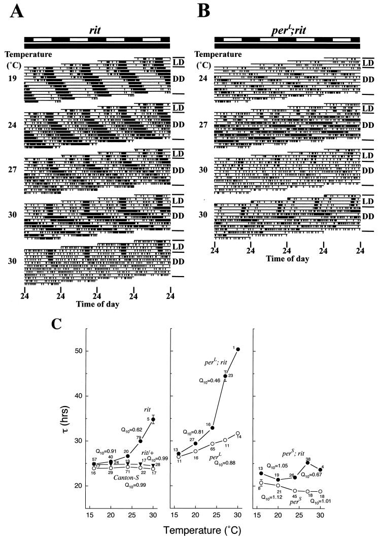
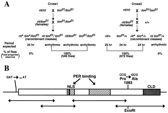
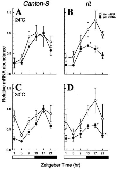
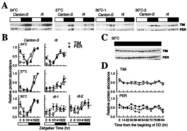
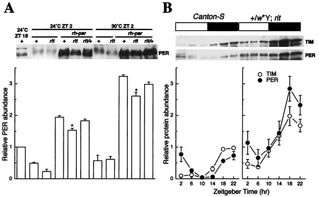
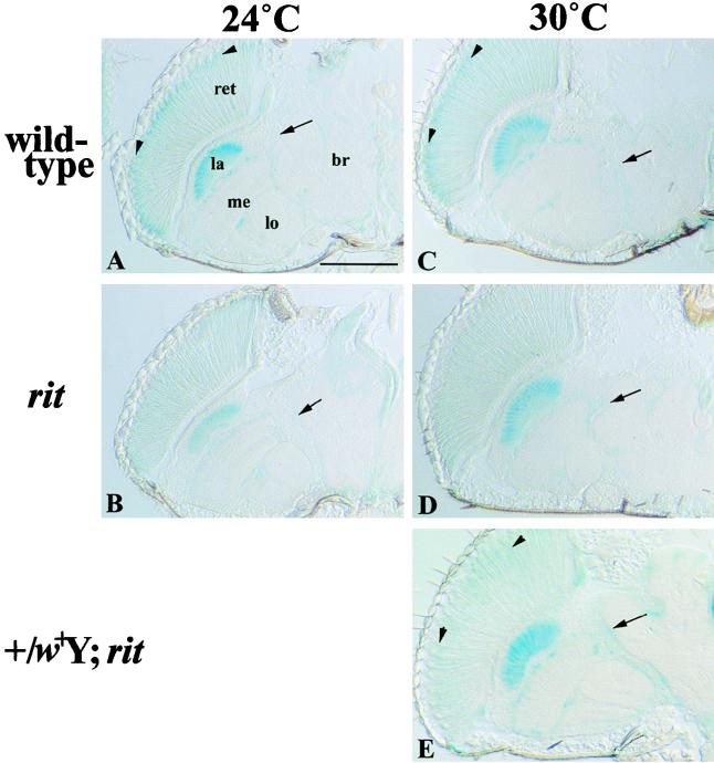
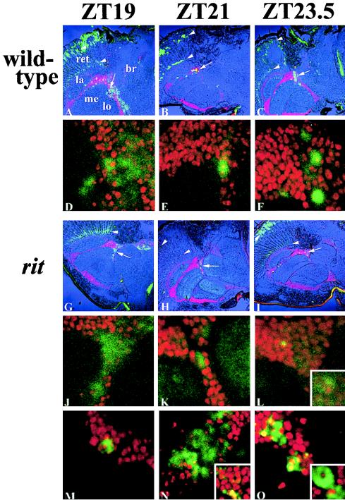
A fundamental feature of circadian clocks is temperature compensation of period. The free-running period of ritsu (timrit) (a novel allele of timeless [tim]) mutants is drastically lengthened in a temperature-dependent manner. PER and TIM protein levels become lower in timrit mutants as temperature becomes higher. This mutation reduces per mRNA but not tim mRNA abundance. PER constitutively driven by the rhodopsin1 promoter is lowered in rit mutants, indicating that timrit mainly affects the per feedback loop at a posttranscriptional level. An excess of per+ gene dosage can ameliorate all rit phenotypes, including the weak nuclear localization of PER, suggesting that timrit affects circadian rhythms by reducing PER abundance and its subsequent transportation into nuclei as temperature increases.


Figures

FIG. 1
FIG. 1
rit lengthens circadian periods as temperature increases. (A and B) Locomotor activity records at various temperatures for rit (A) and perL; rit (B) flies. Flies were held in LD12:12 for the first 3 days and then kept in DD. Light regimens (white bars, light; black bars, dark) are indicated above the tetraplotted (4 days) actograms. (C) Changes in period at various temperatures. Mean values at 20, 28, and 30°C were calculated to combine data at 19 and 20, 27 and 28, and 29 and 30°C, respectively. ○, strains carrying no mutation on the second chromosome (Canton-S, perL, and perS); ●, strains carrying the rit mutation (rit, perL; rit and perS; rit). ▾, heterozygous rit/+ strain. Vertical bars show standard errors of the means. Numbers beside symbols represent the numbers of rhythmic flies. Q10 values are separately represented when the changes in period are different between the temperature ranges below and above 24°C.
FIG. 2
FIG. 2
Duplication of the per+ and tim+ genes restores the extra long period in rit flies. (A) The rit phenotype is restored by the per+ gene translocation in a dosage-dependent manner. +/w+Y; rit is the rit male carrying the per+ gene translocation on the Y chromosome; C(1)DX represents females carrying the attached X chromosome. (B) Complementation test with tim01. tim01 does not complement rit, and the tim+ gene translocation restores the rit phenotype. The per+ gene translocation also restores the phenotype shown in rit/tim flies. (C) The rit phenotype is restored by the tim duplication. Dp(2;Y)odd4.13 and Dp(2;Y)odd2.31 carry the tim+ gene translocation on the Y chromosome. Numbers beside symbols represent the numbers of flies showing rhythmicity at each temperature.
FIG. 3
FIG. 3
rit is a novel allele of tim. (A) Mating schemes to test whether recombination occurs between rit and tim01. All genotypes and their expected period in the second generation, assuming that recombination occurs, are listed. Rhythmicities were judged by chi-square periodogram analysis (43) ranging from 19 to 29 h. The arrhythmic category includes flies showing an extra long period over the circadian range. See Results for details. (B) Schematic representation of the coding region in the timrit cDNA. The coding sequence is indicated by a box. The arrow lines indicate PCR fragments amplified for sequencing. Amino acid numbering is as specified by Myers et al. (29); domains indicated by closed boxes are based on the study by Saez and Young (37). Met, translation start; NLS, nuclear localization signal; CLD, cytoplasmic localization domain.
FIG. 4
FIG. 4
Daily patterns of per and tim mRNA cyclings under LD. RNase protection assays were performed on total RNAs from wild-type and rit flies entrained in LD12:12 at 24°C (A and B) and at 30°C (C and D). See Materials and Methods for details. Values at each point are means of three to eight experiments. Vertical bars show standard errors of the means. Light regimens are indicated (white bars, light; black bars, dark). We defined the lights-on point as ZT0 and the lights-off point as ZT12. Asterisks indicate that the mean value for rit flies is significantly different from that for wild-type flies (t test, P < 0.05).
FIG. 5
FIG. 5
PER and TIM abundance in wild-type and rit flies at various temperatures. Adult head homogenates were obtained from flies entrained at 24, 27, and 30°C and subjected to Western blot analysis using anti-TIM antibody followed by anti-PER antibody as described in Materials and Methods. TIM and PER bands in panel A were quantified by densitometry. Measurements obtained at each point were normalized by the maximum value for wild-type flies under each condition. (B) Mean abundances of TIM and PER. Values at each point were means of three to nine experiments. Vertical bars show standard errors of the means. Asterisks indicate that the mean value for rit flies is significantly different from that for wild-type flies (t test, P < 0.05). Data for rit flies obtained at 30°C were classified into two groups (bottom row in panel B; see also Results for details). (C and D) Under constant darkness at 30°C, no or very weak cycling is indicated by the data from 8 h of sampling in both proteins. Experiments were independently done two and three times for TIM and PER, respectively (D). Light regimens are indicated (white symbols, light; black symbols, dark).
FIG. 6
FIG. 6
rit affects PER abundance at a posttranscriptional level. (A) Constitutive expression of PER, using the rh1-per fusion gene. Flies were collected at ZT2 except for one wild-type strain collected at ZT18 as a control. PER bands detected by Western blot analysis using anti-PER antibody were quantified and were normalized by the PER level of the wild type. +, wild-type (Canton-S); rit/+, heterozygous rit; rh-per, protein extracts were obtained from flies carrying the rh-per construct. Lysates were obtained from the following strains: wild type as a control (lane 1), wild type (lanes 2 and 7), rit (lanes 3 and 8), rh-per (lanes 4 and 9), rit; rh-per (lanes 5 and 10), and rit/+; rh-per (lanes 6 and 12). Values at each point were means of four experiments. Asterisks indicate that the mean value for rit; rh-per flies is significantly different from that for rh-per flies at the same temperature (t test, P < 0.05). Vertical bars show standard errors of the means. (B) Daily fluctuation of PER and TIM in rit flies with an excess per gene dosage. Adult head homogenates were obtained from flies entrained at 30°C. Values at each point were normalized by the maximum value for the wild type. Values at each point were means of four experiments. Vertical bars show standard errors of the means. Light regimens are indicated (white bars, light; black bars, dark).
FIG. 7
FIG. 7
Nuclear localization of PER is impaired in rit flies. Horizontal section of fly heads stained with X-Gal (A to D and E). The spatial pattern of per gene expression was monitored by using the per-lacZ fusion gene. Horizontal head sections in wild-type and rit flies kept at 24°C (A and B) or 30°C (C and D) were stained for 2 h at 37°C. per was expressed in retina (ret), optic lobes (lamina [la], medulla [me], and lobula [lo]), and central brain (br) in wild-type flies (A and C). Nuclei in photoreceptor cells are clearly stained (arrowheads) in wild-type but not rit flies at both 24 and 30°C. Staining of LNs is indicated by black arrows. The expression level in the PER–β-Gal fusion protein in rit flies is lower than that in wild-type flies. Such a weak PER localization in nuclei was rescued by the excess of per+ gene dosage in +/w+Y; rit even at 30°C (E). The scale bar represents 80 μm.
FIG. 8
FIG. 8
PER localization in lateral neurons at 30°C. Horizontal sections of fly heads obtained at ZT19, -21, and -23.5 were stained with anti-PER antibody coupled to FITC (green color). Counterstaining of nuclei was done with propidium iodide (red color) after RNase treatment. When PER staining overlaps nuclear staining, yellow color is observed. For symbols, see the legend to Fig. 7. In wild-type flies (A to C), PER signals are comparable to those seen with X-Gal staining in Fig. 7C. LNs at a magnification of ×8 are represented in panels D to F. PER signal is observed in the cytoplasm at ZT19 and in nuclei at both ZT21 and ZT23.5. In rit flies (G to I), staining similar to that in wild-type flies can be observed, although the PER signal is weaker especially in the LNs (J to L). Thus, images in which PER signals were amplified are illustrated (M to O). PER is in the cytoplasm at ZT19 (J and M). At ZT21, there are three patterns of PER staining surrounding a nucleus (center and right upper corner in panel N), overlapping in a nucleus of the center of a broad PER staining area (middle left in panel N), and just overlapping in nuclei (inset in panel N). At ZT23.5, PER enters nuclei (O) or stays in the cytoplasm (inset in panel O).

References

    1. Allada R, White N E, So W V, Hall J C, Rosbash M. A mutant Drosophila homolog of mammalian Clock disrupts circadian rhythms and transcription of period and timeless. Cell. 1998;93:791–804. - PubMed
    1. Baylies M K, Vosshall L B, Sehgal A, Young M W. New short period mutations of the Drosophila clock gene per. Neuron. 1992;9:575–581. - PubMed
    1. Cooper M K, Hamblen-Coyle M J, Liu X, Rutila J E, Hall J C. Dosage compensation of the period gene in Drosophila melanogaster. Genetics. 1994;138:721–732. - PMC - PubMed
    1. Cote G G, Brody S. Circadian rhythms in Drosophila melanogaster: analysis of period as a function of gene dosage at the per (period) locus. J Theor Biol. 1986;121:487–503. - PubMed
    1. Curtin K D, Huang Z J, Rosbash M. Temporally regulated nuclear entry of the Drosophila period protein contributes to the circadian clock. Neuron. 1995;14:365–372. - PubMed

Publication types

MeSH terms

LinkOut - more resources