. 1999 Jun;19(6):4355-65.
doi: 10.1128/MCB.19.6.4355.

Cooperative interaction between GATA-4 and GATA-6 regulates myocardial gene expression

Affiliations

Cooperative interaction between GATA-4 and GATA-6 regulates myocardial gene expression

F Charron et al. Mol Cell Biol. 1999 Jun.

Abstract

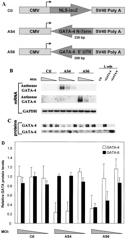
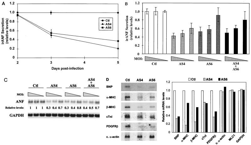
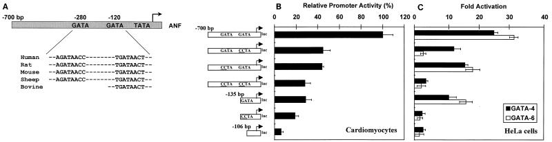
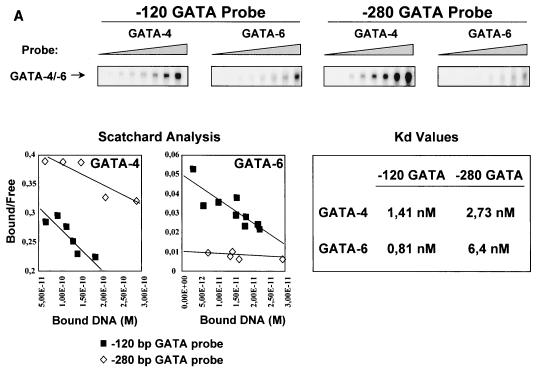
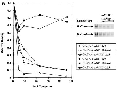
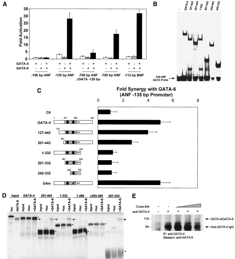
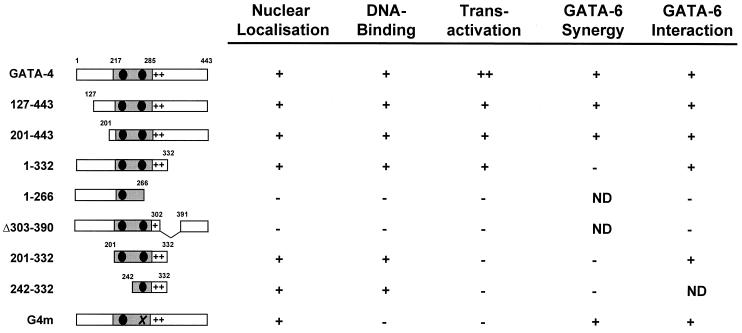
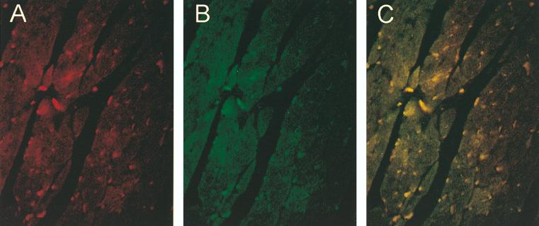
Two members of the GATA family of transcription factors, GATA-4 and GATA-6, are expressed in the developing and postnatal myocardium and are equally potent transactivators of several cardiac promoters. However, several in vitro and in vivo lines of evidence suggest distinct roles for the two factors in the heart. Since identification of the endogenous downstream targets of GATA factors would greatly help to elucidate their exact functions, we have developed an adenovirus-mediated antisense strategy to specifically inhibit GATA-4 and GATA-6 protein production in postnatal cardiomyocytes. Expression of several endogenous cardiac genes was significantly down-regulated in cells lacking GATA-4 or GATA-6, indicating that these factors are required for the maintenance of the cardiac genetic program. Interestingly, transcription of some genes like the alpha- and beta-myosin heavy-chain (alpha- and beta-MHC) genes was preferentially regulated by GATA-4 due, in part, to higher affinity of GATA-4 for their promoter GATA element. However, transcription of several other genes, including the atrial natriuretic factor and B-type natriuretic peptide (ANF and BNP) genes, was similarly down-regulated in cardiomyocytes lacking one or both GATA factors, suggesting that GATA-4 and GATA-6 could act through the same transcriptional pathway. Consistent with this, GATA-4 and GATA-6 were found to colocalize in postnatal cardiomyocytes and to interact functionally and physically to provide cooperative activation of the ANF and BNP promoters. The results identify for the first time bona fide in vivo targets for GATA-4 and GATA-6 in the myocardium. The data also show that GATA factors act in concert to regulate distinct subsets of genes, suggesting that combinatorial interactions among GATA factors may differentially control various cellular processes.


Figures

FIG. 1
FIG. 1
Cardiomyocytes are efficiently infected by adenovirus. Ventricular cardiomyocytes isolated from 4-day neonatal rats were mock infected (A) or infected at MOIs of 1 (B), 4 (C), and 16 (D) with the Ctl adenovirus, which expresses an NLS-lacZ gene. Twenty hours later, cells were assayed for β-galactosidase activity.
FIG. 2
FIG. 2
Characterization of AS4 and AS6. (A) Schematic representation of the constructs used to generate the recombinant adenoviruses. NLS-lacZ, a 358-bp fragment from the extreme N-terminal portion of GATA-4, or a 359-bp fragment from the 5′ UTR of GATA-6 was cloned downstream of the CMV promoter and upstream of an SV40 poly(A) sequence in order to generate the recombinant adenoviruses. (B) Transgene expression is dose dependent. Ventricular cardiomyocytes were infected at MOIs of 16, 4, and 1 (corresponding to the progressively narrowing triangle) with Ctl, AS4, and AS6. RNA was isolated 3 days postinfection. Northern blot analysis showed that both antisense transgenes were efficiently expressed in a dose-dependent manner. (C) AS4 and AS6 are specific and efficient at decreasing GATA-4 and GATA-6 protein levels. Ventricular cardiomyocytes were infected as described above, and nuclear extracts were isolated 3 days postinfection and analyzed by Western blotting. L cells overexpressing GATA-4 or GATA-6 were used as controls for the specificity of the antibodies. (D) Quantification of the effects of AS4 and AS6 on GATA-4 and GATA-6 protein levels. The data represent the means of two independent Western blots performed as described for panel C and quantified by densitometry. At an MOI of 4, GATA-4 levels were decreased specifically by AS4 (80% reduction), while GATA-6 levels were reduced specifically by AS6 (60% reduction).
FIG. 3
FIG. 3
In vivo transcriptional targets for GATA-4 and GATA-6 in postnatal cardiomyocytes. (A) irANF secretion is decreased in a time-dependent manner by AS4 and AS6. Ventricular cardiomyocytes were infected at an MOI of 4 with Ctl, AS4, and AS6. Secreted irANF was assayed in the cardiomyocyte culture medium after a 24-h accumulation period at 2, 3, and 5 days postinfection. Since the effect of the antisense DNA on irANF secretion is clearly visible at 3 days postinfection, subsequent analyses were performed at this time point. (B) irANF secretion is decreased in a dose-dependent manner by AS4, AS6, and AS4 plus AS6. Ventricular cardiomyocytes were infected at MOIs of 16, 4, and 1 (corresponding to the progressively narrowing triangle) with Ctl, AS4, AS6, and AS4 plus AS6. (C) ANF mRNA levels are decreased in a dose-dependent manner by AS4, AS6, and AS4 plus AS6. Ventricular cardiomyocytes were infected as for panel B, and total RNA was analyzed by Northern blotting. The ANF mRNA levels are expressed relative to that of Ctl-infected cardiomyocytes. The GAPDH mRNA levels were unaffected. (D) Many cardiac genes are decreased by AS4 and AS6. Ventricular cardiomyocytes were infected at an MOI of 4 with Ctl, AS4, and AS6, and Northern blot analysis was performed on poly(A)+ RNA (left). Relative mRNA levels were quantified with a PhosphorImager (right). Note how α-MHC and β-MHC mRNAs were preferentially down-regulated by AS4, whereas cardiac (c.) α-actin, myosin light-chain 1 (MLC1), and GAPDH mRNA levels were not affected by AS4 or AS6.
FIG. 4
FIG. 4
ANF is a direct transcriptional target for both GATA-4 and GATA-6. (A) Alignment of sequences of human, rat, mouse, sheep, and bovine (for which only a partial sequence is available) ANF promoters. Note that two consensus WGATAR elements, a proximal one at −120 bp and a distal one at −280 bp, are entirely conserved across species. (B) The proximal and distal GATA elements are major contributors of ANF promoter activity in neonatal cardiomyocytes. Wild-type and mutated ANF promoters fused to the luciferase (luc) reporter gene were transiently transfected into ventricular cardiomyocytes isolated from 4-day neonatal rats. Luciferase activity was assayed 36 h posttransfection. The results are expressed relative to the −700 bp ANF promoter activity. (C) GATA-4 and GATA-6 transactivate the ANF promoter. HeLa cells were cotransfected with a GATA-4 or GATA-6 expression vector and wild-type or mutated ANF promoters fused to the luciferase reporter gene. Luciferase activity was assayed 36 h posttransfection. The results are expressed as fold activation by GATA-4 or GATA-6. In all cases, the data represent the mean ± standard deviation of two or three independent experiments carried out in duplicate.
FIG. 5
FIG. 5
GATA-4 and GATA-6 bind GATA elements with different affinities. (A) EMSAs were performed with increasing amounts of radiolabeled probe (−120-bp GATA or −280-bp GATA) and a constant amount of nuclear extracts from L cells overexpressing GATA-4 or GATA-6. GATA binding is shown in the upper panel. Scatchard analysis were performed on the binding data, and the relative affinities (Kd values) are shown. (B) GATA-4 has higher affinity than GATA-6 for the −265-bp α-MHC GATA element. EMSAs were performed with nuclear extracts from L cells overexpressing GATA-4 or GATA-6 incubated with a radiolabeled −120-bp ANF GATA element and increasing amounts of an unlabeled competitor (−120-bp ANF GATA, −120-bp ANF GATAmut, or −265-bp α-MHC GATA; top right panel). Relative bindings were quantitated and plotted as a function of the amount of unlabeled competitor (left).
FIG. 5
FIG. 5
GATA-4 and GATA-6 bind GATA elements with different affinities. (A) EMSAs were performed with increasing amounts of radiolabeled probe (−120-bp GATA or −280-bp GATA) and a constant amount of nuclear extracts from L cells overexpressing GATA-4 or GATA-6. GATA binding is shown in the upper panel. Scatchard analysis were performed on the binding data, and the relative affinities (Kd values) are shown. (B) GATA-4 has higher affinity than GATA-6 for the −265-bp α-MHC GATA element. EMSAs were performed with nuclear extracts from L cells overexpressing GATA-4 or GATA-6 incubated with a radiolabeled −120-bp ANF GATA element and increasing amounts of an unlabeled competitor (−120-bp ANF GATA, −120-bp ANF GATAmut, or −265-bp α-MHC GATA; top right panel). Relative bindings were quantitated and plotted as a function of the amount of unlabeled competitor (left).
FIG. 6
FIG. 6
GATA-4 and GATA-6 functionally and physically interact to activate cardiac promoters. (A) GATA-4 and GATA-6 cooperatively activate cardiac promoters. ANF and BNP reporter vectors were cotransfected with 200 ng of GATA-4 and GATA-6 expression vectors in HeLa cells. Luciferase activity was assayed 36 h posttransfection. The results are expressed as fold activation by GATA-4 and/or GATA-6. (B) DNA binding by the GATA-4 mutants. EMSAs were performed on the −120-bp ANF GATA element, using various GATA-4 mutants overexpressed in L cells. The results are summarized in Fig. 7. (C) Mapping of the domain(s) required for synergy between GATA-4 and GATA-6. HeLa cells were cotransfected with GATA-6 and various GATA-4 mutant expression vectors. Luciferase activity was assayed 36 h posttransfection. The results are expressed as fold synergy of the −135-bp ANF promoter, which is defined by the activation of the −135-bp ANF promoter by both GATA-4 and GATA-6 divided by the sum of the activation by GATA-4 and GATA-6 alone. (D) The zinc fingers and the basic region of GATA-4 are sufficient for physical interaction with GATA-6. Polyhistidine-tagged GATA-4 and wild-type GATA-6 were in vitro cotranscribed and cotranslated in the presence of radiolabeled methionine. The reaction mixture was then incubated with a nickel resin in order to pull down His-tagged GATA-4 from the mixture. Interacting proteins were resolved by SDS-PAGE. Luciferase (luc) was used as a negative control for interaction. The various His-tagged GATA-4 proteins are indicated with asterisks, and the interacting GATA-6 proteins are marked by arrows. Note that due to the His tag, GATA-4 has a slightly lower electrophoretic mobility than GATA-6. (E) GATA-4 and GATA-6 interact in vivo in postnatal cardiomyocytes. Cardiomyocyte whole-cell extracts were cross-linked by using glutaraldehyde for 15, 30, and 150 min, followed by immunoprecipitation (IP) with an anti-GATA-4 antibody. The immunoprecipitates were resolved by SDS-PAGE, and the GATA-4/GATA-6 complexes were revealed by Western blotting using an anti-GATA-6 antibody. Positions of molecular weight standards (in kilodaltons) are indicated on the left. The strong signal is due to the GATA-4 antibody immunoglobulin G (IgG) heavy chains. Note that the GATA-4/GATA-6 complex migrates at about 110 kDa, which corresponds to the sum of the molecular masses of GATA-4 and GATA-6.
FIG. 7
FIG. 7
Summary of GATA-4 functional domains. The GATA-4 constructs used in this study are shown, along with some of their functional properties. ND, not determined.
FIG. 8
FIG. 8
GATA-4 and GATA-6 colocalize in postnatal cardiomyocytes. Immunofluorescence studies were performed on heart sections from neonatal mice, using a specific anti-GATA-4 antibody (A) and a specific anti-GATA-6 antibody (B). (C) Superposition of panels A and B indicates that GATA-4 and GATA-6 colocalize in most postnatal cardiomyocytes.

References

    1. Agah R, Frenkel P A, French B A, Michael L H, Overbeek P A, Schneider M D. Gene recombination in postmitotic cells. Targeted expression of Cre recombinase provokes cardiac-restricted, site-specific rearrangement in adult ventricular muscle in vivo. J Clin Investig. 1997;100:169–179. - PMC - PubMed
    1. Antakly T, Raquidan D, O’Donnel D, Katnick L. Regulation of glucocorticoid receptor expression. I. Use of a specific radioimmunoassay and antiserum to a synthetic peptide of the N-terminal domain. Endocrinology. 1990;126:1821–1828. - PubMed
    1. Ardati A, Nemer M. A nuclear pathway for α1-adrenergic receptor signaling in cardiac cells. EMBO J. 1993;12:5131–5139. - PMC - PubMed
    1. Argentin S, Ardati A, Tremblay S, Lihrmann I, Robitaille L, Drouin J, Nemer M. Developmental stage-specific regulation of atrial natriuretic factor gene transcription in cardiac cells. Mol Cell Biol. 1994;14:777–790. - PMC - PubMed
    1. Argentin S, Sun Y-L, Lihrmann I, Schmidt T J, Drouin J, Nemer M. Distal cis-acting promoter sequences mediate glucocorticoid stimulation of cardiac atrial natriuretic factor gene transcription. J Biol Chem. 1991;266:23315–23322. - PubMed

Publication types

MeSH terms

LinkOut - more resources