. 1999 Jun;19(6):4366-78.
doi: 10.1128/MCB.19.6.4366.

KAP-1 corepressor protein interacts and colocalizes with heterochromatic and euchromatic HP1 proteins: a potential role for Krüppel-associated box-zinc finger proteins in heterochromatin-mediated gene silencing

Affiliations

KAP-1 corepressor protein interacts and colocalizes with heterochromatic and euchromatic HP1 proteins: a potential role for Krüppel-associated box-zinc finger proteins in heterochromatin-mediated gene silencing

R F Ryan et al. Mol Cell Biol. 1999 Jun.

Abstract

Krüppel-associated box (KRAB) domains are present in approximately one-third of all human zinc finger proteins (ZFPs) and are potent transcriptional repression modules. We have previously cloned a corepressor for the KRAB domain, KAP-1, which is required for KRAB-mediated repression in vivo. To characterize the repression mechanism utilized by KAP-1, we have analyzed the ability of KAP-1 to interact with murine (M31 and M32) and human (HP1alpha and HP1gamma) homologues of the HP1 protein family, a class of nonhistone heterochromatin-associated proteins with a well-established epigenetic gene silencing function in Drosophila. In vitro studies confirmed that KAP-1 is capable of directly interacting with M31 and hHP1alpha, which are normally found in centromeric heterochromatin, as well as M32 and hHP1gamma, both of which are found in euchromatin. Mapping of the region in KAP-1 required for HP1 interaction showed that amino acid substitutions which abolish HP1 binding in vitro reduce KAP-1 mediated repression in vivo. We observed colocalization of KAP-1 with M31 and M32 in interphase nuclei, lending support to the biochemical evidence that M31 and M32 directly interact with KAP-1. The colocalization of KAP-1 with M31 is sometimes found in subnuclear territories of potential pericentromeric heterochromatin, whereas colocalization of KAP-1 and M32 occurs in punctate euchromatic domains throughout the nucleus. This work suggests a mechanism for the recruitment of HP1-like gene products by the KRAB-ZFP-KAP-1 complex to specific loci within the genome through formation of heterochromatin-like complexes that silence gene activity. We speculate that gene-specific repression may be a consequence of the formation of such complexes, ultimately leading to silenced genes in newly formed heterochromatic chromosomal environments.


Figures

FIG. 1
FIG. 1
Schematic diagram illustrating the architecture of the KAP-1/TIF1α family of transcriptional regulatory proteins. (A) The conserved motifs include the RING finger, B boxes (B1 and B2), coiled-coil, PHD (also known as the LAP domain), and bromodomain (Bromo). Note the overall similar architectures among this family of proteins defined by the layout of the various domains. The putative HP1BD (black boxes) is conserved in each protein; the nuclear receptor interaction domain (NRID) is conserved only in TIF1α. Regions of significantly enriched amino acids, serine-glycine-proline (SGP) and serine-threonine-alanine-glycine-proline (STAGP), are also spatially conserved in this family. The minimal KRAB binding domain comprises the RBCC domain and is marked with a striped bar above the hKAP-1 protein. Also indicated is the region of hKAP-1 expressed as a recombinant protein and used to raise anti-KAP1 polyclonal antisera (αKAP-1 Ab). (B) Amino acid alignment of the putative HP1BD of KAP-1 and mTIF1α. Mutation of valines 681 and 682 of mTIF1α to glutamic acid (VV 681,682 EE) were previously observed to abolish mHP1α binding to mTIF1α (30). The corresponding sequences are from data bank entries 78773 (hKAP-1) and 78219 (mTIF1α).
FIG. 2
FIG. 2
The HP1 family of human and mouse heterochromatin proteins. The regions of conservation which are diagnostic for the HP1 family of proteins include the NH2-terminal chromodomain (black boxes) and the COOH-terminal chromo shadow domain (grey boxes). On the right is a summary of the immunoreactivities for the various antibodies (Ab) available to recombinant heterochromatin proteins derived from humans and mice. NT, not tested. The corresponding sequences are from the following data bank entries: hHP1α, 60277; hHP1β, 23197; hHP1γ, 26312; mHP1α, 99641; M31, 56690; and M32, 56683.
FIG. 3
FIG. 3
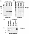
The KAP-1 protein binds to both human and murine HP1 family proteins. (A) Analysis of the GST fusion proteins affinity purified as a soluble protein from E. coli extracts and used in the binding assays. Approximately 5 μg of each protein was electrophoresed on an SDS–10% polyacrylamide gel and stained with Coomassie blue. Equivalent quantities of protein (5 μg) were used for each binding reaction as determined by Coomassie blue staining and quantification against BSA. MW, molecular weight markers. (B) hKAP-1 was prepared by in vitro transcription-translation (IVT) of a full-length human cDNA, and the [35S]methionine-labeled protein (arrow) was used in binding reactions with the indicated GST fusion proteins. The input lane represents the total amount of KAP-1 in each reaction mixture. No binding was observed in reactions containing GST alone. (C) A HeLa whole-cell extract containing endogenous KAP-1 was used in binding reactions with the indicated GST fusion protein. GST-KRAB represents a positive control for KAP-1 interaction, and GST-KRAB(DV) is a mutant with reduced affinity for KAP-1, which served as a negative control for KAP-1 interaction. Retention of KAP-1 by the GST fusion protein was detected by Western blot analysis using affinity-purified anti-KAP-1 antibodies. The input lane represents 10% of the KAP-1 in each binding reaction as detected by Western blot analysis of the extract.
FIG. 4
FIG. 4
Localization of the HP1BD in KAP-1. COS-1 cells were transfected with the indicated expression constructs containing different regions of KAP-1, and extracts from these cells were assayed for the ability of KAP-1 to bind GST-hHP1α. All KAP-1 molecules which included the putative HP1BD bound to GST-hHP1α, as indicated by the plus signs. For notation, see the legend to Fig. 1.
FIG. 5
FIG. 5
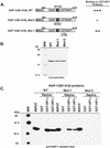
Amino acid substitutions in the HP1BD abolish HP1 binding in vitro. (A) The indicated wild-type (WT) or mutated segment of KAP-1 (for notation, see the legend to Fig. 1) was expressed in bacteria and purified by nickel chelate chromatography under native conditions. These highly purified proteins (B) were used in GST binding assays using the recombinant proteins shown in Fig. 3A. After the binding reactions and extensive washing, the amount of bound KAP-1 protein was determined by Western blot analysis using affinity-purified anti-KAP-1 sera (C). The input lane represents the total amount of recombinant KAP-1 added to each binding reaction mixture. No binding was detected for GST alone. Note that the recombinant Mut2 protein, which possesses the RV-EE mutation in the putative HP1BD, completely abolishes binding to both hHP1α and hHP1γ. MW, molecular weight markers.
FIG. 6
FIG. 6
Purified recombinant HP1 proteins can bind a DNA–GAL4-KRAB–KAP-1 ternary complex. A gel shift assay was performed by incubating a highly purified recombinant 6HisGAL4-KRAB domain protein with KAP-1 derived from a CNE, the indicated GST fusion protein (increasing concentrations from left to right indicated by open triangles: 50, 100, and 200 ng each of GST, GST-M31, GST-M32, GST-hHP1α, and GST-hHP1γ), and a γ-32P-labeled GAL4-UAS oligonucleotide probe. The complexes were separated by nondenaturing PAGE, and DNA-protein interactions were detected by autoradiography. The symbols on the left denote the following complexes: arrow, DNA–6HisGAL4-KRAB; single arrowhead, DNA–6HisGAL4-KRAB–KAP-1 ternary complex; double arrowhead, DNA–6HisGAL4-KRAB–KAP-1–HP1 quaternary complex. The free probe (FP) was allowed to run off the bottom of the gel to allow for better separation of the complexes at the top of the gel.
FIG. 7
FIG. 7
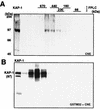
Cell-derived KAP-1 complexes increase in apparent size in the presence of the heterochromatin protein M32. (A) A [35S]methionine-labeled CNE was subjected to gel filtration (Superose 6), and the column fractions were analyzed for KAP-1 by immunoprecipitation. (B) A nonradioactive CNE was incubated with purified GST-M32 protein, and the mixture was subjected to gel filtration as for panel A. The column fractions were incubated with glutathione-Sepharose beads and washed, and the retained KAP-1 was detected in a Western blot assay using affinity-purified anti-KAP-1 sera. Lane 1 is a sample of the unfractionated CNE containing endogenous KAP-1 as a positive control and size marker. Indicated above the gels are the positions of the molecular mass standards used to calibrate the Superose 6 column, which were determined by Coomassie blue staining of duplicate gels.
FIG. 8
FIG. 8
Colocalization of endogenous KAP-1 with the heterochromatin protein M31 in interphase nuclei. All images were derived from the staining of a single population of cells simultaneously for both antigens and viewed by confocal microscopy. (A to F) Each row illustrates an independent random field of asynchronously growing NIH 3T3 fibroblasts to provide a representative view of the various staining patterns observed for both KAP-1 and M31. Staining of individual antigens is identified by the color given in the upper corners of each image. The merged images are shown on the far right for each set. As illustrated, staining for both antigens was exclusively nuclear, with very little background staining in the cytoplasm. KAP-1 staining demonstrated a grainy, speckled pattern throughout the nucleus which was largely excluded from nucleoli. M31 staining displayed a strong association with large heterochromatic granules. (G to I) High-magnification single-cell analysis of KAP-1 and M31 subnuclear distribution. Each panel shows a single nucleus from the field displayed in panels D to F. Note the discrete pericentromeric heterochromatin and nucleoli, as detected by M31 (56), and the concentration of KAP-1 around these structures (I).
FIG. 9
FIG. 9
Colocalization of endogenous KAP-1 with the heterochromatin protein M32 in interphase nuclei. All images were derived from the staining of a single population of cells simultaneously for both antigens and viewed by confocal microscopy. (A to C) Random fields of asynchronously growing NIH 3T3 fibroblasts. (D to F) KAP-1 and hHP1γ staining in human HEp-2 cells, as indicated in the lower right corner of each image (arrows point to two apparent daughter cells). Note the speckled pattern for M32 which is largely excluded from nucleoli and pericentromeric heterochromatin. In several cells, a significant overlap was observed between KAP-1 and M32 staining (C, F, and I). The most dramatic representation of this colocalization is portrayed in panels G to I, representing a high-magnification single-cell analysis of KAP-1 and M32 subnuclear distribution in a NIH 3T3 nucleus, where the green and red speckles show a near complete overlap, yielding a uniform yellow in the merged image (I).
FIG. 10
FIG. 10
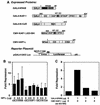
Modulation of KRAB/KAP-1 repression activity by exogenous hHP1α and a dominant negative KAP-1. (A) Plasmids used for transfection. The indicated GAL4-DNA binding domain fusion proteins were expressed from the SV40 enhancer/promoter (SV40ep) in mammalian cells. The full-length hHP1α contained a FLAG epitope tag at the amino terminus. The fragment of KAP-1 (aa 423 to 584) which contains the HP1BD was expressed as an NH2-terminal histidine fusion protein from a CMV vector. The SV40 promoter-based luciferase (Luc) reporter plasmid contains five synthetic GAL4-UAS sites and was used in all transfections. CC, coiled coil; Br, bromodomain; RD1, repression domain 1; CD, chromodomain; CSD, chromo shadow domain; pA, poly(A) site. (B) One microgram of each GAL4 expression plasmid was cotransfected into NIH 3T3 cells with increasing amounts of the hHP1α plasmid. Fold repression was calculated by comparing normalized luciferase activities with that of cells transfected in the absence of a GAL4 effector protein. The error bars represent the standard errors observed for three independent transfections, each performed in duplicate. (C) A GAL4-KRAB expression plasmid was cotransfected with increasing amounts of CMV–KAP-1 (aa 423 to 584), which encodes the putative HP1BD. Note that GAL4-KRAB repression activity is almost completely abolished in the presence of 4 μg of the CMV–KAP-1 (aa 423 to 584) plasmid, suggesting that the HP1BD is titrating endogenous HP1 proteins, thus relieving GAL4-KRAB-mediated repression via a squelching mechanism.
FIG. 11
FIG. 11
Mutations in the HP1BD reduce the intrinsic repression activity of the KAP-1 protein. (A) The plasmids used for transfection include GAL4 fusions to the wild-type (WT) KAP-1 or the Mut1 and Mut2 versions depicted in Fig. 5. Each plasmid (for notation, see the legend to Fig. 10) was tested for stable expression via transfection into COS-1 cells followed by immunoprecipitation analyses using both anti-KAP-1 and anti-GAL4 sera (B). These plasmids were then used in transfection assays with the 5XGAL4-UAS-SV40 luciferase reporter plasmid (Fig. 10A). (C) The wild-type (WT) GAL4–KAP-1 protein displayed potent repression of the reporter plasmid. However, the heterologous Mut2 protein, which does not bind HP1 proteins (Fig. 5) is reduced in repression activity, while the heterologous Mut1 protein displayed an intermediate effect on KAP-1-mediated repression. MW, molecular weight markers.
FIG. 12
FIG. 12
Summary of protein-protein interactions identified in the KRAB-ZFP–KAP-1 repression pathway. We propose that the KRAB-ZFP family of transcriptional repressors function in part as sequence-specific DNA binding proteins which recruit the KAP-1 corepressor to a target gene. This interaction is dependent on the RBCC domain of KAP-1. Together, the HP1BD, PHD, and bromodomain comprise the surfaces which mediate gene silencing via interaction with the indicated potential partners. This work has defined members of the HP1 family of heterochromatin proteins as likely downstream effectors of KAP-1 which may mediate the assembly of stable, higher-order silenced domains in the eukaryotic nucleus. For notation, see the legend to Fig. 10. pol, polymerase.

Similar articles

Cited by

References

    1. Aasland R, Stewart A F. The chromo shadow domain, a second chromo domain in heterochromatin-binding protein 1, HP1. Nucleic Acids Res. 1995;23:3163–3173. - PMC - PubMed
    1. Albagli O, Dhordain P, Deweindt C, Lecocq G, Leprince D. The BTB/POZ domain: a new protein-protein interaction motif common to DNA- and actin-binding proteins. Cell Growth Differ. 1995;6:1193–1198. - PubMed
    1. Auble D T, Hansen K E, Mueller C G, Lane W S, Thorner J, Hahn S. Mot1, a global repressor of RNA polymerase II transcription, inhibits TBP binding to DNA by an ATP-dependent mechanism. Genes Dev. 1994;8:1920–1934. - PubMed
    1. Baniahmad A, Leng X, Burris T P, Tsai S Y, Tsai M J, O’Malley B W. The tau 4 activation domain of the thyroid hormone receptor is required for release of a putative corepressor(s) necessary for transcriptional silencing. Mol Cell Biol. 1995;15:76–86. - PMC - PubMed
    1. Bellefroid E J, Poncelet D A, Lecocq P J, Revelant O, Martial J A. The evolutionarily conserved Kruppel-associated box domain defines a subfamily of eukaryotic multifingered proteins. Proc Natl Acad Sci USA. 1991;88:3608–3612. - PMC - PubMed

Publication types

MeSH terms