. 1999 Jun;19(6):4379-89.
doi: 10.1128/MCB.19.6.4379.

Ajuba, a novel LIM protein, interacts with Grb2, augments mitogen-activated protein kinase activity in fibroblasts, and promotes meiotic maturation of Xenopus oocytes in a Grb2- and Ras-dependent manner

Affiliations

Ajuba, a novel LIM protein, interacts with Grb2, augments mitogen-activated protein kinase activity in fibroblasts, and promotes meiotic maturation of Xenopus oocytes in a Grb2- and Ras-dependent manner

R K Goyal et al. Mol Cell Biol. 1999 Jun.

Abstract

LIM domain-containing proteins contribute to cell fate determination, the regulation of cell proliferation and differentiation, and remodeling of the cell cytoskeleton. These proteins can be found in the cell nucleus, cytoplasm, or both. Whether and how cytoplasmic LIM proteins contribute to the cellular response to extracellular stimuli is an area of active investigation. We have identified and characterized a new LIM protein, Ajuba. Although predominantly a cytosolic protein, in contrast to other like proteins, it did not localize to sites of cellular adhesion to extracellular matrix or interact with the actin cytoskeleton. Removal of the pre-LIM domain of Ajuba, including a putative nuclear export signal, led to an accumulation of the LIM domains in the cell nucleus. The pre-LIM domain contains two putative proline-rich SH3 recognition motifs. Ajuba specifically associated with Grb2 in vitro and in vivo. The interaction between these proteins was mediated by either SH3 domain of Grb2 and the N-terminal proline-rich pre-LIM domain of Ajuba. In fibroblasts expressing Ajuba mitogen-activated protein kinase activity persisted despite serum starvation and upon serum stimulation generated levels fivefold higher than that seen in control cells. Finally, when Ajuba was expressed in fully developed Xenopus oocytes, it promoted meiotic maturation in a Grb2- and Ras-dependent manner.


Figures

FIG. 1
FIG. 1
Nucleotide and derived amino acid sequences of murine Ajuba cDNA. Amino acids are numbered on the left, and nucleotides are numbered on the right. The three LIM domains are underlined. Potential SH3 recognition motifs are in bold type. A putative NES is underlined with a broken line.
FIG. 2
FIG. 2
Northern blot analysis for Ajuba mRNA expression in cell lines and tissues. Total RNA (12 μg) from each tissue was subjected to formaldehyde-agarose gel electrophoresis, transferred to a Zetabind membrane, and hybridized with a 32P-labeled Ajuba partial cDNA probe corresponding to the pre-LIM domain. The arrows identify the 3-kb Ajuba mRNA.
FIG. 3
FIG. 3
Ajuba cDNA encodes a protein of 55 kDa specifically recognized by anti-Ajuba immune serum. The cDNA of Ajuba, clone 3B, was subcloned into pBS K/S; 1 mg of linearized plasmid was transcribed and translated in the presence of [35S]methionine. In lane 1, 1/15 of final reaction (rxn) volume was loaded; in lane 2, 1/4 of the translation reaction was diluted to 1 ml in lysis buffer and 5 μl of preimmune serum was added, followed by protein A-agarose; in lane 3, 1/4 of translation product was diluted to 1 ml in lysis buffer and 5 μl of affinity purified anti-Ajuba immune serum was added, followed by protein A-agarose. Samples were run on an SDS–8% polyacrylamide gel under reducing conditions, the gel was dried, and autoradiography was performed. The arrowhead on the right indicates the mobility of Ajuba protein product. Mobility of molecular weight standards is indicated in kilodaltons on the left.
FIG. 4
FIG. 4
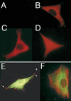
Immunofluorescence analysis of NIH 3T3 fibroblasts expressing Ajuba isoforms. Immunofluorescence was performed as described in Materials and Methods. (A to D) Single immunofluorescence with anti-Myc antiserum; (E) dual immunofluorescence with anti-Myc (green) and antipaxillin (red) antisera; (F) dual immunofluorescence with anti-Myc antiserum (green) and rhodamine-conjugated phalloidin (red). Cells in panel A have been transfected with control empty expression vectors; cells in panel B contain Myc-tagged wild-type Ajuba; cells in panel C contain Myc-tagged pre-LIM Ajuba; cells in panel D contain Myc-tagged LIM Ajuba; cells in panels E stably express Myc-tagged wild-type Ajuba and were transiently transfected with a paxillin-containing plasmid prior to immunofluorescence; cells in panel F contain Myc-tagged Ajuba. Arrows in panel E identify sites of focal adhesions to the slides. Panel E has been slightly overexposed to visualize endogenous paxillin. In all other panels, exposure times are equivalent.
FIG. 5
FIG. 5
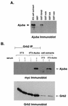
Ajuba associates with Grb2 in vitro and in vivo in a serum-dependent manner. (A) ES cell extracts (ca. 10 million cells per lane) were incubated with approximately 5 μg of the indicated fusion proteins. Bound products were isolated with glutathione-agarose beads and resolved on an SDS–8% polyacrylamide gel under reducing conditions, transferred to nitrocellulose, and immunoblotted with anti-Ajuba antiserum. The arrow on the left identifies the mobility of endogenous Ajuba in ES cell extracts (lane 1). (B) 3T3 fibroblasts (lanes 1 and 2) or 3T3 fibroblasts containing Myc-Ajuba (lanes 3 and 4) were incubated overnight in serum-free medium and then either lysed (−, lanes 1 and 3) or incubated with medium containing 50% serum for 15 min at 37°C and then lysed (+, lanes 2 and 4). Extracts from approximately 20 million cells were immunoprecipitated with anti-Grb2 antiserum (lanes 1 to 4), resolved by SDS-PAGE, transferred to nitrocellulose, and immunoblotted with anti-Myc antiserum (upper panel). Lanes 5 and 6 are cell extracts from approximately 0.5 million 3T3 and 3T3.myc-Ajuba cells, respectively. Subsequently the blot was stripped and reprobed with anti-Grb2 antiserum. Proteins were visualized by enhanced chemiluminescence. Arrows on the right indicate the expected mobilities of Myc-Ajuba and Grb2.
FIG. 6
FIG. 6
The pre-LIM domain of Ajuba and the SH3 domains of Grb2 mediate the association of Ajuba and Grb2. (A) Extracts from approximately 10 million 3T3.PreLIM Ajuba cells (lanes 2 to 5) or 3T3.LIM Ajuba cells (lanes 7 to 10) were incubated with approximately 5 μg of the indicated fusion proteins. Bound products were isolated with glutathione-agarose beads and resolved on an SDS–8% polyacrylamide gel under reducing conditions, transferred to nitrocellulose, and immunoblotted with anti-Myc antiserum (upper panel). Subsequently the blot was stripped and reprobed with anti-GST antiserum to indicate the level of fusion proteins present in each sample (lower panel). Lane 1 is a detergent-soluble extract from ca. 0.5 million 3T3 cells containing the Myc-tagged pre-LIM domain of Ajuba. The arrow on the left identifies the expected mobility of the Myc-pre-LIM protein. Lane 6 is an extract from 3T3 cells containing Myc-tagged LIM domains of Ajuba. The arrow on the right identifies the expected mobility of the Myc-LIM only protein. (B) Extracts from approximately 10 million F9 cells were incubated with approximately 5 μg of the indicated fusion proteins (lanes 2 to 5). Bound products were isolated with glutathione-agarose beads and resolved on an SDS–8% polyacrylamide gel under reducing conditions, transferred to nitrocellulose, and immunoblotted with anti-Grb2 antiserum (upper panel). Subsequently the blot was stripped and reprobed with anti-GST antiserum to indicate the level of fusion protein present in each sample (lower panel). Lane 1 is a detergent-soluble extract from ca. 0.5 million F9 cells. (C) Extracts from approximately 10 million 3T3.Pre-LIM Ajuba cells were incubated with approximately 5 μg of the indicated fusion proteins (lanes 2 to 9). Bound products were isolated with glutathione-agarose beads and resolved on an SDS–8% polyacrylamide gel under reducing conditions, transferred to nitrocellulose, and immunoblotted with anti-Myc antiserum (upper panel). Subsequently the blot was stripped and reprobed with anti-GST antiserum to indicate the level of fusion protein present in each sample (lower panel). Lane 1 is a detergent-soluble extract from ca. 0.5 million 3T3 cells containing Myc–pre-LIM Ajuba.
FIG. 7
FIG. 7
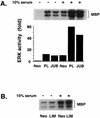
Ajuba expression in 3T3 cells results in an augmentation of MAP kinase activity. Cells were incubated in serum-free medium overnight and then either lysed (−) or stimulated for 15 min at 37°C with medium containing 10% serum and then lysed (+). The protein concentration of each sample was determined, extracts containing equal amounts of protein were immunoprecipitated with antiserum against ERK, and in vitro kinase reactions were performed with MBP as the substrate. Products were resolved by SDS-PAGE under reducing conditions, the gel was dried, and autoradiography was performed. Gels were scanned, and the relative quantities of phosphorylated MBP were determined. (A) Lanes 1 and 4, control 3T3.Neo cells; lanes 2 and 5, 3T3.PreLIM Ajuba cells; lanes 3 and 6, 3T3.Ajuba cells. (B) Lanes 1 and 3, control 3T3.Neo cells; lanes 2 and 4, 3T3.LIM cells.
FIG. 8
FIG. 8
Ajuba expression in Xenopus oocytes promotes meiotic maturation in a Grb2- and Ras-dependent manner. (A) Mature Xenopus oocytes were injected with 50 ng of in vitro-transcribed mRNA as described in Materials and Methods. After recovery, healthy oocytes were cultured in medium containing 3 μM progesterone; the percentage of oocytes undergoing GVBD was scored after 8 h. In all samples, between 40 and 50 oocytes were scored. For each set, multiple experiments were performed and a representative set is shown. The mRNAs injected for each column are listed below the graph. (B) The first group (columns 1 to 4) of oocytes were treated with insulin, and the percentage undergoing GVBD was scored at 20 h posttreatment. The second group (columns 5 to 8) were treated with progesterone, and the percentage undergoing GVBD was scored 8 h after treatment. In all samples, between 40 and 50 oocytes were scored. For each set, multiple experiments were performed and a representative set is shown. The mRNAs injected for each column are listed below the graph.
FIG. 9
FIG. 9
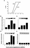
Ajuba expression in oocytes results in earlier onset of meiotic maturation (GVBD) coinciding with earlier activation of MAP kinase activity. Batches of oocytes were injected with water (control) or Ajuba mRNA. After recovery, healthy oocytes were cultured in medium containing 3 μM progesterone. Oocytes were scored hourly for the presence of GVBD until a maximal level was reached (A). The percentage of oocytes that had undergone GVBD at 10 h was set as 100%. Results at each time point are presented as a percentage of the maximal level. At selected time points, oocytes were picked and lysed. (B and C) MAP kinase activity determined by immunoblotting with an antiserum that recognizes activated MAP kinase. (B) Ajuba-injected oocytes; (C) control injected oocytes. The relative amount of Ajuba protein expression was determined by immunoblotting with anti-Myc antiserum (D). ERK protein level was determined by immunoblotting with anti-ERK antiserum (E). Panels D and E show results from Ajuba-injected oocytes.

Similar articles

Cited by

References

    1. Alexandropoulos K, Cheng G, Baltimore D. Proline-rich sequences that bind to src homology 3 domains with individual specificities. Proc Natl Acad Sci USA. 1995;92:3110–3114. - PMC - PubMed
    1. Andresson T, Ruderman J V. The kinase Eg2 is a component of the xenopus oocyte progesterone-activated signaling pathway. EMBO J. 1998;17:5627–5637. - PMC - PubMed
    1. Arber S, Halder G, Caroni P. Muscle LIM protein, a novel essential regulator of myogenesis, promotes myogenic differentiation. Cell. 1994;79:221–231. - PubMed
    1. Beckerle M C. Identification of a new protein localized at sites of cell-substratum adhesion. J Cell Biol. 1988;103:1679–1687. - PMC - PubMed
    1. Beckerle M C. Zyxin: zinc fingers at sites of cell adhesion. BioEssays. 1997;19:949–957. - PubMed

Publication types

MeSH terms

Associated data