. 1999 Jun;19(6):4423-30.
doi: 10.1128/MCB.19.6.4423.

Structural and functional heterogeneity of nuclear bodies

Affiliations

Structural and functional heterogeneity of nuclear bodies

D B Bloch et al. Mol Cell Biol. 1999 Jun.

Abstract

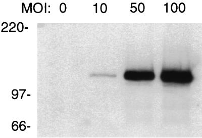
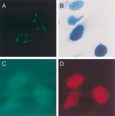
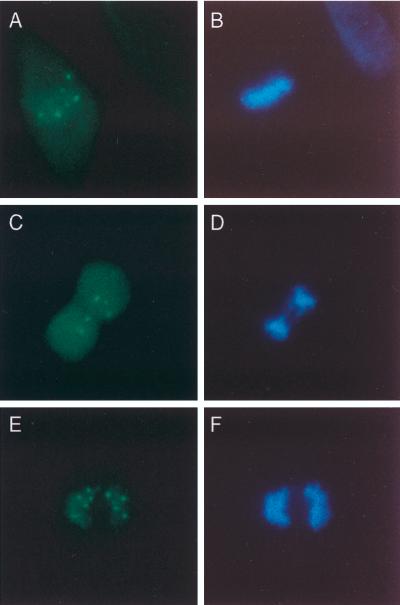
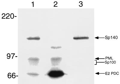
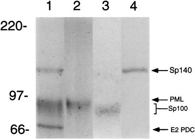
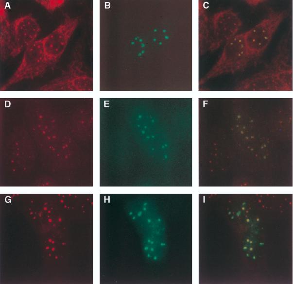
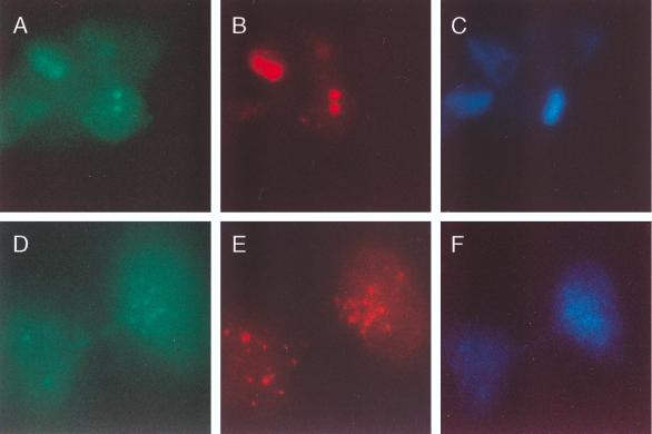
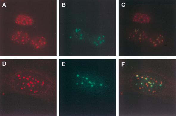
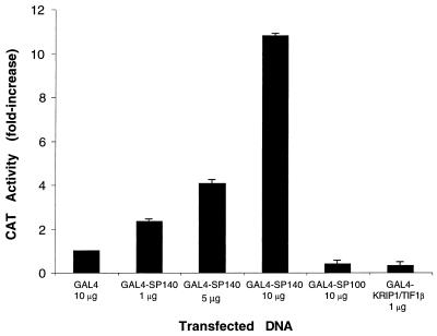
The nuclear body is a cellular structure that appears to be involved in the pathogenesis of acute promyelocytic leukemia and viral infection. In addition, the nuclear body is a target of autoantibodies in patients with the autoimmune disease primary biliary cirrhosis. Although the precise function of the nuclear body in normal cellular biology is unknown, this structure may have a role in the regulation of gene transcription. In a previous investigation, we identified a leukocyte-specific, gamma interferon (IFN-gamma)-inducible autoantigen designated Sp140. The objectives of the present study were to investigate the cellular location of Sp140 with respect to the nuclear-body components PML and Sp100 and to examine the potential role of Sp140 in the regulation of gene transcription. We used adenovirus-mediated gene transfer to express Sp140 in human cells and observed that the protein colocalized with PML and Sp100 in resting cells and associated with structures containing PML during mitosis. In cells infected with the adenovirus expressing Sp140 and incubated with IFN-gamma, the number of PML-Sp100 nuclear bodies per cell increased but immunoreactive Sp140 was not evenly distributed among the nuclear bodies. Sp140 associated with a subset of IFN-gamma-induced PML-Sp100 nuclear bodies. To examine the potential effect of Sp140 on gene transcription, a plasmid encoding Sp140 fused to the DNA-binding domain of GAL4 was cotransfected into COS cells with a chloramphenicol acetyltransferase (CAT) reporter gene containing five GAL4-binding sites and a simian virus 40 enhancer region. The GAL4-Sp140 fusion protein increased the expression of the reporter gene. In contrast, Sp100 fused to the GAL4 DNA-binding domain inhibited CAT activity in transfected mammalian cells. The results of this study demonstrate that Sp140 associates with a subset of PML-Sp100 nuclear bodies in IFN-gamma-treated cells and that Sp140 may activate gene transcription. Taken together, these observations suggest that the nuclear bodies within a cell may be heterogeneous with respect to both composition and function.


Figures

FIG. 1
FIG. 1
Immunoblot of Ad.Sp140-infected and control HEp-2 cells. Rat anti-Sp140 antiserum was used to detect Sp140 in uninfected HEp-2 cells or cells incubated with Ad.Sp140 at MOIs of 10, 50, and 100 PFU/cell. There was a dose-dependent increase in the level of Sp140 in infected HEp-2 cells.
FIG. 2
FIG. 2
Cellular distribution of Sp140 or β-galactosidase expressed by an adenovirus vector. (A) When rat anti-Sp140 antiserum was used, a typical nuclear-body staining pattern was observed in Ad.Sp140-infected HEp-2 cells. (B) To demonstrate that the adenovirus vector did not direct the encoded protein to the nuclear body, Ad.βgal was used to infect HEp-2 cells. β-Galactosidase was observed diffusely throughout the nucleus of these cells. (C and D) To confirm that the replication-deficient adenovirus vector did not alter the cellular location of nuclear-body components, cells were infected with Ad.βgal and then stained with monoclonal antibody directed against β-galactosidase and with human serum containing antibodies directed against PML and Sp100. Staining for β-galactosidase was observed diffusely throughout the nucleus (C, green). Staining for PML and Sp100 revealed the same nuclear body pattern (D, red) as that seen in uninfected cells (results not shown).
FIG. 3
FIG. 3
Sp140 associates with chromosomes in dividing cells. To determine the cellular location of Sp140 during mitosis, cells were infected with Ad.Sp140 and stained with rat anti-Sp140 antiserum (green) and DAPI (blue), which selectively binds to DNA. Sp140-containing nuclear bodies localized near chromosomes during metaphase (A and B), anaphase (C and D), and telophase (E and F).
FIG. 4
FIG. 4
Immunoblot of Ad.Sp140-infected HEp-2 cells with sera from patients with primary biliary cirrhosis or with rat anti-Sp140 antiserum. Antibodies in serum from patient K142 with primary biliary cirrhosis (lane 1) reacted with Sp140 (140 kDa), PML (90 kDa), Sp100 (∼80 kDa), and E2 PDC (70 kDa). Antibodies in serum from patient F111 (lane 2) reacted with PML, Sp100, E2 PDC, and an unidentified 120-kDa protein but did not react with Sp140. Rat anti-Sp140 antiserum (lane 3) reacted only with the 140-kD protein. Note that we and others have observed that Sp100 migrates as an ∼80-kDa protein (31, 36).
FIG. 5
FIG. 5
Immunoblot of Ad.Sp140-infected HEp-2 cells demonstrating the specificity of affinity-purified human antibodies. Antibodies in K142 serum (lane 1) reacted with Sp140 (140 kDa), PML (90 kDa), Sp100 (∼80 kDa), and E2 PDC (70 kDa). In contrast, anti-PML antibodies (lane 2) and anti-Sp100 antibodies (lane 3), affinity purified from human serum, reacted only with the corresponding proteins. Rat anti-Sp140 antibodies (lane 4) reacted only with the 140-kDa protein.
FIG. 6
FIG. 6
Immunofluorescence microscopy of Ad.Sp140-infected HEp-2 cells. (A to C) Cells were incubated with serum from primary biliary cirrhosis patient F111 (A, red) and rat anti-Sp140 antiserum (B, green). (D to F) Affinity-purified anti-Sp100 antibodies reacted with nuclear bodies in Ad.Sp140-infected cells (D), and Sp140 was detected within these structures (E). (G to I) Anti-Sp140 antibodies (H) also colocalized with affinity-purified anti-PML antibodies (G) in Ad.Sp140-infected cells. Colocalization of green and red fluorescence yields a yellow image (C, F, and I). To confirm the species specificity of the secondary antibodies used in this study, Ad.Sp140-infected cells were stained with normal rat serum and serum from primary biliary cirrhosis patient F111 and were subsequently incubated with both secondary antibodies. In these cells red but not green nuclear bodies were observed. In addition, infected cells were stained with rat anti-Sp140 antiserum and normal human serum and were subsequently incubated with both secondary antibodies. In these cells green but not red nuclear bodies were observed (data not shown).
FIG. 7
FIG. 7
Sp140 colocalizes with PML in dividing cells. To determine the cellular location of Sp40 during mitosis, HeLa cells were infected with Ad.Sp140 and stained with antibodies directed against Sp140 and PML. (A and B) Sp140 (green, A) localized to PML-containing aggregates (red, B) near the chromosomal mass in cells in metaphase. (D and E) In addition, Sp140 (green, D) associated with PML-containing nuclear bodies (E) in late anaphase. (C and F) DAPI was used to determine the stage of cellular division.
FIG. 8
FIG. 8
Immunofluorescence microscopy of IFN-γ-treated, Ad.Sp140-infected cells. HEp-2 cells were infected with Ad.Sp140 and incubated with IFN-γ for 48 h. (A and B) They were subsequently fixed and stained with affinity-purified anti-PML antibodies (A) and rat anti-Sp140 antiserum (B). (D and E) In addition, cells were stained with affinity-purified anti-Sp100 antibodies (D) and rat anti-Sp140 antiserum (E). (C and F) Colocalization of green and red fluorescence yields a yellow image. As expected, IFN-γ treatment increased the number of PML-Sp100-containing nuclear bodies (see, for comparison, Fig. 6D and G). The number of Sp140-containing nuclear bodies did not increase.
FIG. 9
FIG. 9
Sp140 functions as a transcriptional enhancer when tethered to DNA. The GAL4 DNA-binding domain or GAL4 DNA-binding domain fusions of Sp140, Sp100, or KRIP-1/TIF1β were transfected into COS cells together with a reporter plasmid. When 1, 5, and 10 μg of pBXG-Sp140 were used, there was a dose-dependent increase in CAT activity. In contrast, 10 μg of pBXG-Sp100 and 1 μg of pBXG-KRIP-1/TIF1β markedly inhibited CAT activity. CAT reporter activity was expressed as a percentage of the activity obtained with the GAL4 DNA-binding domain control. Transfections were performed in triplicate, and 0.5 μg of reporter plasmid was used in each transfection. The total amount of plasmid DNA was the same in each transfection. Results are presented as means and standard errors of the means. To control for transfection efficiency, a plasmid encoding growth hormone was cotransfected with the reporter plasmid and the growth hormone levels in the tissue culture medium were measured 48 h after transfection. There was no significant difference in transfection efficiency of pBXG, pBXG-Sp140, pBXG-Sp100, or pBXG-KRIP-1/TIF1β.

References

    1. Ahn J-H, Hayward G S. The major immediate-early proteins IE1 and IE2 of human cytomegalovirus colocalize with and disrupt PML-associated nuclear bodies at very early times in infected permissive cells. J Virol. 1997;71:4599–4613. - PMC - PubMed
    1. Ahn M-J, Nason-Burchenal K, Moasser M M, Dmitrovsky E. Growth suppression of acute promyelocytic leukemia cells having increased expression of the non-rearranged alleles; RARα or PML. Oncogene. 1995;10:2307–2314. - PubMed
    1. Ascoli C A, Maul G G. Identification of a novel nuclear domain. J Cell Biol. 1991;112:785–795. - PMC - PubMed
    1. Bloch D B, de la Monte S M, Guigaouri P, Filippov A, Bloch K D. Identification and characterization of a leukocyte-specific component of the nuclear body. J Biol Chem. 1996;46:29198–29204. - PubMed
    1. Borrow J, Goddard A, Sheer D, Solomon E. Molecular analysis of acute promyelocytic leukemia breakpoint cluster region on chromosome 17. Science. 1990;249:1577–1580. - PubMed

Publication types

MeSH terms

LinkOut - more resources