. 1999 Jun;19(6):4431-42.
doi: 10.1128/MCB.19.6.4431.

Collagenase 3 is a target of Cbfa1, a transcription factor of the runt gene family involved in bone formation

Affiliations

Collagenase 3 is a target of Cbfa1, a transcription factor of the runt gene family involved in bone formation

M J Jiménez et al. Mol Cell Biol. 1999 Jun.

Abstract

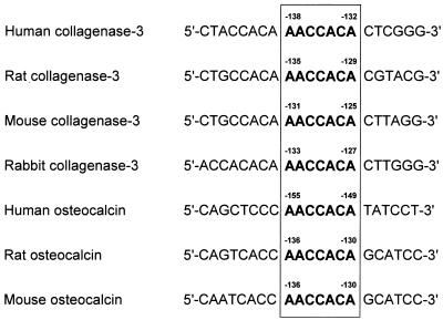
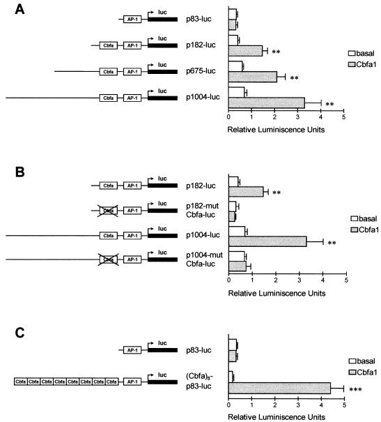
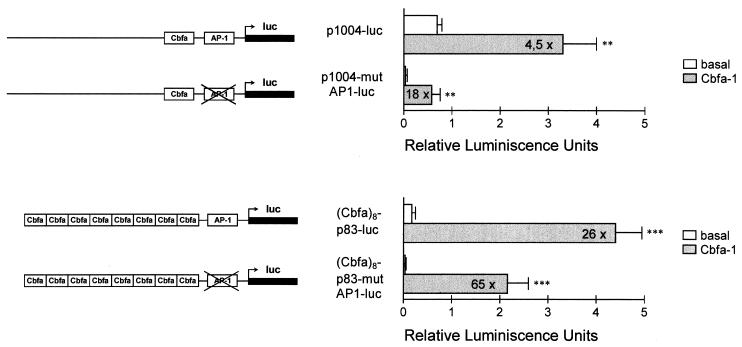
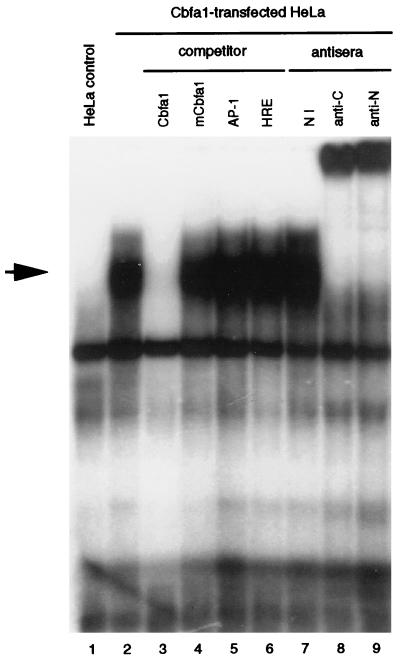
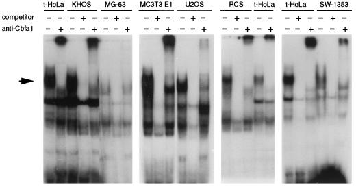
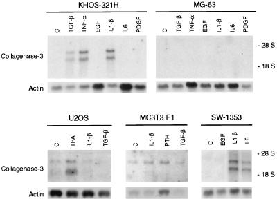
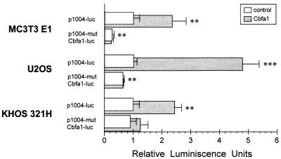
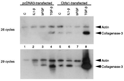
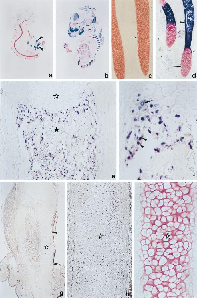
Collagenase 3 (MMP-13) is a recently identified member of the matrix metalloproteinase (MMP) gene family that is expressed at high levels in diverse human carcinomas and in articular cartilage from arthritic patients. In addition to its expression in pathological conditions, collagenase 3 has been detected in osteoblasts and hypertrophic chondrocytes during fetal ossification. In this work, we have evaluated the possibility that Cbfa1 (core binding factor 1), a transcription factor playing a major role in the expression of osteoblastic specific genes, is involved in the expression of collagenase 3 during bone formation. We have functionally characterized a Cbfa motif present in the promoter region of collagenase 3 gene and demonstrated, by cotransfection experiments and gel mobility shift assays, that this element is involved in the inducibility of the collagenase 3 promoter by Cbfa1 in osteoblastic and chondrocytic cells. Furthermore, overexpression of Cbfa1 in osteoblastic cells unable to produce collagenase 3 leads to the expression of this gene after stimulation with transforming growth factor beta. Finally, we show that mutant mice deficient in Cbfa1, lacking mature osteoblasts but containing hypertrophic chondrocytes which are also a major source of collagenase 3, do not express this protease during fetal development. These results provide in vivo evidence that collagenase 3 is a target of the transcriptional activator Cbfa1 in these cells. On the basis of these transcriptional regulation studies, together with the potent proteolytic activity of collagenase 3 on diverse collagenous and noncollagenous bone and cartilage components, we proposed that this enzyme may play a key role in the process of bone formation and remodeling.


Figures

FIG. 1
FIG. 1
Nucleotide sequence comparison between human, rat, mouse, and rabbit collagenase 3 promoter regions and human, rat, and mouse osteocalcin promoter regions around the Cbfa element (boxed).
FIG. 2
FIG. 2
Functional analysis of the Cbfa element in the collagenase 3 promoter. (A) HeLa cells were cotransfected with several collagenase 3 promoter deletions fused to firefly luciferase (luc) reporter gene constructs and with pCMV-Cbfa1 (grey bars) or pcDNA3 (white bars) as the effector plasmid. Plasmid pRL-TK was used as an internal control of transfection efficiency as described in Materials and Methods. Values represent firefly luciferase-to-Renilla luciferase ratios. (B) Similarly, two Cbfa mutant constructs of different lengths (p1004-mutCbfa-luc and p182-mutCbfa-luc) were analyzed for transcriptional activity and compared to the wild-type constructs, in the presence or absence of Cbfa1. (C) Analysis of a plasmid containing eight copies of Cbfa element from the collagenase 3 promoter cloned upstream of the −83 promoter segment. Data are expressed as means ± standard errors of at least three independent experiments. Asterisks indicate significant differences from the control (∗, P < 0.05; ∗∗, P < 0.01; ∗∗∗, P < 0.001).
FIG. 3
FIG. 3
Analysis of the role played by the AP-1 element in collagenase 3 promoter activation by Cbfa1 in HeLa cells. The complete collagenase 3 promoter construct or a construct containing eight copies of the Cbfa element linked to the p83-luc promoter and containing a double mutation in the AP-1 element (p1004-mutAP1-luc and Cbfa8-p83-mutAP1-luc) was transfected in HeLa cells, as described in the legend to Fig. 2 and assayed for luciferase (luc) activity. The corresponding wild-type constructs were also transfected in parallel experiments. Data are expressed as means ± standard errors of at least three independent experiments. Asterisks indicate significant differences from the control (∗∗, P < 0.01; ∗∗∗, P < 0.001).
FIG. 4
FIG. 4
Electrophoretic mobility shift assay demonstrating specific binding of Cbfa1 to the Cbfa element in the collagenase 3 promoter. A complex (marked by an arrow) appears when nuclear extracts from HeLa cells transfected with pCMV-Cbfa1 are incubated with a labeled collagenase 3 promoter Cbfa element (lane 2). This complex is absent in control cells transfected with pcDNA3 (lane 1). To demonstrate the specificity of this binding, 20-fold molar excesses of different unlabeled probes (Cbfa [lane 3], mutant Cbfa1 [mCbfa1; lane 4], AP-1 [lane 5], and HRE [lane 6]) were added to the binding reaction as competitors. To assess the presence of Cbfa1 in the complexes, nuclear extracts were incubated with nonimmune serum (lane 7) or with specific antibodies against Cbfa1 (anti-αA1C17 [lane 8] and anti-αA1N35 [lane 9] [42]).
FIG. 5
FIG. 5
Analysis of the presence of Cbfa1 binding activity in six different bone-derived cell lines. The binding assays were performed with KHOS 321H, U2OS, MC3T3 E1, MG-63, RCS, or SW1353 nuclear extracts and Cbfa1-transfected HeLa nuclear extracts (t-HeLa) as a reference. A 20-fold molar excess of unlabeled Cbfa oligonucleotide was used as competitor where indicated. Antiserum against Cbfa1 was added to the reaction mixture as indicated (the same results were obtained with anti-αA1N35 and anti-αA1C17).
FIG. 6
FIG. 6
Northern blot analysis of collagenase 3 expression in osteoblastic KHOS 321H, MG-63, U2OS, and MC3T3 E1 and chondrocytic SW1353 cell lines. Northern blot analysis was performed with 10 μg of total RNA from KHOS 312H, MG-63, U2OS, MC3T3 E1, or SW1353 cells incubated for 24 h in the presence of EGF (10 ng/ml), IL-1β (10 ng/ml), IL-6 (20 ng/ml), PTH (10−8 M), platelet-derived growth factor (PDGF; 10 ng/ml), TPA (10−6 M), TGF-β (5 ng/ml), TNF-α (20 ng/ml), or vehicle alone (C). Filters were hybridized with a collagenase 3 cDNA probe and with a β-actin probe to verify RNA loading.
FIG. 7
FIG. 7
Functional analysis in osteoblastic cell lines of the Cbfa element present in the collagenase 3 promoter. MC3T3 E1, U2OS, and KHOS 321H cells were cotransfected with the collagenase 3 promoter construct p1004-luc or p1004-mutCbfa-luc fused to firefly luciferase reporter gene and with pCMV-Cbfa1 (grey bars) or pcDNA3 (white bars) as an effector plasmid. Plasmid pRL-TK was used as an internal control of transfection efficiency as described in Materials and Methods. Values represent firefly luciferase-to-Renilla luciferase ratios, normalized so that a relative activity of 1 was assigned to the basal activity of the wild-type construct in every cell line. Data are expressed as means ± standard errors of at least three independent experiments. Asterisks indicate significant differences from the control (∗∗, P < 0.01; ∗∗∗, P < 0.001).
FIG. 8
FIG. 8
Effect of Cbfa1 and TGF-β on collagenase 3 expression in MG-63 osteosarcoma cells. Cells were transiently transfected with pcDNA3 (lanes 1 to 4) or pCMV-Osf2/Cbfa1 (lanes 5 to 8). Transfected cells were stimulated with vehicle alone, IL-1β (5 ng/ml), bFGF (5 ng/ml), or TGF-β (5 ng/ml) for 24 h. RNA from the stimulated transfected cells was further prepared and used for RT-PCR as described in Materials and Methods. Aliquots of samples were taken at 26 and 29 cycles of amplification. Samples were separated in agarose gel, transferred to nylon filters, and hybridized with collagenase 3 and actin probes. The results shown are from a representative experiment and were consistently reproducible in several independent experiments.
FIG. 9
FIG. 9
Collagenase 3 expression in Cbfa1-deficient mice. (a and b) Saggital sections of 18.5-dpc Cbfa1−/− (a) and wild-type (b) embryos showing alkaline phosphatase activity (dark blue) and stained with nuclear fast red. An intense alkaline phosphatase activity is observed in the skeletal tissues of the wild-type embryo. This activity is virtually absent in the skeletal tissues of the Cbfa1-deficient embryo, composed only of cartilage (stained in red), although it is present in epithelial cells from the small intestine (arrowheads). (c and d) Higher magnifications of ribs from embryos shown in panels a and b, respectively. In the wild-type embryo (d), cartilage templates are ossified at the central region, as revealed by an intense alkaline phosphatase activity (arrowhead), and show hyaline cartilage at the edges (arrow). By contrast, ribs from Cbfa1-deficient embryos (c) are devoid of alkaline phosphatase activity and appear mainly formed by hypertrophic chondrocytes. (e) In situ hybridization of a wild-type embryo with collagenase 3 antisense probe in a saggital section of the proximal half of the tibia. Labeling is found in both hypertrophic chondrocytes from the cartilage (white star) and osteoblastic cells localized along forming bone trabeculae present in the bone marrow cavity (black star). (f) Higher magnification of a region of the tibia from panel e, showing specific labeling in distal hypertrophic chondrocytes (arrow) and small cells from the periosteal collar (arrowheads). (g) In situ hybridization of Cbfa1-deficient embryos with collagenase 3 antisense probe. No specific signal is found, although long bones show a central part (arrowheads) occupied mainly by hypertrophic chondrocytes (star). (h) Higher magnification of the central region of the bone template shown in panel g. Hypertrophic chondrocytes are devoid of specific signal. (i) Parallel section of the bone presented in panel h stained with fast red and showing cells having morphological features of hypertrophic chondrocytes (star). Original magnifications: a and b, ×2.8; c and d, ×40; e, ×100; f, ×225; g, ×16; h and i, ×256.

Similar articles

Cited by

References

    1. Airola K, Johansson N, Kariniemi A L, Kähäri V M, Saarialho-Kere U K. Human collagenase-3 is expressed in malignant squamous epithelium of the skin. J Investig Dermatol. 1997;109:225–231. - PubMed
    1. Angel P, Baumann I, Stein B, Delius H, Rahmsdorf H J, Herrlich P. 12-O-Tetradecanoyl-phorbol-13-acetate induction of the human collagenase gene is mediated by an inducible enhancer element located in the 5′-flanking region. Mol Cell Biol. 1987;7:2256–2266. - PMC - PubMed
    1. Anglard P, Melot T, Guérin E, Thomas G, Basset P. Structure and promoter characterization of the human stromelysin-3 gene. J Biol Chem. 1995;270:20337–20344. - PubMed
    1. Aronson B D, Fisher A L, Blechman K, Caudy M, Gergen J P. Groucho-dependent and -independent repression activities of Runt domain proteins. Mol Cell Biol. 1997;17:5581–5587. - PMC - PubMed
    1. Balbín M, López-Otín C. Hormonal regulation of the human pepsinogen C gene in breast cancer cells: identification of a cis-acting element mediating its induction by androgens, glucocorticoids, and progesterone. J Biol Chem. 1996;271:15175–15181. - PubMed

Publication types

MeSH terms

LinkOut - more resources