. 1999 Jun;19(6):4452-64.
doi: 10.1128/MCB.19.6.4452.

Fli-1, an Ets-related transcription factor, regulates erythropoietin-induced erythroid proliferation and differentiation: evidence for direct transcriptional repression of the Rb gene during differentiation

Affiliations

Fli-1, an Ets-related transcription factor, regulates erythropoietin-induced erythroid proliferation and differentiation: evidence for direct transcriptional repression of the Rb gene during differentiation

A Tamir et al. Mol Cell Biol. 1999 Jun.

Abstract

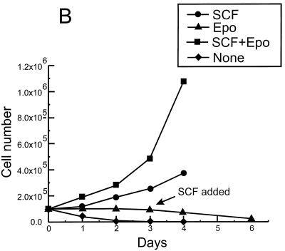
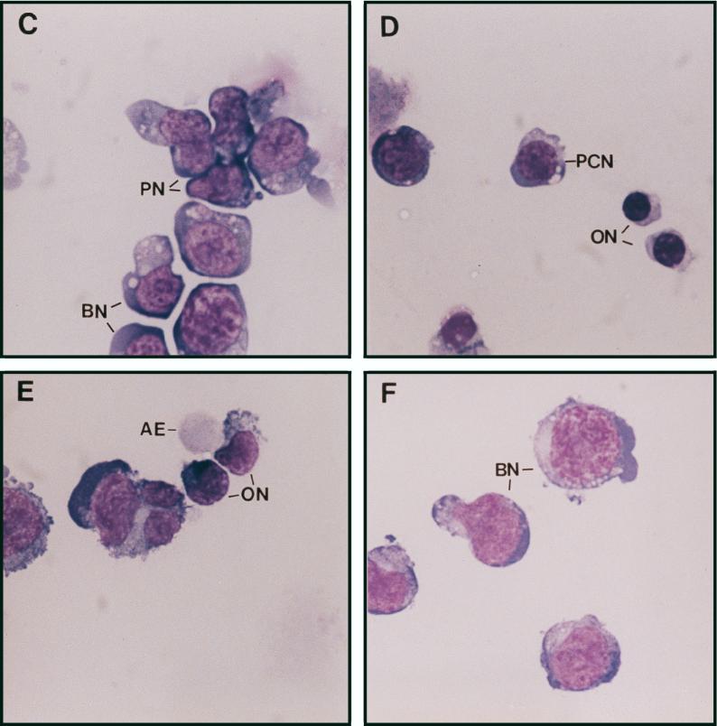
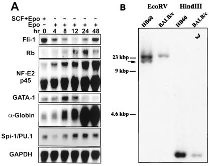
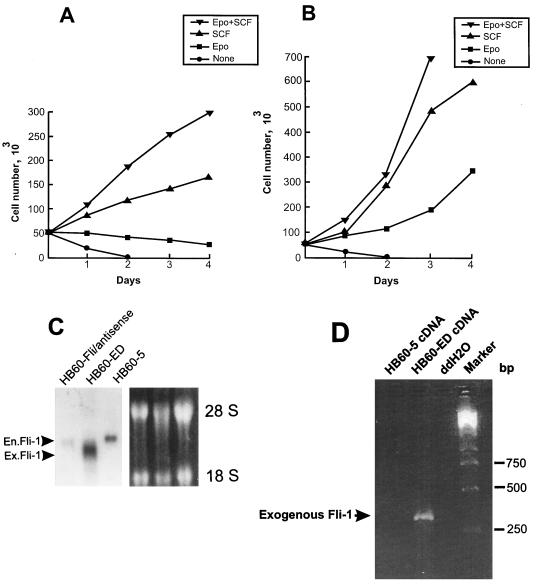
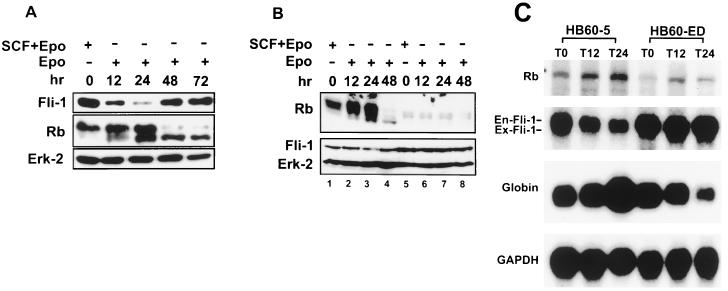
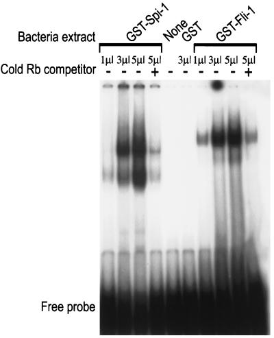
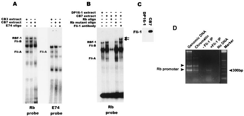
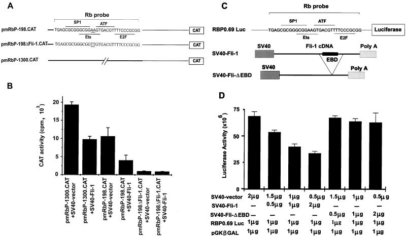
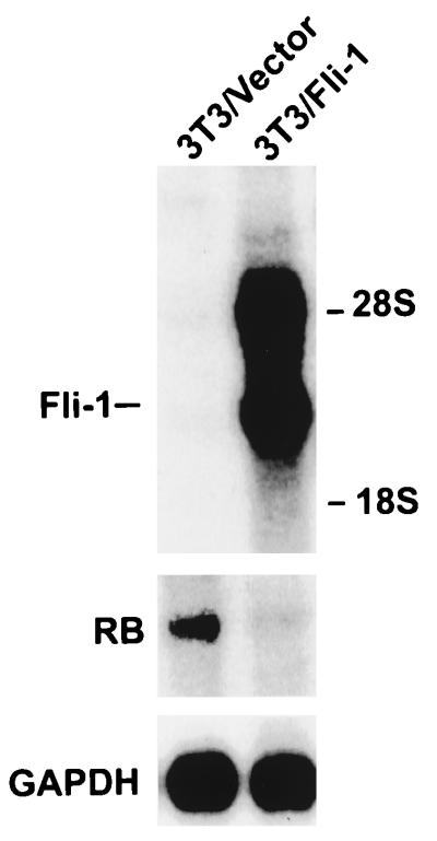
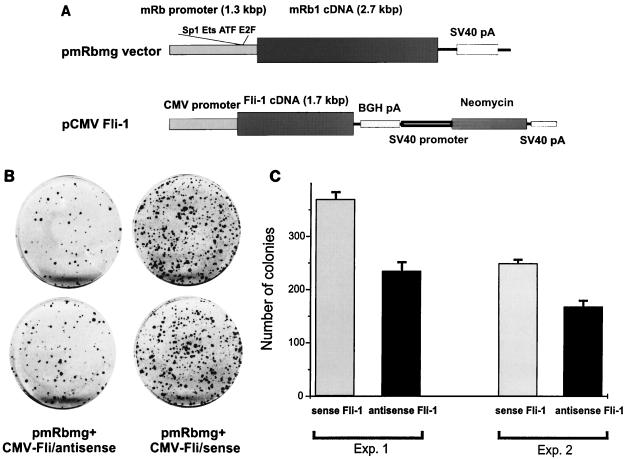
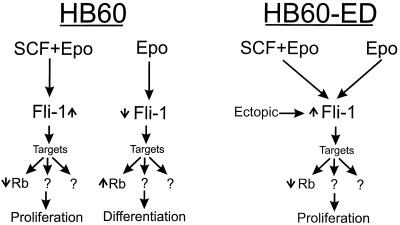
Erythropoietin (Epo) is a major regulator of erythropoiesis that alters the survival, proliferation, and differentiation of erythroid progenitor cells. The mechanism by which these events are regulated has not yet been determined. Using HB60, a newly established erythroblastic cell line, we show here that Epo-induced terminal erythroid differentiation is associated with a transient downregulation in the expression of the Ets-related transcription factor Fli-1. Constitutive expression of Fli-1 in HB60 cells, similar to retroviral insertional activation of Fli-1 observed in Friend murine leukemia virus (F-MuLV)-induced erythroleukemia, blocks Epo-induced differentiation while promoting Epo-induced proliferation. These results suggest that Fli-1 modulates the response of erythroid cells to Epo. To understand the mechanism by which Fli-1 regulates erythropoiesis, we searched for downstream target genes whose expression is regulated by this transcription factor. Here we show that the retinoblastoma (Rb) gene, which was previously shown to be involved in the development of mature erythrocytes, contains a Fli-1 consensus binding site within its promoter. Fli-1 binds to this cryptic Ets consensus site within the Rb promoter and transcriptionally represses Rb expression. Both the expression level and the phosphorylation status of Rb are consistent with the response of HB60 cells to Epo-induced terminal differentiation. We suggest that the negative regulation of Rb by Fli-1 could be one of the critical determinants in erythroid progenitor cell differentiation that is specifically deregulated during F-MuLV-induced erythroleukemia.


Figures

FIG. 1
FIG. 1
Establishment of the erythroblastic cell line HB60. Duplicate cultures (106) of the F-MuLV-induced primary erythroleukemia cell line HB60-t (A) or its derivative cell line HB60 (B) were incubated in the presence or absence of recombinant SCF (100 ng/ml) and/or Epo (0.1 U/ml) for the indicated times. The number of viable cells was determined by trypan blue dye exclusion. The arrow indicates the time at which SCF was added to the Epo-treated culture of HB60 cells, which did not lead to proliferation. (C to E) The clonal HB60-5 cells were grown in the presence of either SCF or Epo. At day 3 of incubation, the cells were harvested and stained with Wright’s stain. HB60-5 cells grown in the presence of SCF plus Epo exhibit the features of pronormoblast (PN) and basophilic normoblast (BN), which define the earliest recognizable stages of erythroid differentiation (C). HB60-5 cells grown in the presence of Epo (D and E) show a wider range of maturation stages, including polychromatophilic normoblast (PCN), orthochromatic normoblast (ON), normoblast (N), and anucleated erythrocyte (AE). HB60-ED cells expressing the exogenous Fli-1 (F) have morphological features of undifferentiated basophilic normoblast (BN) similar to the SCF-Epo-treated cells.
FIG. 1
FIG. 1
Establishment of the erythroblastic cell line HB60. Duplicate cultures (106) of the F-MuLV-induced primary erythroleukemia cell line HB60-t (A) or its derivative cell line HB60 (B) were incubated in the presence or absence of recombinant SCF (100 ng/ml) and/or Epo (0.1 U/ml) for the indicated times. The number of viable cells was determined by trypan blue dye exclusion. The arrow indicates the time at which SCF was added to the Epo-treated culture of HB60 cells, which did not lead to proliferation. (C to E) The clonal HB60-5 cells were grown in the presence of either SCF or Epo. At day 3 of incubation, the cells were harvested and stained with Wright’s stain. HB60-5 cells grown in the presence of SCF plus Epo exhibit the features of pronormoblast (PN) and basophilic normoblast (BN), which define the earliest recognizable stages of erythroid differentiation (C). HB60-5 cells grown in the presence of Epo (D and E) show a wider range of maturation stages, including polychromatophilic normoblast (PCN), orthochromatic normoblast (ON), normoblast (N), and anucleated erythrocyte (AE). HB60-ED cells expressing the exogenous Fli-1 (F) have morphological features of undifferentiated basophilic normoblast (BN) similar to the SCF-Epo-treated cells.
FIG. 2
FIG. 2
Fluctuation in the expression of Fli-1 during Epo-induced differentiation. (A) HB60-5 cells (5 × 10⁶) were grown in the presence of Epo or Epo plus SCF for the indicated times. Total RNAs (20 μg) prepared from these cells were Northern blotted, transferred to nitrocellulose, and sequentially hybridized with cDNA probes for the following genes: Fli-1, GATA-1, NF-E2 p45, Rb, α-globin, Spi-1/PU.1, and GAPDH. (B) Ten-microgram aliquots of genomic DNA extracted from the HB60 cells and normal BALB/c spleen cells were digested with the indicated restriction enzymes, Southern blotted, and hybridized with Spi-1 probe A. The arrow shows the position of the rearranged band.
FIG. 3
FIG. 3
Effect of overexpression of Fli-1 on proliferation of HB60-5 cells by Epo. HB60-5 cells (5 × 10⁶) were transfected with either the sense or antisense CMV-Fli-1 expression vector (Fig. 8A), and the pools of transfected cells were incubated in the presence of growth factors as indicated. (A) Growth rate of the Fli-1 antisense-transfected HB60-5 cells. (B) Growth rate of the Epo-dependent HB60-ED cells, which are derived from the Fli-1 sense-transfected HB60-5 cells. (C) Analysis of expression of exogenous Fli-1 mRNA in transfected HB60-5 cells. mRNA extracted from HB60-5 cells, the pools of Fli-1 antisense-transfected HB60-5 cells, and HB60-ED cells were Northern blotted and hybridized with Fli-1 cDNA probe. The position of the exogenous Fli-1 (Ex.Fli-1) band, which is slightly smaller than endogenous Fli-1 (En.Fli-1) transcript, is shown by an arrowhead. The ethidium bromide-stained gel shows equal RNA loading. (D) Expression of the exogenous Fli-1 in HB60-ED cells was verified by PCR analysis using two primers corresponding to Fli-1 and transcription termination sequences from the pRc/CMV construct. The PCR products were separated on a 2% agarose gel and stained with ethidium bromide. The arrow shows the location of the 304-bp amplified fragment. A PCR using no cDNA(ddH2O) was used as a negative control.
FIG. 4
FIG. 4
Negative correlation between the expression levels of Fli-1 and Rb proteins and mRNAs. (A) HB60-5 cells were cultured in the presence of SCF-Epo or Epo alone for the indicated times. Cells were lysed, separated on an SDS-acrylamide gel, and subjected to Western blotting using antibodies against the Rb and Fli-1 proteins. Equal loading was determined by blotting the same filter with an anti-mitogen-activated protein kinase (Erk-2) antibody. (B) HB60-5 (lanes 1 to 4) and HB60-ED (lanes 5 to 8) cells were treated with SCF-Epo or Epo alone for the indicated times and Western blotted with Rb, Fli-1, or Erk-2 antibodies as described above. (C) Two-microgram aliquots poly(A)+ mRNA isolated from HB60-5 or HB60-ED cells treated for the indicated times with Epo were Northern blotted and sequentially hybridized with Rb, Fli-1, α-globin, or GAPDH cDNA. The positions of endogenous (En-Fli-1) and exogenous (Ex-Fli-1) Fli-1 mRNAs are shown on the left.
FIG. 5
FIG. 5
DNA binding activity of recombinant Fli-1 and Spi-1/PU.1 proteins to the Rb promoter. Lysates of bacterial cells (1, 3, or 5 μl) expressing GST–Fli-1, GST–Spi-1/PU.1, or GST were incubated with the 32P-labeled Rb oligonucleotides. Binding reactions were performed in the presence of the nonspecific competitor poly(dI-dC) and the presence (+) or absence (−) of cold specific Rb competitor DNA. Complexes were resolved on a 5% polyacrylamide gel. As a control, binding with no protein (None) was performed.
FIG. 6
FIG. 6
Binding of Fli-1 to the Ets site in the Rb promoter. Nuclear extracts (2 μg) prepared from the erythroleukemia cell lines CB3, CB7, and DP16-1 were incubated with 32P-labeled Rb probe (A and B) or E74 probe (A) in the absence or presence of 100-fold excess cold DNA competitor or Fli-1 antibody as indicated. The position of the supershifted Fli-1 complex is indicated by arrows. The nature of major bands in panels A and B is unknown. (C) Total cellular extracts (20 μg) prepared from the cell lines CB7 and DP16-1 were Western blotted and hybridized with anti-Fli-1 polyclonal antibodies. (D) In vivo association of Fli-1 with the Rb promoter by formaldehyde cross-linking. PCR was performed on chromatin fragments isolated after immunoprecipitation with or without Fli-1 antibody or as a control on total genomic or chromatin isolated from DP27-17 erythroleukemic cells. The lower arrowhead on the left marks the position of the 330-bp fragment corresponding to the Rb promoter, while the upper arrowhead marks the position of a 450-bp nonspecific fragment. No DNA, no DNA in the PCR; Marker, 100-bp DNA ladder.
FIG. 7
FIG. 7
Suppression of the Rb promoter by Fli-1. (A) The pmRbP-1300.CAT and pmRbP-198.CAT constructs have been described elsewhere (78). The pmRbPΔFli-1.CAT construct was generated by converting neighboring AA nucleotides to TT in the core Ets binding site and are double underlined. The overlapping recognition sequences for transcription factors RBF-1, SP1, ATF, and E2F as well as the sequences used in the EMSA (Rb probe) are indicated. (B) The Rb promoter-CAT constructs were cotransfected into Rb-negative C33A cells with SV40-Fli-1 or SV40 vector alone, and the levels of CAT production were determined 3 days later. CAT levels were normalized for the levels of β-Gal. (C) The RBP0.69 Luc construct has been previously described. SV40-Fli-ΔEBD is a derivative of SV40-Fli-1 plasmid in which the EBD was deleted by removing the internal NcoI fragment from the Fli-1 cDNA. (D) The Rb promoter construct was cotransfected into C33A cells with the indicated amount of SV40-Fli-1, SV40-Fli-ΔEBD, SV40 vector, and pGKβGAL, and luciferase levels were measured 2 days later. Luciferase activity was normalized for the level of β-Gal.
FIG. 8
FIG. 8
Suppression of Rb by Fli-1 in fibroblasts. Two-microgram aliquots of poly(A)+ mRNA isolated from pooled 3T3 cells transfected with either SV40-Fli-1/Pgk-neo (3T3/Fli-1) or vector alone (pECE)/Pgk-neo (3T3/Vector) were Northern blotted and sequentially hybridized with Rb, Fli-1, or GAPDH probe.
FIG. 9
FIG. 9
Suppression of the Rb promoter by Fli-1 in SAOS-2 cells. (A) The murine Rb and Fli-1 genes, driven by the Rb (pmRbmg) and CMV promoters, respectively. BGH, bovine growth hormone. (B) Duplicate cultures of SAOS-2 cells cotransfected with the pmRbmg construct and either CMV-Fli sense or CMV-Fli antisense. (C) Two additional cotransfection experiments using new plasmid preparations of the pmRbmg and CMV-Fli-1 constructs used for panel B.
FIG. 10
FIG. 10
Model depicting the role of Fli-1 during proliferation and differentiation of erythroblasts. Epo induces downregulation of Fli-1 (↓) in HB60 cells, which results in terminal differentiation (see text). Upregulation of Fli-1 (↑) by ectopic expression (HB60-ED cells) and the addition of SCF, with or without Epo, to the culture of HB60 cells inhibits differentiation and promotes proliferation. These observations suggest the Fli-1 replaces the proliferative effect of SCF signaling in erythroblasts. Moreover, they indicate that Fli-1 functions as a switch mechanism which alters the responsiveness of erythroblast to undergo differentiation or proliferation by ectopic expression. This response is mediated through the regulation of several target genes. Rb is one of these target genes that is negatively regulated by Fli-1. High expression of Rb as a consequence of low Fli-1 could be one of the events involved in erythroid differentiation.

Similar articles

Cited by

References

    1. Andrews N C, Faller D V. A rapid micropreparation technique for extraction of DNA-binding proteins from limiting numbers of mammalian cells. Nucleic Acids Res. 1991;19:2499. - PMC - PubMed
    1. Ben-David Y, Giddens E G, Letwin K, Bernstein A. Erythroleukemia induction by Friend murine leukemia virus: insertional activation of a new member of the ets gene family, Fli-1, closely linked to c-ets-1. Genes Dev. 1991;5:908–918. - PubMed
    1. Ben-David Y, Prideaux V R, Chow V, Benchimol S, Bernstein A. Inactivation of the p53 oncogene by internal deletion or retroviral integration in erythroleukemic cell lines induced by Friend leukemia virus. Oncogene. 1988;3:179–185. - PubMed
    1. Bergh G, Ehinger M, Olofsson T, Baldetorp B, Johnsson E, Brycke H, Lindgren G, Olsson I, Gullberg U. Altered expression of the retinoblastoma tumor-suppressor gene in leukemic cell lines inhibits induction of differentiation but not G1-accumulation. Blood. 1997;89:2938–2950. - PubMed
    1. Chabot B, Stephenson D, Chapman V M, Besmer P, Bernstein A. The proto-oncogene c-kit encoding a transmembrane tyrosine kinase receptor maps to the mouse W locus. Nature. 1988;335:88–89. - PubMed

Publication types

MeSH terms