. 1999 Jun;19(6):4480-94.
doi: 10.1128/MCB.19.6.4480.

Yeast VSM1 encodes a v-SNARE binding protein that may act as a negative regulator of constitutive exocytosis

Affiliations

Yeast VSM1 encodes a v-SNARE binding protein that may act as a negative regulator of constitutive exocytosis

V Lustgarten et al. Mol Cell Biol. 1999 Jun.

Erratum in

  • Mol Cell Biol. 2008 Dec;28(24):7534

Abstract

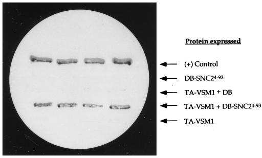
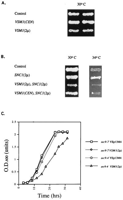
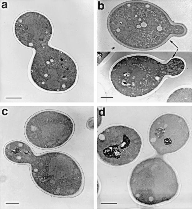
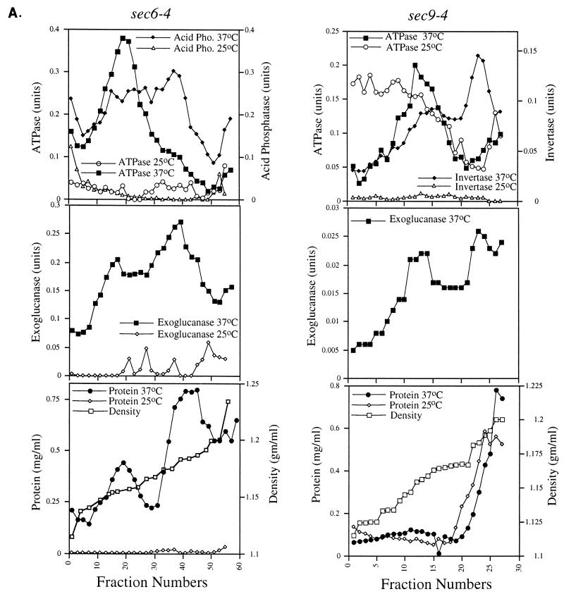
We have screened for proteins that interact with v-SNAREs of the late secretory pathway in the yeast Saccharomyces cerevisiae. A novel protein, designated Vsm1, binds tightly to the Snc2 v-SNARE in the two-hybrid system and can be coimmunoprecipitated with Snc1 or Snc2 from solubilized yeast cell extracts. Disruption of the VSM1 gene results in an increase of proteins secreted into the medium but does not affect the processing or secretion of invertase. In contrast, VSM1 overexpression in cells which bear a temperature-sensitive mutation in the Sec9 t-SNARE (sec9-4 cells) results in the accumulation of non-invertase-containing low-density secretory vesicles, inhibits cell growth and the secretion of proteins into the medium, and blocks rescue of the temperature-sensitive phenotype by SNC1 overexpression. Yet, VSM1 overexpression does not affect yeast bearing a sec9-7 allele which, in contrast to sec9-4, encodes a t-SNARE protein capable of forming a stable SNARE complex in vitro at restrictive temperatures. On the basis of these results, we propose that Vsm1 is a novel v-SNARE-interacting protein that appears to act as negative regulator of constitutive exocytosis. Moreover, this regulation appears specific to one of two parallel exocytic paths which are operant in yeast cells.


Figures

FIG. 1
FIG. 1
The gene product of YER143w (Vsm1) interacts with Snc24-93 in the yeast two-hybrid system. Yeast Y153 cells transformed with various plasmids were tested for β-galactosidase on nitrocellulose filters. Yeast expressing the Gal4 DNA binding domain alone (DB) or fused to Snc24-93 (DB-SNC24-93) were transformed with a plasmid that expresses the transactivating domain of Gal4 (TA) fused to the YER143w (VSM1) gene product (TA-VSM1), or with a control plasmid, and were patched onto selective medium. After 2 days, the cells were replica plated onto nitrocellulose filters, freeze-fractured in liquid nitrogen, and assayed visually for β-galactosidase activity (see Materials and Methods). A positive (+) control consisted of yeast expressing the Gal4 DB fused to the retinoblastoma gene product Rb and the Gal4 TA fused to the PP1α phosphatase.
FIG. 2
FIG. 2
The amino acid sequence of Vsm1 (YER143w). Numbers correspond to amino acid residues.
FIG. 3
FIG. 3
Overexpression of VSM1 specifically inhibits the growth of sec9-4 cells and blocks rescue by SNC1 overexpression. (A) sec9-4 cells were transformed with plasmids expressing VSM1 in single copy [VSM1(CEN)] and multicopy [VSM1(2μm)] or with a control plasmid (Control). Cells were patched onto selective medium prior to being replica plated onto fresh plates and allowed to grow for 2 days at 30°C. (B) sec9-4 cells bearing plasmids expressing VSM1 in single copy [VSM1(CEN)] and multicopy [VSM1(2μm)] or a control plasmid were transformed with a plasmid expressing SNC1 under the control of a constitutive promoter [SNC1(2μm)]. Control cells bearing an empty vector were transformed either with the SNC1 plasmid [SNC1(2μm)] or with a second empty vector (Control). Cells were patched onto selective medium prior to being replica plated onto fresh plates and allowed to grow for 2 days at 30 and 34°C. (C) sec9-4 and sec9-7 yeast strains were transformed either with a control plasmid (YEp13M4) or with a plasmid expressing VSM1 in multicopy [VSM1(2μm)]. Cells were grown to saturation under selective conditions, seeded at a density of 0.02 OD600 units/ml in fresh medium, and allowed to grow till saturation at 26°C. OD600 was measured at different times.
FIG. 4
FIG. 4
Overexpression of VSM1 in sec9-4 cells results in the accumulation of secretory vesicles at the bud tip. sec9-4 cells bearing a control plasmid (A) or a multicopy plasmid which expresses VSM1 (B) were grown to log phase and fixed for thin sectioning and electron microscopy. Thin sections of wild-type cells (W303-1a) (C) and vsm1Δ cells (VL2) (D) are also shown for comparison. Bars, 1 μm.
FIG. 5
FIG. 5
Vsm1 is a membrane-associated protein that localizes to the plasma membrane. (A) Vsm1 exists as a protein doublet in yeast extracts. Cell lysates were prepared from wild-type yeast (lane 1), vsm1Δ yeast expressing HA-tagged Vsm1 from single-copy (CEN) (lane 2) or multicopy (2μm) (lanes 3 and 4) plasmids, and vsm1Δ yeast lacking plasmids (lane 5) and were electrophoresed on SDS-polyacrylamide gels. In lanes 1 to 3 and 5, 50 μg of protein was added; 25 μg was added in lane 4. In addition, lane 6 contained 1 μg of affinity-purified recombinant His6-tagged Vsm1. Detection was performed with a polyclonal anti-Vsm1 antiserum (1:5,000). (B) Vsm1 associates with membranes and is enriched in the P100 fraction. sec6-4 cells maintained at 26°C or temperature shifted (2 h) to 37°C were fractionated as described in Materials and Methods. Aliquots of the various fractions were electrophoresed on SDS–10% polyacrylamide gels and blotted onto nitrocellulose membranes. Quantitative Western analysis was performed with specific antibodies (Ab) (anti-Gas1 [1:10,000], anti-Vsm1 [1:5,000], anti-Sso [1:5,000], and anti-Snc [1:1,000]) and 125I-labeled protein A (1 μCi/blot). In the left panel, 50-μg protein aliquots from the different fractions obtained from cells maintained at 26°C were electrophoresed; 25-μg aliquots of protein were used in the experiment shown in the middle panel. In the right panel, aliquots of the S10 fraction were treated with 1.5 M NaCl–200 mM glycine buffer (pH 2.5) or with additional lysis buffer (lacking salt or glycine) prior to centrifugation at 100,000 × g to yield the supernatant (sup.) and pellet fractions. Samples (15 μg) were electrophoresed, blotted, and detected for protein as described above. (C) Levels of Vsm1 are elevated in cells lacking vacuolar hydrolase, but not proteosome, activities. Total cell lysates (TCL) were prepared from sec6-4 yeast and strains which bear mutations in the protein degradative pathways (e.g., pre1-1 cells, which are deficient in the 20S proteosome activity, and pep4-3 prb1-1 and pep4-3 prb1Δ cells, which are deficient in the vacuolar hydrolase activity). Aliquots of total cell lysates (50 μg) were electrophoresed, blotted, and probed with anti-Vsm1 antibody (1:5,000). Detection was performed quantitatively, using 125I-labeled protein A (1 μCi). (D) Vsm1 localizes to the plasma membrane. Localization of Vsm1 was performed in wild-type cells by indirect immunofluorescence and confocal microscopy. a, cells expressing HA-tagged Vsm1 (expressed from a single-copy plasmid), detected with an affinity-purified anti-HA antibody (1:1,000) and FITC-labeled second antibody; b, staining of Dpm1 with an affinity-purified anti-Dpm1 antibody (3 μg/ml) and FITC-labeled second antibody; c, staining of HA-tagged Snc1 (expressed from a single-copy plasmid) with an affinity-purified anti-HA antibody (1:1,000) and FITC-labeled second antibody; d, staining of Sso protein with an affinity-purified anti-Sso antibody (1:100) and an FITC-labeled second antibody. Staining of the cell nucleus was performed with propidium iodide (1 μg/ml) and visualized via the rhodamine channel.
FIG. 5
FIG. 5
Vsm1 is a membrane-associated protein that localizes to the plasma membrane. (A) Vsm1 exists as a protein doublet in yeast extracts. Cell lysates were prepared from wild-type yeast (lane 1), vsm1Δ yeast expressing HA-tagged Vsm1 from single-copy (CEN) (lane 2) or multicopy (2μm) (lanes 3 and 4) plasmids, and vsm1Δ yeast lacking plasmids (lane 5) and were electrophoresed on SDS-polyacrylamide gels. In lanes 1 to 3 and 5, 50 μg of protein was added; 25 μg was added in lane 4. In addition, lane 6 contained 1 μg of affinity-purified recombinant His6-tagged Vsm1. Detection was performed with a polyclonal anti-Vsm1 antiserum (1:5,000). (B) Vsm1 associates with membranes and is enriched in the P100 fraction. sec6-4 cells maintained at 26°C or temperature shifted (2 h) to 37°C were fractionated as described in Materials and Methods. Aliquots of the various fractions were electrophoresed on SDS–10% polyacrylamide gels and blotted onto nitrocellulose membranes. Quantitative Western analysis was performed with specific antibodies (Ab) (anti-Gas1 [1:10,000], anti-Vsm1 [1:5,000], anti-Sso [1:5,000], and anti-Snc [1:1,000]) and 125I-labeled protein A (1 μCi/blot). In the left panel, 50-μg protein aliquots from the different fractions obtained from cells maintained at 26°C were electrophoresed; 25-μg aliquots of protein were used in the experiment shown in the middle panel. In the right panel, aliquots of the S10 fraction were treated with 1.5 M NaCl–200 mM glycine buffer (pH 2.5) or with additional lysis buffer (lacking salt or glycine) prior to centrifugation at 100,000 × g to yield the supernatant (sup.) and pellet fractions. Samples (15 μg) were electrophoresed, blotted, and detected for protein as described above. (C) Levels of Vsm1 are elevated in cells lacking vacuolar hydrolase, but not proteosome, activities. Total cell lysates (TCL) were prepared from sec6-4 yeast and strains which bear mutations in the protein degradative pathways (e.g., pre1-1 cells, which are deficient in the 20S proteosome activity, and pep4-3 prb1-1 and pep4-3 prb1Δ cells, which are deficient in the vacuolar hydrolase activity). Aliquots of total cell lysates (50 μg) were electrophoresed, blotted, and probed with anti-Vsm1 antibody (1:5,000). Detection was performed quantitatively, using 125I-labeled protein A (1 μCi). (D) Vsm1 localizes to the plasma membrane. Localization of Vsm1 was performed in wild-type cells by indirect immunofluorescence and confocal microscopy. a, cells expressing HA-tagged Vsm1 (expressed from a single-copy plasmid), detected with an affinity-purified anti-HA antibody (1:1,000) and FITC-labeled second antibody; b, staining of Dpm1 with an affinity-purified anti-Dpm1 antibody (3 μg/ml) and FITC-labeled second antibody; c, staining of HA-tagged Snc1 (expressed from a single-copy plasmid) with an affinity-purified anti-HA antibody (1:1,000) and FITC-labeled second antibody; d, staining of Sso protein with an affinity-purified anti-Sso antibody (1:100) and an FITC-labeled second antibody. Staining of the cell nucleus was performed with propidium iodide (1 μg/ml) and visualized via the rhodamine channel.
FIG. 6
FIG. 6
Vsm1 does not localize to secretory vesicles. (A) Two types of secretory vesicles accumulate in sec6-4 and sec9-4 cells. sec6-4 cells expressing HA-VSM1 from a multicopy plasmid and MYC-SNC2 from a single-copy plasmid were grown to log phase, shifted to low-phosphate-containing medium, and then either maintained at permissive conditions (26°C) or shifted to 37°C to induce vesicle accumulation. sec9-4 cells expressing HA-VSM1 and MYC-SNC2 from single-copy plasmids were grown to log phase, shifted to low-glucose-containing medium, and then either maintained at permissive conditions or shifted to 37°C to induce vesicle accumulation. Secretory vesicles from both strains were resolved by differential centrifugation and separation on 15 to 30% Nycodenz gradients. Aliquots of the fractions obtained by density gradient centrifugation were analyzed for density, protein concentration, and the following enzymatic activities: H+-ATPase, acid phosphatase (Acid Pho.), invertase, and exoglucanase (see Materials and Methods). Enzyme activities are expressed in arbitrary units based on absorbance; acid phosphatase and exoglucanase were measured at 415 nm, ATPase was measured at 820 nm, and invertase was measured at 540 nm. (B and C) HA-Vsm1 does not localize to the secretory vesicle fraction. Aliquots of fractions from the density gradients were electrophoresed, blotted, probed with anti-HA antibody (1:5,000), and detected by chemiluminescence. Samples of total cell lysates (50 μg) from sec6-4 (B) and sec9-4 (C) cells shifted to 37°C or maintained at 26°C were run along with 40-μl aliquots from the gradients (TCL). The solid arrow indicates the position of the low-density peak of vesicles (present at 37°C), while the hatched arrow indicates the high-density peak (present at 37°C). (D) Vsm1 does not localize to the LDSV population that accumulates in snc vbm cells. Aliquots (40 μl) of fractions from the density gradients shown in Fig. 6 of reference were electrophoresed, blotted, probed with anti-Vsm1 antibody (1:5,000), and detected by chemiluminescence. Samples of total cell lysates (50 μg) from snc vbm1 and snc vbm2 cells were run in parallel (TCL). The solid arrow indicates the position of the single peak of vesicles. (E) Western analysis of ER, Golgi, and post-Golgi markers in the Nycodenz gradient fractions of sec9-4 cells. Aliquots (40 μl) of the fractions were electrophoresed, blotted, and probed with the following antisera: anti-Wbp1 (1:6,000), anti-Mnn1 (1:1,500), anti-Sec22 (1:2,500), anti-Emp47 (1:3,000), anti-Snc (1:500), anti-Sec4 (1:1,000), anti-Sso (1:5,000), anti-Gas1 (1:5,000), and anti-Hsp150 (1:1,000). Detection was performed by chemiluminescence. Molecular masses are indicated on the left.
FIG. 6
FIG. 6
Vsm1 does not localize to secretory vesicles. (A) Two types of secretory vesicles accumulate in sec6-4 and sec9-4 cells. sec6-4 cells expressing HA-VSM1 from a multicopy plasmid and MYC-SNC2 from a single-copy plasmid were grown to log phase, shifted to low-phosphate-containing medium, and then either maintained at permissive conditions (26°C) or shifted to 37°C to induce vesicle accumulation. sec9-4 cells expressing HA-VSM1 and MYC-SNC2 from single-copy plasmids were grown to log phase, shifted to low-glucose-containing medium, and then either maintained at permissive conditions or shifted to 37°C to induce vesicle accumulation. Secretory vesicles from both strains were resolved by differential centrifugation and separation on 15 to 30% Nycodenz gradients. Aliquots of the fractions obtained by density gradient centrifugation were analyzed for density, protein concentration, and the following enzymatic activities: H+-ATPase, acid phosphatase (Acid Pho.), invertase, and exoglucanase (see Materials and Methods). Enzyme activities are expressed in arbitrary units based on absorbance; acid phosphatase and exoglucanase were measured at 415 nm, ATPase was measured at 820 nm, and invertase was measured at 540 nm. (B and C) HA-Vsm1 does not localize to the secretory vesicle fraction. Aliquots of fractions from the density gradients were electrophoresed, blotted, probed with anti-HA antibody (1:5,000), and detected by chemiluminescence. Samples of total cell lysates (50 μg) from sec6-4 (B) and sec9-4 (C) cells shifted to 37°C or maintained at 26°C were run along with 40-μl aliquots from the gradients (TCL). The solid arrow indicates the position of the low-density peak of vesicles (present at 37°C), while the hatched arrow indicates the high-density peak (present at 37°C). (D) Vsm1 does not localize to the LDSV population that accumulates in snc vbm cells. Aliquots (40 μl) of fractions from the density gradients shown in Fig. 6 of reference were electrophoresed, blotted, probed with anti-Vsm1 antibody (1:5,000), and detected by chemiluminescence. Samples of total cell lysates (50 μg) from snc vbm1 and snc vbm2 cells were run in parallel (TCL). The solid arrow indicates the position of the single peak of vesicles. (E) Western analysis of ER, Golgi, and post-Golgi markers in the Nycodenz gradient fractions of sec9-4 cells. Aliquots (40 μl) of the fractions were electrophoresed, blotted, and probed with the following antisera: anti-Wbp1 (1:6,000), anti-Mnn1 (1:1,500), anti-Sec22 (1:2,500), anti-Emp47 (1:3,000), anti-Snc (1:500), anti-Sec4 (1:1,000), anti-Sso (1:5,000), anti-Gas1 (1:5,000), and anti-Hsp150 (1:1,000). Detection was performed by chemiluminescence. Molecular masses are indicated on the left.
FIG. 7
FIG. 7
LDSVs accumulate in sec9-4 cells overexpressing VSM1. (A) sec9-4 cells expressing HA-VSM1 from a multicopy plasmid were grown to log phase, shifted to low-glucose-containing medium, and then processed to yield secretory vesicles as described in Materials and Methods. Vesicle-containing membrane fractions were resolved by differential centrifugation and separation on 15 to 30% Nycodenz gradients. Aliquots of the fractions obtained by density gradient centrifugation were analyzed for density, protein concentration, and the following enzymatic activities: H+-ATPase, invertase, and exoglucanase. Enzyme activities are expressed in arbitrary units based on absorbance; exoglucanase was measured at 415 nm, ATPase was measured at 820 nm, and invertase was measured at 540 nm. (B and C) Uranyl acetate-stained membranes from the early (B) and later (C) fractions of the gradient (fractions 4 and 5 and fractions 14 and 15, respectively). Bars represent 100 nm.
FIG. 8
FIG. 8
Vsm1 coimmunoprecipitates with Snc1 and Snc2. Cell lysates prepared from wild-type yeast expressing HA-tagged Snc1 or Snc2 from either single-copy (A) or multicopy (B) expression plasmids were subjected to immunoprecipitation (IP) with anti-HA antibody (ab). Duplicate samples were immunoprecipitated with excess HA peptide (pep) (75 μg). Immunoprecipitates were electrophoresed, blotted, and probed with either anti-Vsm1 (1:5,000) or anti-HA (1:5,000) antibody. Detection was performed by chemiluminescence (A) and by 125I-protein A labeling and autoradiography (B). For panel A, cell lysates were prepared from sec18-1 cells expressing HA-Snc2 that were either shifted to 37°C or maintained at 26°C (room temperature [RT]). Immunoprecipitation and detection were performed in a similar manner. Molecular masses are indicated on the left.

Similar articles

Cited by

References

    1. Aalto M K, Ruohonen L, Hosono K, Keranen S. Cloning and sequencing of the yeast Saccharomyces cerevisiae SEC1 gene localized on chromosome IV. Yeast. 1992;8:587–588. - PubMed
    1. Aalto M K, Ronne H, Keränen S. Yeast syntaxins Sso1p and Sso2p belong to a family of related membrane proteins that function in vesicular transport. EMBO J. 1993;12:4095–4104. - PMC - PubMed
    1. Ames B N. Assay of inorganic phosphate, total phosphate, and phosphatases. Methods Enzymol. 1966;8:115–118.
    1. Araki S, Tamori Y, Kawanishi M, Shinoda H, Masugi J, Mori H, Niki T, Okazawa H, Kubota T, Kasuga M. Inhibition of the binding of SNAP-23 to syntaxin 4 by Munc18c. Biochem Biophys Res Commun. 1997;234:257–262. - PubMed
    1. Barlowe C. Coupled ER to Golgi transport reconstituted with purified cytosolic proteins. J Cell Biol. 1997;139:1097–1108. - PMC - PubMed

Publication types

MeSH terms

Associated data