. 1999 May 17;145(4):659-72.
doi: 10.1083/jcb.145.4.659.

LST1 is a SEC24 homologue used for selective export of the plasma membrane ATPase from the endoplasmic reticulum

Affiliations

LST1 is a SEC24 homologue used for selective export of the plasma membrane ATPase from the endoplasmic reticulum

K J Roberg et al. J Cell Biol. .

Abstract

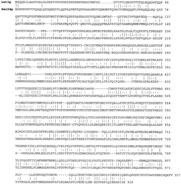
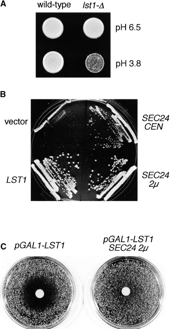
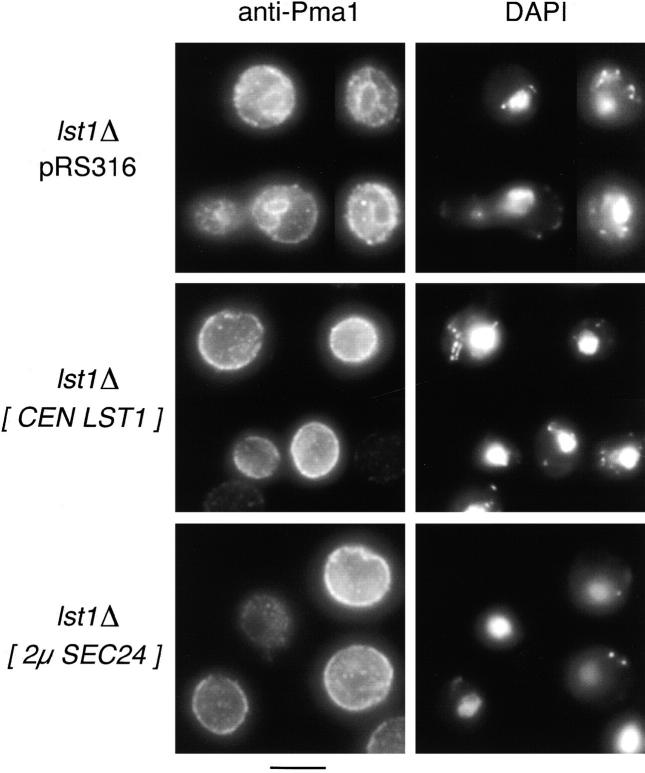
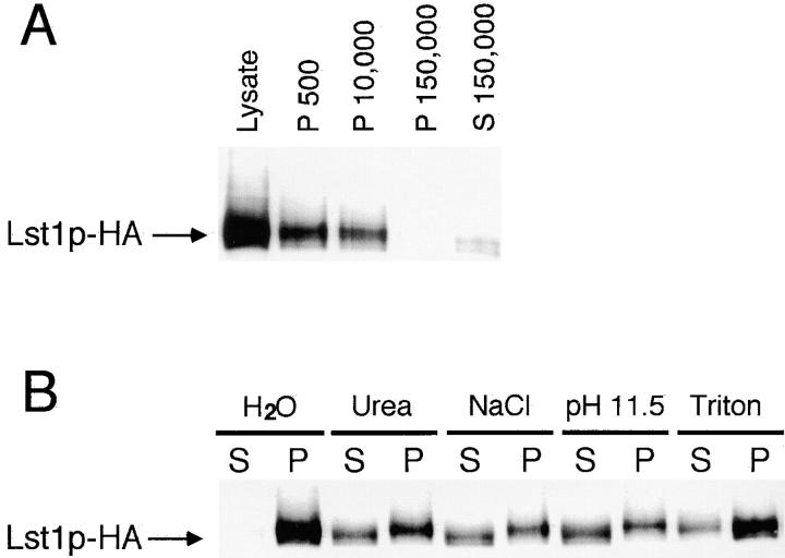
In Saccharomyces cerevisiae, vesicles that carry proteins from the ER to the Golgi compartment are encapsulated by COPII coat proteins. We identified mutations in ten genes, designated LST (lethal with sec-thirteen), that were lethal in combination with the COPII mutation sec13-1. LST1 showed synthetic-lethal interactions with the complete set of COPII genes, indicating that LST1 encodes a new COPII function. LST1 codes for a protein similar in sequence to the COPII subunit Sec24p. Like Sec24p, Lst1p is a peripheral ER membrane protein that binds to the COPII subunit Sec23p. Chromosomal deletion of LST1 is not lethal, but inhibits transport of the plasma membrane proton-ATPase (Pma1p) to the cell surface, causing poor growth on media of low pH. Localization by both immunofluorescence microscopy and cell fractionation shows that the export of Pma1p from the ER is impaired in lst1Delta mutants. Transport of other proteins from the ER was not affected by lst1Delta, nor was Pma1p transport found to be particularly sensitive to other COPII defects. Together, these findings suggest that a specialized form of the COPII coat subunit, with Lst1p in place of Sec24p, is used for the efficient packaging of Pma1p into vesicles derived from the ER.


Figures

Figure 1
Figure 1
Colony-sectoring screen for mutations that are lethal with sec13-1. CKY423 (ade2 ade3 leu2 ura3 sec13-1 [pKR4: SEC13, ADE3]) can lose the plasmid pKR4 when grown at 24°C on YPD, to give ade2 ade3 segregants that form white sectors within a red colony. Mutagenized cells that have acquired an lst mutation cannot grow without the pKR4 plasmid and form nonsectoring, solid red colonies. Of 132 nonsectoring colonies, the sectoring in 57 was restored by transformation with a second SEC13-bearing plasmid (pKR1).
Figure 2
Figure 2
Comparison of LST1 and SEC24 sequences. Identities are indicated by solid lines and similarities are indicated by dotted lines. Overall amino acid identity is 23%.
Figure 3
Figure 3
Functional relationships between LST1 and SEC24. (A) Sensitivity of lst1Δ mutants to acidic medium. Equal numbers of wild-type (CKY443) or lst1Δ::LEU2 (CKY534) cells were spotted onto YPD medium, pH 6.5, or acidic YPD medium (brought to pH 3.8 by the addition of HCl). Plates were photographed after incubation at 37°C for 2 d. (B) A lst1Δ::LEU2 strain (CKY552) was transformed with: vector only, pRS316; LST1 on a centromeric plasmid, pKR17; SEC24 on a centromeric plasmid, pAF70; or SEC24 on a 2μ plasmid, pKR34; and streaked onto YPD medium, pH 3.8. Colonies were photographed after growth at 37°C for 2 d. (C) A wild-type strain (CKY473) was transformed either with a plasmid carrying pGAL1–LST1 (pKR35) and vector control (pRS425), or with pKR35 and SEC24 on a 2μ plasmid (pKR41). Transformants were plated at a density of 800 cells/cm2 on SMM plates containing 2% raffinose and then 3 mg galactose solution was placed on a sterile 1-cm filter on top of the lawn. The plates were photographed after growth at 30°C for 2 d.
Figure 5
Figure 5
Pma1p accumulates the ER in lst1Δ cells and this accumulation is suppressed by overexpression of SEC24. Cells grown in SMM at 30°C were fixed with formaldehyde and then stained for immunofluorescence microscopy with affinity-purified anti-Pma1p antibody and FITC-conjugated secondary antibody. The same fields of cells, stained with DAPI to label the nuclear DNA, are also shown. Top panels, montage of lst1Δ cells (CKY536 carrying the empty vector pRS316); middle panels, genotypically wild-type cells (CKY536 carrying the LST1 plasmid pKR17); bottom panels, lst1Δ cells suppressed by SEC24 (CKY536 carrying the 2μ SEC24 plasmid pKR34). Bar, 5 μm.
Figure 4
Figure 4
Pma1p defects caused by lst1Δ. (A) lst1Δ cells (CKY534) were photographed using differential interference contrast microscopy after growth at 37°C on YPD, pH 3.8. A montage of multibudded cells is shown. Cells of this type comprise ∼10% of a lst1Δ culture, but are never seen in wild-type grown under the same conditions. Bar, 10 μm. (B) Reduced capacity for proton pumping by lst1Δ cells. Wild-type (CKY443) and lst1Δ (CKY536) were grown to exponential phase in YPD medium, pH 6.8, at 37°C. Cells were incubated in water overnight and then suspended in 10 mM glycine buffer at pH 4.0. Proton efflux from the cells after addition of glucose was recorded as a decrease in the pH of the external medium. Based on the average rate of change in pH over the first 5 min after glucose addition, lst1Δ cells exhibited 65% the rate of proton efflux as wild-type.
Figure 4
Figure 4
Pma1p defects caused by lst1Δ. (A) lst1Δ cells (CKY534) were photographed using differential interference contrast microscopy after growth at 37°C on YPD, pH 3.8. A montage of multibudded cells is shown. Cells of this type comprise ∼10% of a lst1Δ culture, but are never seen in wild-type grown under the same conditions. Bar, 10 μm. (B) Reduced capacity for proton pumping by lst1Δ cells. Wild-type (CKY443) and lst1Δ (CKY536) were grown to exponential phase in YPD medium, pH 6.8, at 37°C. Cells were incubated in water overnight and then suspended in 10 mM glycine buffer at pH 4.0. Proton efflux from the cells after addition of glucose was recorded as a decrease in the pH of the external medium. Based on the average rate of change in pH over the first 5 min after glucose addition, lst1Δ cells exhibited 65% the rate of proton efflux as wild-type.
Figure 6
Figure 6
Cell fraction to localize Pma1p in lst1Δ cells. Wild-type (CKY443) and lst1Δ (CKY536) cells were grown in YPD at 24°C and then were shifted to 37°C for 3 h. Cell lysates were fractionated on density gradients of 20–60% sucrose. Relative levels of Pma1p, Gas1p (plasma membrane marker), and Sec61p (ER marker) in each fraction were quantitated by immunoblotting and densitometry. GDPase (Golgi compartment marker) was determined by enzymatic assay.
Figure 7
Figure 7
Transport of invertase is not affected by lst1Δ. Wild-type (CKY540), lst1Δ (CKY542), and sec12-4 (CKY541) strains expressing invertase from the constitutive pTPI1–SUC2 fusion, were grown to exponential phase at 24°C in SMM medium, pH 6.5, without methionine. Wild-type and lst1Δ strains were shifted to 37°C, grown for 3 h, and the sec12-4 (CKY541) strain was shifted to 37°C 5 min before labeling. Cells were pulse-labeled with [35S]methionine and cysteine for 5 min and then chased by the addition of an excess of unlabeled methionine and cysteine. Invertase was immunoprecipitated from labeled extracts and resolved by SDS-PAGE. Positions of the core glycosylated ER form and mature Golgi and secreted forms of invertase are indicated.
Figure 8
Figure 8
Immunolocalization of Lst1p-HA. CKY535 (MATa lst1Δ::LEU2 leu2-3,112 ura3-52 [pKR17HA]) expressing Lst1p-HA from a centromeric plasmid was fixed and labeled with mouse anti-HA, FITC-conjugated anti–mouse antibodies, rabbit anti-Kar2p, and rhodamine-conjugated anti–rabbit antibodies. Nuclear DNA was visualized by DAPI staining. Cell bodies were visualized by differential interference contrast microscopy (DIC). Bar, 1 μm.
Figure 9
Figure 9
The intracellular distribution of Lst1p. (A) Cells expressing Lst1p-HA from a centromeric plasmid (CKY535) were gently lysed and subjected to sequential centrifugation steps, giving 500 g, 10,000 g, and 150,000 g pellet fractions (P) and a 150,000 g supernatant fraction (S). Each sample contains extract from the same number of cells. (B) Cell lysates were treated for 1 h at 4°C with either 2.5 M urea, 500 mM NaCl, 100 mM sodium carbonate (pH 11.5), or 1% Triton X-100. Pellet (P) and supernatant (S) fractions were then separated by centrifugation at 50,000 g. Lst1p-HA was detected by SDS-PAGE and immunoblotting with anti-HA antibody.
Figure 10
Figure 10
Lst1p/–Sec23p complex is membrane associated. (A) Affinity isolation of Lst1p/–Sec23p complexes. GST–Lst1p-HA or GST alone was coexpressed with Sec23p and isolated by affinity to glutathione Sepharose beads. Proteins bound to glutathione beads were loaded in lanes 2 and 4. One-sixth of the total lysate was loaded in lanes 1 and 3. (B) GST–LST1-HA, SEC23, or both were expressed from the GAL1 promoter. Cell lysates were cleared of cell debris by centrifugation at 300 g for 2 min. Pellet (P) and supernatant (S) fractions from cleared cell lysates were separated by centrifugation at 10,000 g for 30 min. An aliquot of the total cleared lysate (T) was removed before centrifugation. An equal number of cell equivalents were loaded for each sample. The GST–Lst1p-HA fusion was detected using anti-HA antibodies. For both A and B, the Sec23p protein was detected using anti-Sec23p antibodies.

References

    1. Abeijon C, Orlean P, Robbins PW, Hirschberg CB. Topography of glycosylation in yeast: characterization of GDP mannose transport and lumenal guanosine diphosphatase activities in Golgi-like vesicles. Proc Natl Acad Sci USA. 1989;86:6935–6939. - PMC - PubMed
    1. Aridor M, Weissman J, Bannykh S, Nuoffer C, Balch WE. Cargo selection by the COPII budding machinery during export from the ER. J Cell Biol. 1998;141:61–70. - PMC - PubMed
    1. Barlowe C, Schekman R. SEC12encodes a guanine-nucleotide- exchange factor essential for transport vesicle budding from the ER. Nature. 1993;365:347–349. - PubMed
    1. Barlowe C, Orci L, Yeung T, Hosobuchi M, Hamamoto S, Salama N, Rexach MF, Ravazzola M, Amherdt M, Schekman R. COPII: a membrane coat formed by Sec proteins that drive vesicle budding from the endoplasmic reticulum. Cell. 1994;77:895–907. - PubMed
    1. Bartel PL, Fields S. Analyzing protein–protein interactions using two-hybrid system. Methods Enzymol. 1995;254:241–263. - PubMed

Publication types

MeSH terms