. 1999 May 17;145(4):673-88.
doi: 10.1083/jcb.145.4.673.

Fusogenic domains of golgi membranes are sequestered into specialized regions of the stack that can be released by mechanical fragmentation

Affiliations

Fusogenic domains of golgi membranes are sequestered into specialized regions of the stack that can be released by mechanical fragmentation

M Dominguez et al. J Cell Biol. .

Abstract

A well-characterized cell-free assay that reconstitutes Golgi transport is shown to require physically fragmented Golgi fractions for maximal activity. A Golgi fraction containing large, highly stacked flattened cisternae associated with coatomer-rich components was inactive in the intra-Golgi transport assay. In contrast, more fragmented hepatic Golgi fractions of lower purity were highly active in this assay. Control experiments ruled out defects in glycosylation, the presence of excess coatomer or inhibitory factors, as well as the lack or consumption of limiting diffusible factors as responsible for the lower activity of intact Golgi fractions. Neither Brefeldin A treatment, preincubation with KCl (that completely removed associated coatomer) or preincubation with imidazole buffers that caused unstacking, activated stacked fractions for transport. Only physical fragmentation promoted recovery of Golgi fractions active for transport in vitro. Rate-zonal centrifugation partially separated smaller transport-active Golgi fragments with a unique v-SNARE pattern, away from the bulk of Golgi-derived elements identified by their morphology and content of Golgi marker enzymes (N-acetyl glucosaminyl and galactosyl transferase activities). These fragments released during activation likely represent intra-Golgi continuities involved in maintaining the dynamic redistribution of resident enzymes during rapid anterograde transport of secretory cargo through the Golgi in vivo.


Figures

Figure 1
Figure 1
Morphometry reveals Golgi fractions with differing degrees of structural integrity and cisternal stacking. Random sampling of cross sections of the fixed and filtered Golgi fractions; Gi (A), GE (B), and WNG fractions (C). The designation of Golgi apparatus as having single cisternae (1), or in stacks of two (2), three (3), or four (4) cisternae is indicated on the micrographs. Bars, 400 nm. Quantitative evaluation (histograms) was based on the analysis of 315 Golgi flattened cisternal profiles of the Gi fraction, 496 profiles of the GE fraction, and 1,617 profiles of the WNG fraction. As seen from the magnification bar, Golgi cisternae are considerably longer in the WNG fraction.
Figure 2
Figure 2
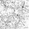
WNG fraction visualized by tannic acid staining. Two types of coats are readily observed: bristle-like (large arrows) and fuzz-coated structures (short arrows) usually of slightly greater diameter than Golgi flattened cisternae. A peroxisomal core contaminant (P) is indicated. Bar, 400 nm.
Figure 3
Figure 3
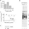
Sedimentation analysis of WNG, GE, and Gi Golgi fractions. The different Golgi fractions (Gi, GE, WNG) were adjusted to 0.25 M sucrose in their respective buffers loaded onto a 2-ml 0.4-M sucrose cushion (buffered) and centrifuged in a SW60 centrifuge rotor under various g max · min conditions by varying time and/or speed. The pelleted fractions were evaluated for their GalT content as described in Materials and Methods.
Figure 4
Figure 4
The WNG fraction is a defective acceptor in the Golgi cell-free transport assay. (A) CHO, Gi, GE, and WNG fractions have different acceptor activities. Each 25-μl Golgi cell-free transport assay received identical amounts of Golgi fraction (0.8 μg protein) and the extent of [3H]-GlcNAc incorporated into a specific membrane protein (the VSV-G protein) was determined. Near identical transport activities of the same Golgi fractions were found when BFA was included at 20 μg/ml (not shown). The galactosyl transferase–specific activity (GalT sp. act.) of the Gi, GE, and WNG fractions was 7.6, 10.1, and 24.3 mU, respectively. (B) Glycosylation of endogenous glycopeptide acceptors of Golgi fractions was assessed with UDP-[3H]- GlcNAc as sugar donor and Golgi fractions as endogenous acceptors. Approximately 4 μg cell fraction protein corresponding to ∼55,000 dpm of trichloroacetic acid–precipitable radioactivity was applied to each lane and separated on a 5–15% gel by SDS-PAGE. Fluorographic exposure was for 2 mo. Molecular mass markers in kilodaltons are indicated on the right. (C) Mixing experiments reveal that the acceptor WNG fraction neither generates a diffusible inhibitory factor nor consumes a diffusible limiting factor in the Golgi cell-free transport assays. Transport was determined as for A. The indicated amounts of WNG fraction (GalT sp. act., 13.8 mU) were added to transport assays containing either 0.25 (○) or 0.5 (▪) μg of the Gi fraction (GalT sp. act., 7.6 mU). (D) An identical amount of Gi and WNG fraction (45 μg protein) was applied to each lane, and p58 content in each fraction was evaluated by immunoblot analysis.
Figure 5
Figure 5
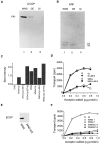
Coatomer is not responsible for the lower activity of WNG membranes. Identical amounts of each Golgi fraction (30 μg) were analyzed for β-COP (A) and ARF (B) content by immunoblot with the polypeptides indicated in kilodaltons. GalT sp. act. of the WNG, GE, and Gi fractions was 11.5, 7.7, and 7.6 mU, respectively. (C) Modulation of β-COP content of Golgi fractions by addition of cytosol, Brefeldin A, and/or GTPγS for 1 h at 37°C, and then pelleted through a sucrose cushion. The extent of membrane-associated β-COP was evaluated by Western blotting, followed by densitometry. The higher level of coatomer in reactions containing WNG membranes likely reflects in part the higher content of Golgi enzymes in those fractions. (D) Brefeldin A does not activate WNG. Transport assays containing the indicated amounts of Gi and WNG fractions were carried out in the presence or absence of 20 μg/ ml BFA for 2 h at 30°C. (E) Removal of β-COP by 0.3 M KCl. The β-COP content of the WNG and WNGKCl fractions were evaluated by immunoblotting. Equal amounts of protein (12.5 μg) were applied to each lane. (F) Both WNG and WNGKCl fractions are inactive acceptors. The indicated amounts of Gi, WNG, and WNGKCl were added to assays and the extent of transport in 1 h was determined as described for A. The GalT sp. act. of the WNG fractions was 14.4 and 13.3 mU and for the WNGKCl fractions was 16.3 and 13.3 mU for experiments 1 and 2, respectively.
Figure 6
Figure 6
Harsh homogenization in sucrose-imidazole buffer leads to disruption of Golgi structural integrity. (A) The parent low speed pellet (1,570 g max × 10 min) from a WNG preparation was treated with a rotating Teflon® pestle of the Potter-Elvehjem homogenizer in STM or SI buffer. Pellet (P) and supernatant (S) fractions (1,570 g max for 10 min) were analyzed for GalT-specific activity and total protein content. (B) Activation by disruption. Standard WNG fractions and WNG fractions disrupted by harsh homogenization in sucrose-imidazole (WNGdis) were prepared in parallel as described in Materials and Methods. The indicated amounts of the each Golgi fraction were tested for acceptor activity as described for Fig. 4 A. The GalT sp. act. of the WNG fractions were 11.7 and 11.9 mU, while those of the WNGdis fractions were 6.7 and 7.7 mU. The different GalT specific activity of the WNGdis and WNG fractions is likely a consequence of the enhanced protein content liberated as a consequence of the sucrose imidazole treatment (A, right). (C) Electron microscopy reveals that the WNGdis fraction is partially unstacked. Three separate pairs of WNG and WNGdis fractions were fixed and embedded, and the number of cisternae in individual Golgi profiles was assessed. The results shown correspond to the analysis of 122 and 376 profiles for the WNG and WNGdis preparations, respectively. (D) Random view of a filtered WNGdis fraction with single (1) and double (2) saccular profiles indicated. Bar, 400 nm.
Figure 7
Figure 7
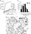
Imidazole-mediated unstacking of Golgi cisternae. Purified WNG fractions were gently incubated in 4 mM imidazole, pH 7.4, 0.25 M sucrose, and recovered as described in Materials and Methods. (A) Partial unstacking does not enhance acceptor activity. The indicated amounts of the parent and imidazole (I)-treated WNG fractions (WNGI) were compared with Gi fractions in the transport assay as described in Fig. 2 A. The GalT sp. act. for the WNG fractions were 11.6 and 12.5 mU, and for the WNGI fractions, 12.6 and 17.5 mU. (B) A representative micrograph of the WNGI fraction showing single (1) as well as double (2) cisternae. Although the cisternal membranes reveal greater access and free surfaces, there remains partial association with adjacent cisternae. Bar, 400 nm. (C) Morphometry of randomly sampled WNG or WNGI fractions reveals an increase in single cisternae after incubation in imidazole. A total of 89 (WNG) and 223 (WNGI) Golgi profiles were assessed as described in Materials and Methods.
Figure 8
Figure 8
Morphometry of Golgi apparatus in hepatic and CHO fractions. (A) Saccular lengths were measured with a MOP III image analyzer and scored as their cumulative frequency. The dotted line represents the median cisternal length of the WNG fraction. (B) Correlation of acceptor activity with fragmentation. The frequencies were obtained from the intercepts with the dashed line of A, while the percent maximum transport was determined from the extent of transport measured with 0.8 μg of each fraction relative to that observed with a similar amount of the Gi fraction (100%). Simple linear regression (solid line) between saccular length and Golgi acceptor activity reveals a significant relationship with a coefficient of simple correlation (r = 0.91). To restrict the analysis to liver Golgi fractions, the point for CHO membranes (open triangle) was not used for this analysis, although the coefficient of regression remains unaltered if the point is included. (C) The VSV-G–containing donor Golgi fraction is fragmented and reveals a large number of short single saccular profiles (1). Bar, 400 nm. (D) Quantitation of 207 donor CHO Golgi profiles revealed 82% to be single cisternae.
Figure 9
Figure 9
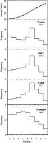
Rate-zonal separation of fusogenic active components of WNGdis. WNGdis preparations were loaded onto step sucrose gradients and analyzed by rate-zonal centrifugation as described in Materials and Methods. Each fraction was analyzed for sucrose and protein content, and GalT, NAGT, and transport activity. All assays were performed in duplicate under nonsaturating conditions. The number of fractionations is indicated in parentheses.
Figure 10
Figure 10
Transport-active components of WNGdis. The specific activity of transport competent components of the WNGdis fraction was calculated by dividing the values of transport activity (Fig. 9, bottom) over that of protein (Fig. 9, second panel). The highest concentration of transport-active acceptors is found in fraction 4. Similar results were obtained when normalizing transport activity to content of NAGT I.
Figure 11
Figure 11
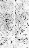
Morphology of WNG- dis rate-zonal subfractions. The subfractions from each portion of the gradient (fractions 1–8) of a typical fractionation (averaged in Fig. 9) were processed for random-sampling EM as described in Materials and Methods. A gradual increase in size in structures are found through fractions 1–8. Stacks made up of two saccules (double arrowheads) are found in fractions 6–8. Small vesicular profiles are also seen (small arrows), but throughout the gradient. Flattened cisternal elements of various lengths (arrowheads) are also frequent, especially in fractions 3–5 (arrowheads). A fortuitous section of a profile in fraction 6 reveals a continuity between two flattened cisternae and a lipoprotein-filled vesicular profile (*). Bar, 400 nm.
Figure 12
Figure 12
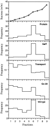
Distribution of GS15, GS28, and Vti1-rp2 in rate-zonal subfractions. A gradient was generated as for Fig. 9 and analyzed for content of GS15, GS28, and Vti1-rp2 by immunoblotting. Equal volumes of each fraction from the sucrose gradient (45 μl each) were evaluated. GS15 was visualized by chemiluminescence, while GS25 and Vti1-rp2 were detected using 125I-labeled goat anti–mouse or 125I goat anti–rabbit antibodies, respectively, followed by x-ray film exposure for 3 d. The mobilities of the respective antigens are indicated at left.
Figure 13
Figure 13
Rate-zonal centrifugation of Golgi v-SNARES in WNGdis subfractions. The gradient generated for Fig. 12 was analyzed for content of sucrose, total protein, GalT activity, and cell-free transport. The distribution of GS28 and Vti1-rp2 obtained by quantitative analysis of the immunoblot in Fig. 12 using a PhosphoImager (Fujix BAS1000 analyzer using a BASIII plate, Fuji) is also shown. Western blotting was repeated twice with a similar distribution observed.

References

    1. Acharya U, Jacobs R, Peters J-M, Watson N, Farquhar MG, Malhotra V. The formation of Golgi stacks from vesiculated Golgi membranes requires two distinct fusion events. Cell. 1995;82:895–904. - PubMed
    1. Balch WE, Dunphy WG, Braell WA, Rothman JE. Reconstitution of the transport of protein between successive compartments of the Golgi measured by the coupled incorporation of N-acetylglucosamine. Cell. 1984;39:405–416. - PubMed
    1. Bannykh SI, Balch WE. Membrane dynamics at the endoplasmic reticulum-Golgi interface. J Cell Biol. 1997;138:1–4. - PMC - PubMed
    1. Balch WE, Rothman JE. Characterization of protein transport between successive compartments of the Golgi apparatus: asymmetric properties of donor and acceptor activities in a cell-free system. Arch Biochem Biophys. 1985;240:413–425. - PubMed
    1. Baudhuin P, Evrard P, Berthet J. Electron microscopic examination of subcellular fractions. J Cell Biol. 1967;32:181–191. - PMC - PubMed

Publication types