. 1999 May 17;145(4):727-40.
doi: 10.1083/jcb.145.4.727.

Changes in the balance of phosphoinositide 3-kinase/protein kinase B (Akt) and the mitogen-activated protein kinases (ERK/p38MAPK) determine a phenotype of visceral and vascular smooth muscle cells

Affiliations

Changes in the balance of phosphoinositide 3-kinase/protein kinase B (Akt) and the mitogen-activated protein kinases (ERK/p38MAPK) determine a phenotype of visceral and vascular smooth muscle cells

K Hayashi et al. J Cell Biol. .

Abstract

The molecular mechanisms behind phenotypic modulation of smooth muscle cells (SMCs) remain unclear. In our recent paper, we reported the establishment of novel culture system of gizzard SMCs (Hayashi, K., H. Saga, Y. Chimori, K. Kimura, Y. Yamanaka, and K. Sobue. 1998. J. Biol. Chem. 273: 28860-28867), in which insulin-like growth factor-I (IGF-I) was the most potent for maintaining the differentiated SMC phenotype, and IGF-I triggered the phosphoinositide 3-kinase (PI3-K) and protein kinase B (PKB(Akt)) pathway. Here, we investigated the signaling pathways involved in de-differentiation of gizzard SMCs induced by PDGF-BB, bFGF, and EGF. In contrast to the IGF-I-triggered pathway, PDGF-BB, bFGF, and EGF coordinately activated ERK and p38MAPK pathways. Further, the forced expression of active forms of MEK1 and MKK6, which are the upstream kinases of ERK and p38MAPK, respectively, induced de-differentiation even when SMCs were stimulated with IGF-I. Among three growth factors, PDGF-BB only triggered the PI3-K/PKB(Akt) pathway in addition to the ERK and p38MAPK pathways. When the ERK and p38MAPK pathways were simultaneously blocked by their specific inhibitors or an active form of either PI3-K or PKB(Akt) was transfected, PDGF-BB in turn initiated to maintain the differentiated SMC phenotype. We applied these findings to vascular SMCs, and demonstrated the possibility that the same signaling pathways might be involved in regulating the vascular SMC phenotype. These results suggest that changes in the balance between the PI3-K/PKB(Akt) pathway and the ERK and p38MAPK pathways would determine phenotypes of visceral and vascular SMCs. We further reported that SMCs cotransfected with active forms of MEK1 and MKK6 secreted a nondialyzable, heat-labile protein factor(s) which induced de-differentiation of surrounding normal SMCs.


Figures

Figure 1
Figure 1
Effects of PDGF-BB, bFGF, EGF, and IGF-I on a phenotype of cultured gizzard SMCs. (A) Expression of caldesmon mRNAs (top panel; h-caldesmon mRNA [h-CaD, 4.8 kb] and l-caldesmon mRNA [l-CaD, 4.1 kb]) and calponin mRNA (middle panel; 1.8 kb) in SMCs stimulated with indicated growth factors. The bottom panel shows the 28S rRNA stained by methylene blue. Gizzard SMCs were cultured on laminin-coated plates in DME supplemented with 0.2% BSA for 24 h, and then stimulated with the same medium containing PDGF-BB (20 ng/ml), bFGF (10 ng/ml), EGF (10 ng/ml), or IGF-I (2 ng/ml) for 3 or 5 d. Caldesmon and calponin mRNAs in freshly isolated SMCs (Pre) and cultured SMCs under various conditions were analyzed by Northern blotting. (B) Comparison of cell morphology of SMCs under PDGF-BB, bFGF, EGF, or IGF-I–stimulated conditions for 5 d. The results are shown from four independent experiments.
Figure 1
Figure 1
Effects of PDGF-BB, bFGF, EGF, and IGF-I on a phenotype of cultured gizzard SMCs. (A) Expression of caldesmon mRNAs (top panel; h-caldesmon mRNA [h-CaD, 4.8 kb] and l-caldesmon mRNA [l-CaD, 4.1 kb]) and calponin mRNA (middle panel; 1.8 kb) in SMCs stimulated with indicated growth factors. The bottom panel shows the 28S rRNA stained by methylene blue. Gizzard SMCs were cultured on laminin-coated plates in DME supplemented with 0.2% BSA for 24 h, and then stimulated with the same medium containing PDGF-BB (20 ng/ml), bFGF (10 ng/ml), EGF (10 ng/ml), or IGF-I (2 ng/ml) for 3 or 5 d. Caldesmon and calponin mRNAs in freshly isolated SMCs (Pre) and cultured SMCs under various conditions were analyzed by Northern blotting. (B) Comparison of cell morphology of SMCs under PDGF-BB, bFGF, EGF, or IGF-I–stimulated conditions for 5 d. The results are shown from four independent experiments.
Figure 2
Figure 2
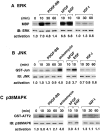
Characterization of the downstream signaling pathways triggered by PDGF-BB, bFGF, EGF, and IGF-I in SMCs. Gizzard SMCs were plated on laminin and cultured in DME supplemented with 0.2% BSA for 24 h, and then stimulated with the following growth factors for the indicated times: 20 ng/ml PDGF-BB, 10 ng/ml bFGF, 10 ng/ml EGF, 2 ng/ml IGF-I, or 10 μg/ml anisomycin. The cells were lysed and the extracts were assayed for the kinase activities of ERK (A), JNK (B), p38MAPK (C), PI3-K (D), and PKB(Akt) (E) as described in Materials and Methods. The left-hand lanes (0 min) in A–E were the controls without stimulation. Immunoblottings were performed to determine the amounts of kinase proteins in the cell lysates. The top and middle panels show the results of kinase assays and immunoblottings (IB), respectively. The relative activation rates normalized to respective control are shown in the bottoms. The representative results are shown from five independent experiments.
Figure 2
Figure 2
Characterization of the downstream signaling pathways triggered by PDGF-BB, bFGF, EGF, and IGF-I in SMCs. Gizzard SMCs were plated on laminin and cultured in DME supplemented with 0.2% BSA for 24 h, and then stimulated with the following growth factors for the indicated times: 20 ng/ml PDGF-BB, 10 ng/ml bFGF, 10 ng/ml EGF, 2 ng/ml IGF-I, or 10 μg/ml anisomycin. The cells were lysed and the extracts were assayed for the kinase activities of ERK (A), JNK (B), p38MAPK (C), PI3-K (D), and PKB(Akt) (E) as described in Materials and Methods. The left-hand lanes (0 min) in A–E were the controls without stimulation. Immunoblottings were performed to determine the amounts of kinase proteins in the cell lysates. The top and middle panels show the results of kinase assays and immunoblottings (IB), respectively. The relative activation rates normalized to respective control are shown in the bottoms. The representative results are shown from five independent experiments.
Figure 3
Figure 3
The effect of blocking the ERK and p38MAPK signalings on the expression of caldesmon and calponin mRNAs in gizzard SMCs under PDGF-BB–stimulated conditions. (A) Inhibition of PDGF-BB–induced activation of ERK and p38MAPK by PD98059 and/or SB203589. The SMCs plated on laminin were cultured in DME supplemented with 0.2% BSA for 24 h, and then pretreated with either PD98059 (30 μM) or SB203580 (20 μM), or both drugs for 1 h, followed by PDGF-BB (20 ng/ml) stimulation in the presence of vehicle alone or indicated drugs for 10 min. The representative data are shown from three independent experiments. (B) The expression of caldesmon and calponin mRNAs was analyzed by Northern blotting as shown in Fig. 1 A. Total RNAs were isolated from 3-d–cultured SMCs plated on laminin under indicated conditions, and then analyzed by Northern blotting using caldesmon and calponin cDNA fragments as probes. Culture conditions were as follows: IGF-I (2 ng/ml), PDGF-BB (20 ng/ml), PDGF-BB (20 ng/ml) in the presence of either PD98059 (30 μM) or SB203580 (20 μM) or both drugs. The results are shown from four independent experiments.
Figure 4
Figure 4
Rescue of PDGF-BB–induced SMC de-differentiation by blocking the ERK and p38MAPK pathways. Gizzard SMCs plated on laminin were stimulated with 2 ng/ml IGF-I (a and d), 20 ng/ml PDGF-BB (b and e), or 20 ng/ml PDGF-BB in the presence of both PD98059 (30 μM) and SB203580 (20 μM) (c and f) for 3 d. Ligand-induced contractility was monitored by the addition of carbachol (1 mM) for 1 min. Photographs show cultured SMCs before (a, b, and c) and after (d, e, and f) carbachol treatment. The data are presented from five independent experiments.
Figure 5
Figure 5
Regulation of the caldesmon promoter activity mediated through the PI3-K/PKB(Akt) pathway and the ERK and p38MPAK pathways. (A) The PI3-K activity in nonstimulated SMCs transfected with control plasmid (pCMV5) and expression plasmid of c-Myc–tagged active PI3-K p110α subunit, pCMV5p110αact (PI3-Kact). Gizzard SMCs transfected with indicated plasmids were cultured under nonstimulated conditions, and the PI3-K activity was determined by immunoprecipitation with anti–c-Myc monoclonal antibody, followed by in vitro kinase assays as described in Materials and Methods. (B) The PKB(Akt) activity in SMCs transfected with control plasmid, pCS2+MT, expression plasmid of c-Myc–tagged wild-type PKB(Akt), pCS2+MT-PKB(Akt)wt, or expression plasmid of c-Myc–tagged active PKB(Akt), pCS2+MT-PKB(Akt)act. The SMCs were cultured under nonstimulated conditions for 2 d after transfection, and then half of the cultures were stimulated by 2 ng/ml IGF-I with or without treatment of 20 μM LY294002. PKB(Akt) assays were carried out as described above. In A and B, the top and bottom panels are the results of kinase assay and immunoblotting (IB) to determine the amounts of kinase proteins in the cell lysates, respectively. The kinase activities are shown from three independent experiments. (C) Effects of PDGF-BB– or IGF-I–triggered signalings on the caldesmon promoter activity. The promoter construct of caldesmon, GP3CAT, was transfected into 3-d–cultured SMCs under the following conditions: DME supplemented with 0.2% BSA alone or BSA plus 20 ng/ml PDGF-BB. The GP3CAT (2 μg) was cotransfected with RSV-luciferase (1 μg) and control plasmid (1 μg), expression plasmid of c-Myc–tagged active PI3-K p110α (PI3-Kact, 1 μg), or expression plasmid of c-Myc– tagged active PKB(Akt) (PKB(Akt)act, 1 μg), respectively. After transfection, SMCs were stimulated with 2 ng/ml IGF-I, 2 ng/ml IGF-I plus 20 μM LY294002, 20 ng/ml PDGF-BB, or 20 ng/ml PDGF-BB plus PD98059 (30 μM) and SB203580 (20 μM). The promoter activity was assayed at 48 h after transfection as described in Materials and Methods. The relative promoter (CAT) activities were normalized to the activity in culture SMCs under IGF-I–stimulated conditions, which was defined as 100%. Each value represents the average ± SD of three independent experiments. A promoterless control CAT plasmid (pUC0CAT) did not show detectable CAT activity under the same conditions (data not shown).
Figure 6
Figure 6
Inhibition of the caldesmon promoter activity in gizzard SMCs by the forced expression of active MEK1 and MKK6. (A) Effects of active and dominant-negative MEK1 or MKK6 on the kinase activities of ERK (a) and p38MAPK (b) in cultured SMCs under non-stimulated and PDGF-BB– stimulated conditions. The SMCs were cotransfected with 2 μg each of expression plasmid of active or dominant-negative MEK1 and/or MKK6 or control plasmid together with 2 μg Flag-tagged ERK2 or Flag-tagged p38MAPK. The SMCs were cultured under nonstimulated conditions for 2 d, and half of the cultures were stimulated with 20 ng/ml PDGF-BB for 10 min. The cells were lysed and subjected to kinase assays after immunoprecipitation with anti-Flag monoclonal antibody. The top and bottom panels are the results of kinase assay and immunoblotting (IB) to determine the amounts of Flag-tagged kinase proteins in the cell lysates. The representative data are from two independent experiments. (B) Effects of the forced expression of active or dominant-negative MEK1 and/or MKK6 on the caldesmon promoter activity. The SMCs were cotransfected with GP3CAT (2 μg), RSV-luciferase (1 μg), and either or both expression vectors carrying active or dominant-negative MEK1 and/or MKK6 (1 μg). The total amounts of transfected plasmids were adjusted to 5 μg by the addition of control vector, pCS2+. The promoter activities were determined as described in the legend of Fig. 5. The relative promoter activities were normalized to the activity in cultured SMCs under IGF-I–stimulated conditions without expression vectors carrying MEK1 or MKK6, which was defined as 100%. Each value represents the average ± SD of three independent experiments.
Figure 7
Figure 7
Detection of de-differentiation–inducing factor(s) from SMCs cotransfected with active MEK1 and MKK6. (A) Effects of the forced expression of active or dominant-negative MEK1 and/or MKK6 on the endogenous expression of caldesmon and calponin mRNAs. Gizzard SMCs were transfected with 1 μg of indicated expression plasmids, and then cultured under IGF-I–stimulated conditions for 2 d (lanes 1–5) and for 4 d (lanes 7–11). The total amounts of transfected plasmids were adjusted to 2 μg by the addition of pCS2+. The SMCs were also cultured under IGF-I–stimulated conditions (2 ng/ml) without transfection for 2 d (lane 6) and 4 d (lane 12), or under PDGF-BB–stimulated conditions (20 ng/ml) without transfection for 4 d (lane 13). Caldesmon and calponin mRNAs in cultured SMCs were analyzed by Northern blotting as shown in Fig. 1 A. (B) Transfection efficiency and comparison of cell morphology between SMCs transfected with control plasmid (a) and expression plasmids carrying active MEK1 and MKK6 (b). The SMCs were transfected with pCS2+ (2 μg) and pSVβ-galactosidase (1 μg) (a) or with pCS2+MEK1act (1 μg), pCS2+MKK6act (1 μg), and pSVβ-galactosidase (1 μg) (b), and then cultured under IGF-I–stimulated conditions. At 4 d after transfection, β-galactosidase activity was visualized using X-gal as a substrate. (C and D) Dose-dependent effect of transfection with active MEK1 and MKK6 on the SMC phenotype. The SMCs were transfected with the indicated amounts of pCS2+MEK1act and pCS2+MKK6act together with pSVβ-galactosidase (1 μg), and then cultured for 4 d under IGF-I–stimulated conditions. Transfection efficiency was increased in increasing amounts of transfected plasmids as follows: lane 1 in C and top panel in D, <1%; lane 2 in C and middle panel in D, 5%; and lane 3 in C and bottom panel in D, 25%. The expression of caldesmon and calponin mRNAs (C) and cell morphology (D) of transfected SMCs are shown. The representative data are shown from four (A and B) or three (C and D) independent experiments.
Figure 7
Figure 7
Detection of de-differentiation–inducing factor(s) from SMCs cotransfected with active MEK1 and MKK6. (A) Effects of the forced expression of active or dominant-negative MEK1 and/or MKK6 on the endogenous expression of caldesmon and calponin mRNAs. Gizzard SMCs were transfected with 1 μg of indicated expression plasmids, and then cultured under IGF-I–stimulated conditions for 2 d (lanes 1–5) and for 4 d (lanes 7–11). The total amounts of transfected plasmids were adjusted to 2 μg by the addition of pCS2+. The SMCs were also cultured under IGF-I–stimulated conditions (2 ng/ml) without transfection for 2 d (lane 6) and 4 d (lane 12), or under PDGF-BB–stimulated conditions (20 ng/ml) without transfection for 4 d (lane 13). Caldesmon and calponin mRNAs in cultured SMCs were analyzed by Northern blotting as shown in Fig. 1 A. (B) Transfection efficiency and comparison of cell morphology between SMCs transfected with control plasmid (a) and expression plasmids carrying active MEK1 and MKK6 (b). The SMCs were transfected with pCS2+ (2 μg) and pSVβ-galactosidase (1 μg) (a) or with pCS2+MEK1act (1 μg), pCS2+MKK6act (1 μg), and pSVβ-galactosidase (1 μg) (b), and then cultured under IGF-I–stimulated conditions. At 4 d after transfection, β-galactosidase activity was visualized using X-gal as a substrate. (C and D) Dose-dependent effect of transfection with active MEK1 and MKK6 on the SMC phenotype. The SMCs were transfected with the indicated amounts of pCS2+MEK1act and pCS2+MKK6act together with pSVβ-galactosidase (1 μg), and then cultured for 4 d under IGF-I–stimulated conditions. Transfection efficiency was increased in increasing amounts of transfected plasmids as follows: lane 1 in C and top panel in D, <1%; lane 2 in C and middle panel in D, 5%; and lane 3 in C and bottom panel in D, 25%. The expression of caldesmon and calponin mRNAs (C) and cell morphology (D) of transfected SMCs are shown. The representative data are shown from four (A and B) or three (C and D) independent experiments.
Figure 8
Figure 8
Characterization of SMC de-differentiation–inducing factor(s). (A) Gizzard, SMCs were transfected with pCS2+MEK1act (1 μg) and pCS2+MKK6act (1 μg) or with pCS2+ (2 μg), and were cultured for 3 d. Each culture medium was collected; CM1 from SMCs transfected with pCS2+MEK1act and pCS2+MKK6act and CM2 from SMCs transfected with pCS2+. Then, SMCs were cultured under following conditions for 3 d: CM1 (lane 1), CM2 (lane 2), heat-treated CM1 (lane 3), trypsin-treated CM1 (lane 4), follow through (lane 5), and eluted (lanes 6–8) fractions of heparin-affinity column chromatography of CM1, or CM1 plus 1 μM AG1478 (lane 9). Caldesmon and calponin mRNAs in cultured SMCs were analyzed by Northern blotting as shown in Fig. 1 A. (B) Activation of ERK, JNK, and p38MAPK by the conditioned medium. Gizzard SMCs were stimulated under the following conditions: nonstimulation (N), CM1 for 10 min, CM2 for 10 min, PDGF-BB (20 ng /ml) for 10 min (P), or anisomycin (10 μg/ml) for 30 min (A). Then, the cells were lysed and subjected for kinase assays, ERK (top panel), JNK (middle panel), and p38MAPK (bottom panel) as described in Materials and Methods. (A). The data are representative of three (A) or two (B) independent experiments.
Figure 9
Figure 9
Analyses of signaling pathways in regulating the vascular SMC phenotype. Rat vascular SMCs were cultured on laminin in the presence of IGF-I (20 ng/ml) for 2 d, and then stimulated with following conditions for 3 d: 20 ng/ml IGF-I (a and f), 20 ng/ml IGF-I plus 20 μM LY294002 (b and g), 20 ng/ml PDGF-BB (c and h), 20 ng/ml PDGF-BB plus PD98059 (30 μM) and SB203580 (20 μM) (d and i), and the conditioned medium (CM1) from cultured gizzard SMCs transfected with pCS2+MEK1act and pCS2+MKK6act (e and j). Ligand-induced contractility was monitored as described in the legend of Fig. 4. Photographs show SMCs before (a–e) and after (f–j) carbachol treatment. The representative data are shown from five independent experiments.
Figure 10
Figure 10
Distinct signaling pathways are directly involved in the phenotypic determination of visceral and vascular SMCs. Maintenance of a differentiated phenotype of SMCs depends on the PI3-K/PKB(Akt) pathway. In contrast, the coordinate activation of the ERK and p38MAPK pathways induces SMC de-differentiation. IGF-I, which is a potent factor for maintaining the differentiated SMC phenotype, activates the signaling pathway mediated through PI3-K/PKB(Akt), but not MAPKs. Blocking the PI3-K/PKB(Akt) pathway with specific inhibitors of PI3-K, LY249002, or wortmannin, induces SMC de-differentiation. Potent SMC de-differentiation-inducing factors, PDGF-BB, bFGF, and EGF, all activate the ERK and p38MAPK pathways. bFGF and EGF do not enhance the PI3-K/PKB(Akt) pathway, whereas, PDGF-BB does activate it. Thus, PDGF-BB triggers the dual signaling pathways, PI3-K/PKB(Akt) and two MAPKs. When the ERK and p38MAPK pathways were simultaneously blocked by their specific inhibitors, PD98059 and SB203580, PDGF-BB in turn initiates to induce maintaining SMC differentiation. Therefore, the SMC phenotype would be determined by the balance between the strengths of the PI3-K/PKB(Akt) pathway and the ERK and p38MAPK pathways.

Similar articles

Cited by

References

    1. Alessi DR, Cohen P. Mechanism of activation and function of protein kinase B. Curr Opin Genet Dev. 1998;8:55–62. - PubMed
    1. Berrou E, Fontenay-Roupie M, Quarck R, McKenzie FR, Levy-Toledano S, Tobelem G, Bryckaert M. Transforming growth factor β1 inhibits mitogen-activated protein kinase induced by basic fibroblast growth factor in smooth muscle cells. Biochem J. 1996;316:167–173. - PMC - PubMed
    1. Bornfeldt KE, Arnqvist HL, Norstedt G. Regulation of insulin-like growth factor-I gene expression by growth factors in cultured vascular smooth muscle cells. J Endocrinol. 1990;125:381–386. - PubMed
    1. Bornfeldt KE, Rainco EW, Nateano T, Groves LM, Krebs E, Ross R. Insulin-like growth factor-I and platelet-derived growth factor-BB induce directed migration of human arterial smooth muscle cells via signaling pathways that are distinct from those of proliferation. J Clin Invest. 1994;93:1266–1274. - PMC - PubMed
    1. Cercek B, Fishbein MC, Forrester JS, Helfant RH, Fagin JA. Induction of insulin-like growth factor I messenger RNA in rat aorta after balloon denudation. Circ Res. 1990;66:1755–1760. - PubMed

Publication types

MeSH terms

Substances