. 1999 May 17;145(4):757-67.
doi: 10.1083/jcb.145.4.757.

Oxygen stress: a regulator of apoptosis in yeast

Affiliations

Oxygen stress: a regulator of apoptosis in yeast

F Madeo et al. J Cell Biol. .

Abstract

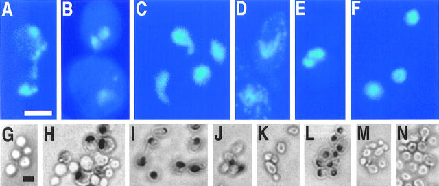
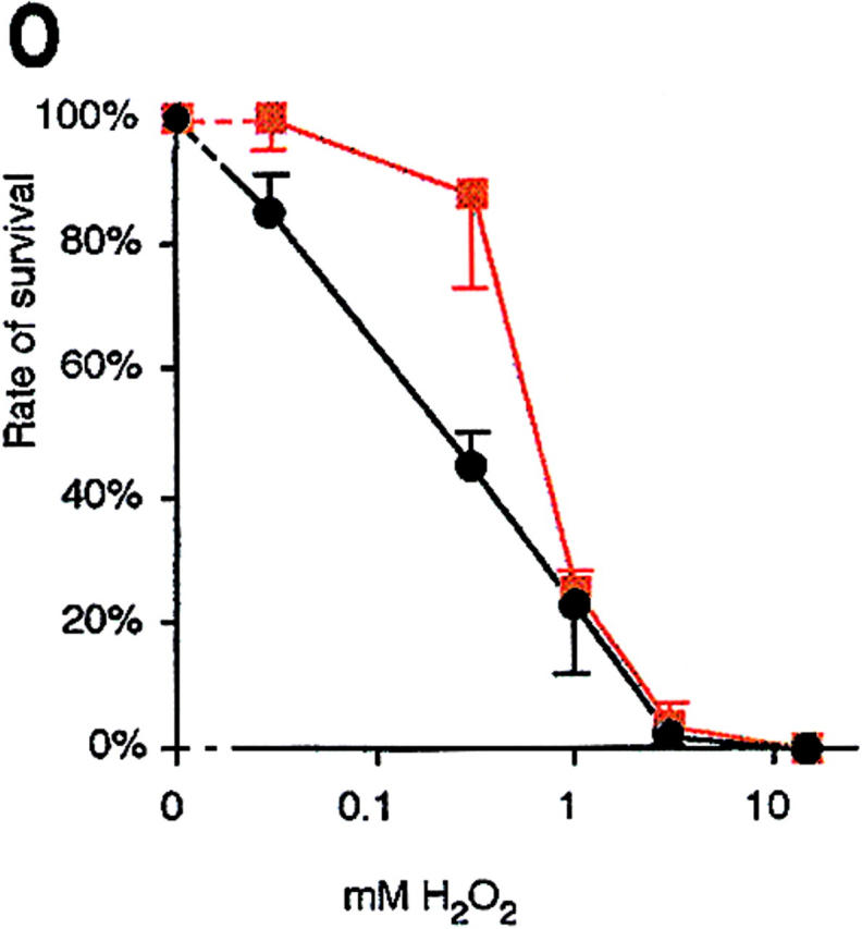
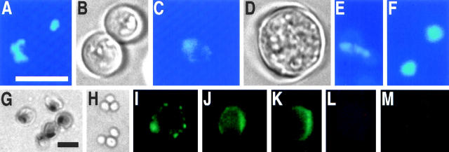
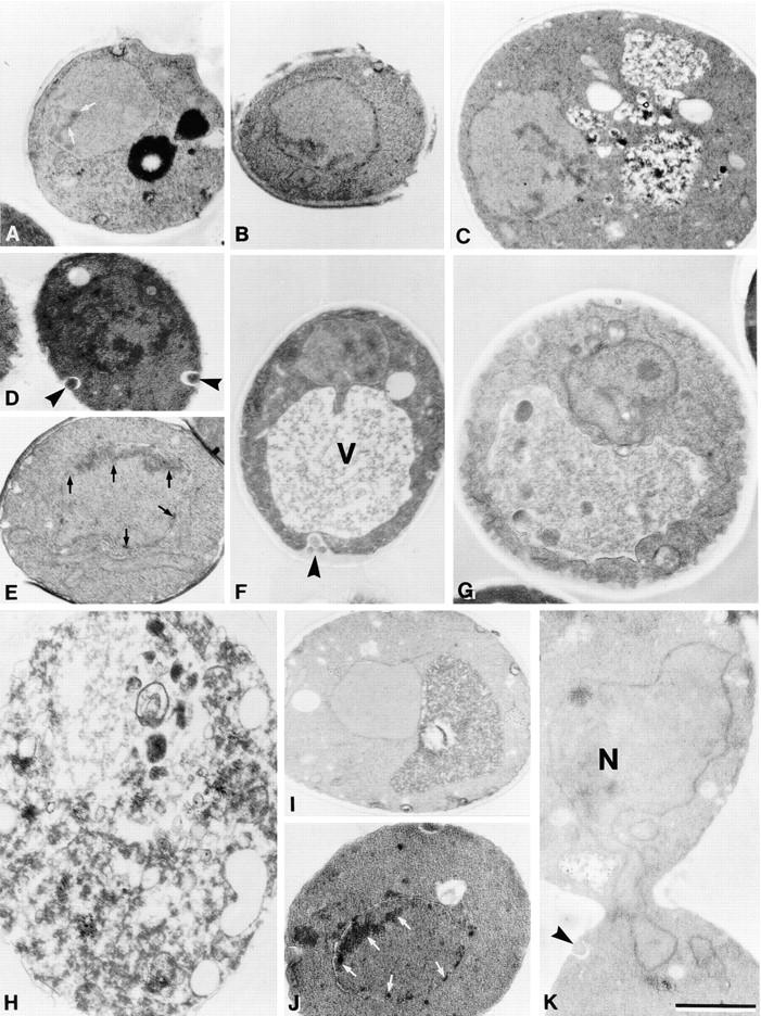
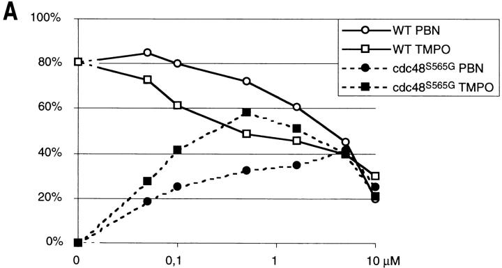
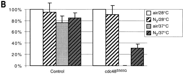
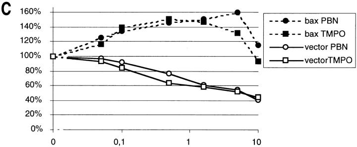
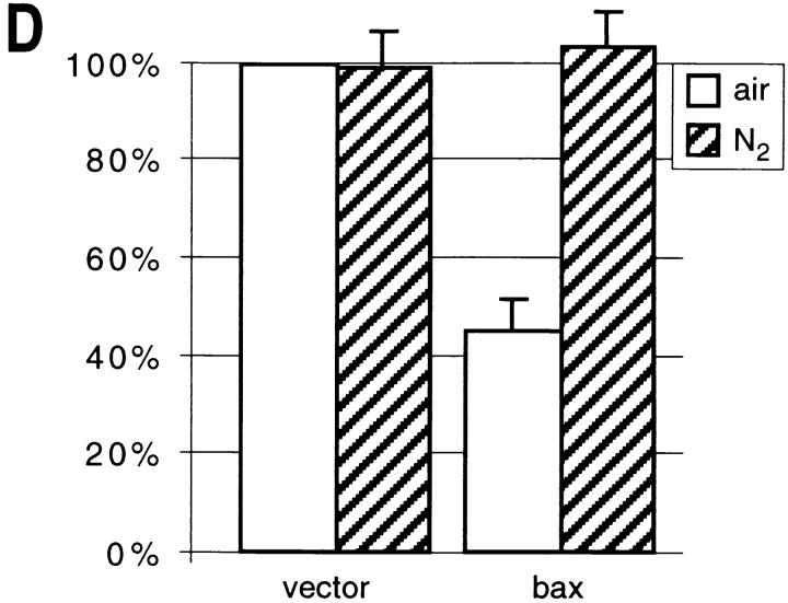
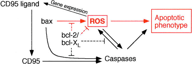
Oxygen radicals are important components of metazoan apoptosis. We have found that apoptosis can be induced in the yeast Saccharomyces cerevisiae by depletion of glutathione or by low external doses of H2O2. Cycloheximide prevents apoptotic death revealing active participation of the cell. Yeast can also be triggered into apoptosis by a mutation in CDC48 or by expression of mammalian bax. In both cases, we show oxygen radicals to accumulate in the cell, whereas radical depletion or hypoxia prevents apoptosis. These results suggest that the generation of oxygen radicals is a key event in the ancestral apoptotic pathway and offer an explanation for the mechanism of bax-induced apoptosis in the absence of any established apoptotic gene in yeast.


Figures

Figure 1
Figure 1
H2O2 induces an apoptotic phenotype in yeast. DAPI-stained wild-type yeast after incubation for 200 min without (F) and with 3 mM H2O2 (A–E) in YEPD. TUNEL reaction after 200 min treatment of exponentially growing cells with 0 mM (G), 1 mM (H), 3 mM (I), 15 mM (J), and 180 mM H2O2 (K), stationary cells treated with 180 mM H2O2 (L), exponentially growing cells treated with 3 mM H2O2 plus 15 μg/ml cycloheximide (M), and exponentially growing cells treated with 15 μg/ml cycloheximide for 230 min (N). Survival (colony formation on YEPD plates, 100% corresponds to the number of plated cells) of wild-type yeast incubated with H2O2 in the absence (black) or presence (red) of 15 μg/ml cycloheximide for 200 min (O). Results were averaged from three experiments. Bars: 5 μm (A–F, G–N).
Figure 1
Figure 1
H2O2 induces an apoptotic phenotype in yeast. DAPI-stained wild-type yeast after incubation for 200 min without (F) and with 3 mM H2O2 (A–E) in YEPD. TUNEL reaction after 200 min treatment of exponentially growing cells with 0 mM (G), 1 mM (H), 3 mM (I), 15 mM (J), and 180 mM H2O2 (K), stationary cells treated with 180 mM H2O2 (L), exponentially growing cells treated with 3 mM H2O2 plus 15 μg/ml cycloheximide (M), and exponentially growing cells treated with 15 μg/ml cycloheximide for 230 min (N). Survival (colony formation on YEPD plates, 100% corresponds to the number of plated cells) of wild-type yeast incubated with H2O2 in the absence (black) or presence (red) of 15 μg/ml cycloheximide for 200 min (O). Results were averaged from three experiments. Bars: 5 μm (A–F, G–N).
Figure 3
Figure 3
A yeast lacking glutathione exhibits the typical markers of apoptosis, nuclear breakage, DNA fragmentation, and exposition of phosphatidylserine. gsh1 mutant (A, C, E) and wild-type control (F shows two cells) grown for 3 d on synthetic medium stained with DAPI; B and D are phase contrast pictures corresponding to A and C. TUNEL test of gsh1 mutant (G) and wild-type control (H) grown for 3 d on synthetic medium. Annexin V binding assay of gsh1 mutant (I–K) and wild-type control (M) grown for 3 d on synthetic medium; L shows the propidium iodide staining corresponding to K. Bars: 10 μm (A–F, I–M); 10 μm (G and H).
Figure 2
Figure 2
Low concentrations of H2O2 and glutathione depletion induce chromatin condensation and margination in S. cerevisiae. Electron micrographs of exponentially grown wild-type strain YPH98 treated with 3 mM H2O2 for 30 min (A), 60 min (B), 120 min (C), or 200 min (D–F), with 3 mM H2O2 plus 15 μg/ml cycloheximide for 200 min (G, nucleolus visible), with 180 mM H2O2 for 200 min (H), untreated control (I), and of gsh1 deletion strain YPH98gsh1 grown on glutathione-free synthetic medium for 3 d (J and K). N, nucleus; V, vacuole; chromatin condensation is marked by arrows, extracellular vesicles (blebs) are marked by arrowheads. Bar, 1 μm.
Figure 4
Figure 4
Yeast mutant KFY437 (allele cdc48 S565G) and yeast-expressing bax accumulate ROS. Rhodamine 123 fluorescence (A–F) and the corresponding phase contrast displays (G–L) after 2 h incubation with dihydrorhodamine 123. Wild-type control (A and G) and mutant KFY437 (B and H) grown at 28°C, wild-type control (C and I) and mutant KFY437 (D and J) grown at 37°C, strain WCG4bax-expressing bax (E and K), strain WCG4bax/bcl-XL expressing bax and bcl-XL (F and L). Fluorescence (M and N) and the corresponding phase contrast displays (Q and R) of wild-type control (M and Q) and mutant KFY437 (N and R) grown at 37°C after 2 h incubation with dichlorodihydrofluorescein diacetate. Fluorescence (O and P) and the corresponding phase contrast displays (S and T) of wild-type control (O and S) and mutant KFY437 (P and T) grown at 37°C after 10 min incubation with dihydroethidium. Flow cytometric analysis of wild-type control (U) and mutant KFY437 (V) after 2 h incubation with dihydrorhodamine 123.
Figure 4
Figure 4
Yeast mutant KFY437 (allele cdc48 S565G) and yeast-expressing bax accumulate ROS. Rhodamine 123 fluorescence (A–F) and the corresponding phase contrast displays (G–L) after 2 h incubation with dihydrorhodamine 123. Wild-type control (A and G) and mutant KFY437 (B and H) grown at 28°C, wild-type control (C and I) and mutant KFY437 (D and J) grown at 37°C, strain WCG4bax-expressing bax (E and K), strain WCG4bax/bcl-XL expressing bax and bcl-XL (F and L). Fluorescence (M and N) and the corresponding phase contrast displays (Q and R) of wild-type control (M and Q) and mutant KFY437 (N and R) grown at 37°C after 2 h incubation with dichlorodihydrofluorescein diacetate. Fluorescence (O and P) and the corresponding phase contrast displays (S and T) of wild-type control (O and S) and mutant KFY437 (P and T) grown at 37°C after 10 min incubation with dihydroethidium. Flow cytometric analysis of wild-type control (U) and mutant KFY437 (V) after 2 h incubation with dihydrorhodamine 123.
Figure 5
Figure 5
DNA strand breakage and chromatin margination in strain KFY437 and in bax-expressing WCG4bax is prevented by free radical spin traps or anaerobic culture conditions. TUNEL reaction of wild-type control (A) and mutant KFY437 (B) grown at 28°C, of wild-type control (C) and mutant KFY437 (D) incubated at 37°C for 4 h, of mutant KFY437 incubated at 37°C for 4 h in the presence of 5 mM N-tert-butyl-α−phenylnitrone (E), of mutant KFY437 incubated at 37°C in a nitrogen atmosphere (F), of bax-expressing WCG4bax (G), of bax-expressing WCG4bax in the presence of 5 mM N-tert-butyl-α−phenylnitrone (H), of bax-expressing WCG4bax incubated in a nitrogen atmosphere (I). Electron micrographs of mutant KFY437 incubated at 37°C for 4 h without addition (J) or in the presence of 5 mM N-tert-butyl-α−phenylnitrone (K), and of bax-expressing WCG4bax without addition (L) or in the presence of 5 mM N-tert-butyl-α−phenylnitrone (M). Bars: (A and J) 10 μm.
Figure 6
Figure 6
Free radical spin traps or anaerobic culture conditions partially suppress temperature sensitivity in strain KFY437 and bax lethality. (A) Strain KFY437 (allele cdc48 S565G) and the corresponding wild-type were adjusted to a cell titer of 2 × 10⁶/ml and incubated with various concentrations of PBN or TMPO for 4 h at 37°C in YEPD. The portion of colony-forming units is plotted against spin trap concentrations. 100% corresponds to the respective strain grown at 28°C without spin traps. (B) Strain KFY437 (allele cdc48 S565G) and the corresponding wild-type were adjusted to a cell titer of 2 × 10⁶/ml in YEPD, incubated with aeration or in a nitrogen stream at 28°C for 30 min, and further incubated at 37 or 28°C for 4 h. Viability was determined as the portion of colony-forming units. 100% corresponds to the respective strain grown aerobically at 28°C. (C) WCG4bax and the corresponding control strain grown on synthetic medium with glucose were transferred to synthetic medium with galactose, diluted to a cell titer of 2 × 10⁶/ml, and incubated for 15 h either with various concentrations of PBN or TMPO. The portion of colony-forming units is plotted against spin trap concentrations. 100% corresponds to the respective strain grown without spin traps. (D) WCG4bax and the corresponding control strain grown on synthetic medium with glucose were transferred to synthetic medium with galactose, diluted to a cell titer of 2 × 10⁶/ml, and incubated for 15 h with aeration or in a nitrogen stream. Viability was determined as the portion of colony-forming units. 100% corresponds to the aerobically grown control strain. The portion of colony-forming units was determined by incubating YEPD plates with 1,000 cells each for 3 d at 28°C and counting visible colonies. Results were averaged from three experiments each. Standard deviations are below 10% for all data points.
Figure 6
Figure 6
Free radical spin traps or anaerobic culture conditions partially suppress temperature sensitivity in strain KFY437 and bax lethality. (A) Strain KFY437 (allele cdc48 S565G) and the corresponding wild-type were adjusted to a cell titer of 2 × 10⁶/ml and incubated with various concentrations of PBN or TMPO for 4 h at 37°C in YEPD. The portion of colony-forming units is plotted against spin trap concentrations. 100% corresponds to the respective strain grown at 28°C without spin traps. (B) Strain KFY437 (allele cdc48 S565G) and the corresponding wild-type were adjusted to a cell titer of 2 × 10⁶/ml in YEPD, incubated with aeration or in a nitrogen stream at 28°C for 30 min, and further incubated at 37 or 28°C for 4 h. Viability was determined as the portion of colony-forming units. 100% corresponds to the respective strain grown aerobically at 28°C. (C) WCG4bax and the corresponding control strain grown on synthetic medium with glucose were transferred to synthetic medium with galactose, diluted to a cell titer of 2 × 10⁶/ml, and incubated for 15 h either with various concentrations of PBN or TMPO. The portion of colony-forming units is plotted against spin trap concentrations. 100% corresponds to the respective strain grown without spin traps. (D) WCG4bax and the corresponding control strain grown on synthetic medium with glucose were transferred to synthetic medium with galactose, diluted to a cell titer of 2 × 10⁶/ml, and incubated for 15 h with aeration or in a nitrogen stream. Viability was determined as the portion of colony-forming units. 100% corresponds to the aerobically grown control strain. The portion of colony-forming units was determined by incubating YEPD plates with 1,000 cells each for 3 d at 28°C and counting visible colonies. Results were averaged from three experiments each. Standard deviations are below 10% for all data points.
Figure 7
Figure 7
ROS participate in the regulation of apoptosis at various levels. Important established regulators of apoptosis. Components and functions present in yeast are marked in red.

Similar articles

Cited by

References

    1. Adams AEM, Pringle JR. Relationship of actin and tubulin distribution to bud growth in wild-type and morphogenetic-mutant Saccharomyces cerevisiae. . J Cell Biol. 1984;98:934–945. - PMC - PubMed
    1. Bischoff JR, Casso D, Beach D. Human p53 inhibits growth in Schizosaccharomyces pombe. . Mol Cell Biol. 1992;12:1405–1411. - PMC - PubMed
    1. Boggs SE, McCormick TS, Lapetina EG. Glutathione levels determine apoptosis in macrophages. Biochem Biophys Res Commun. 1998;247:229–233. - PubMed
    1. Brendel M, Grey M, Maris AF, Hietkamp J, Fesus Z, Pich CT, Dafré AL, Schmidt M, Eckardt-Schupp F, Henriques JA. Low glutathione pools in the original pso3 mutant of Saccharomyces cerevisiaeare responsible for its pleiotropic sensitivity phenotype. Curr Genet. 1998;33:4–9. - PubMed
    1. Budd SL, Castilho RF, Nicholls DG. Mitochondrial membrane potential and hydroethidine-monitored superoxide generation in cultured cerebellar granule cells. FEBS Lett. 1997;415:21–24. - PubMed

Publication types

MeSH terms