. 1999 May 17;145(4):769-82.
doi: 10.1083/jcb.145.4.769.

Location and clonal analysis of stem cells and their differentiated progeny in the human ocular surface

Affiliations

Location and clonal analysis of stem cells and their differentiated progeny in the human ocular surface

G Pellegrini et al. J Cell Biol. .

Abstract

We have analyzed the proliferative and differentiation potential of human ocular keratinocytes. Holoclones, meroclones, and paraclones, previously identified in skin, constitute also the proliferative compartment of the ocular epithelium. Ocular holoclones have the expected properties of stem cells, while transient amplifying cells have variable proliferative potential. Corneal stem cells are segregated in the limbus, while conjunctival stem cells are uniformly distributed in bulbar and forniceal conjunctiva. Conjunctival keratinocytes and goblet cells derive from a common bipotent progenitor. Goblet cells were found in cultures of transient amplifying cells, suggesting that commitment for goblet cell differentiation can occur late in the life of a single conjunctival clone. We found that conjunctival keratinocytes with high proliferative capacity give rise to goblet cells at least twice in their life and, more importantly, at rather precise times of their life history, namely at 45-50 cell doublings and at approximately 15 cell doublings before senescence. Thus, the decision of conjunctival keratinocytes to differentiate into goblet cells appears to be dependent upon an intrinsic "cell doubling clock. " These data open new perspectives in the surgical treatment of severe defects of the anterior ocular surface with autologous cultured conjunctival epithelium.


Figures

Figure 1
Figure 1
The ocular surface. This figure shows areas of the ocular surface from where biopsies were taken. Fornix, limbus, and paracentral cornea are colored in yellow, red, and blue, respectively. The central cornea is indicated by the asterisk. The bulbar region is indicated by the white color. Biopsies were taken from: superior (a) and inferior (b) fornix; superior (c), temporal (d), inferior (e), and nasal (f) bulbar conjunctiva; superior (g), temporal (h), inferior (i), and nasal (l) limbus; paracentral (m) and central (n) cornea.
Figure 2
Figure 2
Histology. Sheets of epithelial cells cultivated from superior limbus (A and C) and superior fornix (B and D) were detached from the culture vessel with the neutral protease Dispase II. Epithelial sheets were either stained with hematoxylin-eosin (A and B) or double-immunostained with K3-specific AE5 mAb and K19-specific RCK108 mAb (C and D). Note that cultured corneal-limbal epithelium is K3+ and K19 (C), whereas cultured conjunctival epithelium is K3 and K19+ (D).
Figure 3
Figure 3
Determination of the colony-forming efficiency. 12 biopsies were taken from different areas (also indicated by lower case letters in parentheses, see Fig. 1) of the ocular surface of a 54-yr-old organ donor woman. 300 cells from each biopsy were plated onto lethally irradiated 3T3-J2 cells. Dishes were stained 12 d later with rhodamine B. Values are expressed as the ratio of the number of colonies over the number of inoculated cells and are indicated in parentheses. Note that conjunctival colony-forming cells are uniformly distributed in forniceal and bulbar conjunctiva, whereas clonogenic cells of the limbus-cornea are segregated in the limbus.
Figure 4
Figure 4
Determination of the number of cell generations. (A) Ocular keratinocytes isolated from the same biopsies analyzed in Fig. 3 (a–n), were serially cultivated until they reached senescence and the number of cell generations was calculated as described in Materials and Methods. (B) Cells obtained from different areas of forniceal and bulbar conjunctiva underwent a comparable number of cell divisions (80–100) before senescence. (C) Corneal-limbal cells endowed with high proliferative capacity were segregated in the limbus. *This value (90.5 ± 7) is the average of cell generations produced by keratinocytes cultivated from superior, temporal, inferior, and nasal limbus.
Figure 5
Figure 5
Determination of the number of progeny generated by conjunctival clones. Clonal analysis and classification of clonal types were performed as described in Materials and Methods. Cultures from 60 conjunctival clones obtained from the same donor were serially cultivated until they reached senescence. The number of cell generations was calculated as described in Materials and Methods. 4 holoclones (H), 43 meroclones (M), and 13 paraclones (P) were analyzed. Blue bars indicate clones from forniceal conjunctiva. Yellow bars indicate clones from bulbar conjunctiva.
Figure 6
Figure 6
Histology and PAS reaction. (A and B) PAS-staining of confluent sheets prepared from secondary cultures of bulbar (A) and limbal (B) keratinocytes. Note that several goblet cells were present in suprabasal layers of cultured conjunctival epithelium (A, at arrows), while none were present in the epithelium generated by limbal-corneal cells (B). (C) Bulbar (yellow lines) and forniceal (blue lines) keratinocytes were serially cultivated and goblet cell content was determined at each cell passage on exponentially growing colonies. Goblet cells were present during the entire life span of the cultures. (D) Forniceal keratinocytes (from a sub-confluent primary culture) were plated (6 × 10³ cells/cm2) in parallel dishes in triplicates. EGF was added after 2 d of cultivation. Cells were trypsinized and counted every 24 h (starting from the third day after plating). Aliquots of the cell suspension were centrifuged on a coverslip and goblet cells were identified and counted (see Materials and Methods). Cells reached confluence 5–6 d after plating. Note that the number of goblet cells increased 25-fold by 1–2 d after confluence. Similar data were obtained with two forniceal and two bulbar cultures. (E–H) Clonal analysis and classification of clonal types were performed as described in Materials and Methods. An aliquot of each of the 339 clones shown in Table II was transferred to a dish and cell were cultivated as described. 3–4 d after plating cultures were fixed and PAS-stained as described. Goblet cells (arrows) are present in cultures generated from a forniceal (E) and a bulbar (F) holoclone. Goblet cells are also present in cultures generated from a forniceal (G) and a bulbar (H) meroclone. PAS-staining of the other clones revealed that 100% and 93% of holoclone- and meroclone-derived cultures, respectively, contained goblet cells. Paraclones were usually goblet negative.
Figure 7
Figure 7
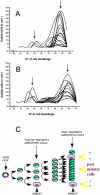
Cell doubling–dependent generation of goblet cells. Quantification of goblet cells during serial cultivation of 26 conjunctival clones. 14 clones, isolated from one donor, are shown in A, and 12 clones generated from a different donor are shown in B. 7 clones were classified as holoclones and 19 clones were classified as meroclones. The x-axis indicates the number of doublings made by each clone. PAS reactions were carried out at each cell passage on exponentially growing colonies. Note that holoclones generated two peaks (arrows in A and B) of goblet cells, at 45–50 cell generations and at 10–20 doublings before senescence. Meroclones generated only one peak of goblet cells at ∼15 doublings before senescence. Analysis of goblet cells began after 20–30 doublings, the preceding interval being devoted to the processing of the biopsy, the isolation of the clones and their growth to suitable large cell population. (C) Schematic description of a model for cell doubling-dependent generation of goblet cells from bipotent conjunctival stem cells. In this model we arbitrarily defined as young transient amplifying cells those cells still able to undergo 35–60 doublings, and old transient amplifying cells those cells undergoing 20–35 cell divisions before senescence. Bipotent conjunctival cells are colored in green and pink. Unipotent epithelial cells are indicated by the uniform green color. Differentiated goblet cells are colored in pink. Both young and old transient amplifying cells can generate goblet cells, at precise times of their life history.

References

    1. Barrandon Y. The epidermal stem cell: an overview. Dev Biol. 1993;4:209–215.
    1. Barrandon Y, Green H. Cell size as a determinant of the clone-forming ability of keratinocytes. Proc Natl Acad Sci USA. 1985;82:5390–5394. - PMC - PubMed
    1. Barrandon Y, Green H. Three clonal types of keratinocytes with different capacities for multiplication. Proc Natl Acad Sci USA. 1987;84:2302–2306. - PMC - PubMed
    1. Christiano AM, Uitto J. Molecular complexity of the cutaneous basement membrane zone. Revelations from the paradigms of epidermolysis bullosa. Exp Dermatol. 1996;5:1–11. - PubMed
    1. Compton CC, Gill JM, Bradford DA, Regauer S, Gallico GG, O'Connor NE. Skin regenerated from cultured epithelial autografts on full-thickness burn wounds from 6 d to 5 yr after grafting. A light, electron microscopic and immunohistochemical study. Lab Invest. 1989;60:600–612. - PubMed

Publication types

Grants and funding