. 1999 May 17;145(4):809-23.
doi: 10.1083/jcb.145.4.809.

Localization of core spindle pole body (SPB) components during SPB duplication in Saccharomyces cerevisiae

Affiliations

Localization of core spindle pole body (SPB) components during SPB duplication in Saccharomyces cerevisiae

I R Adams et al. J Cell Biol. .

Abstract

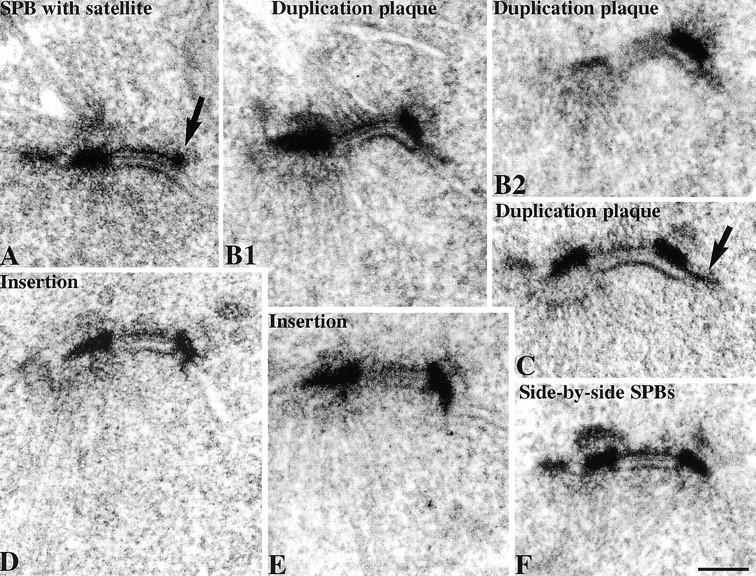
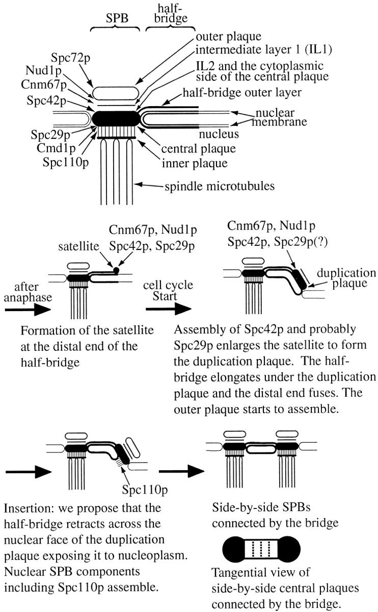
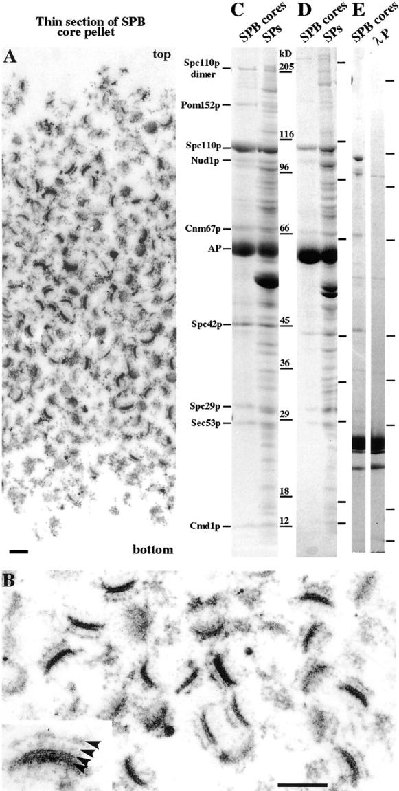
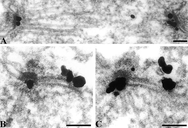
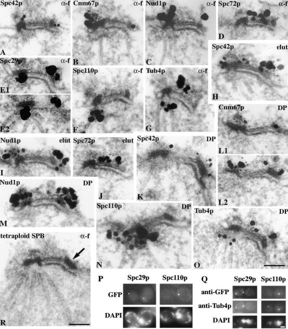
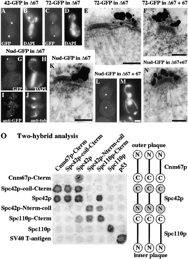
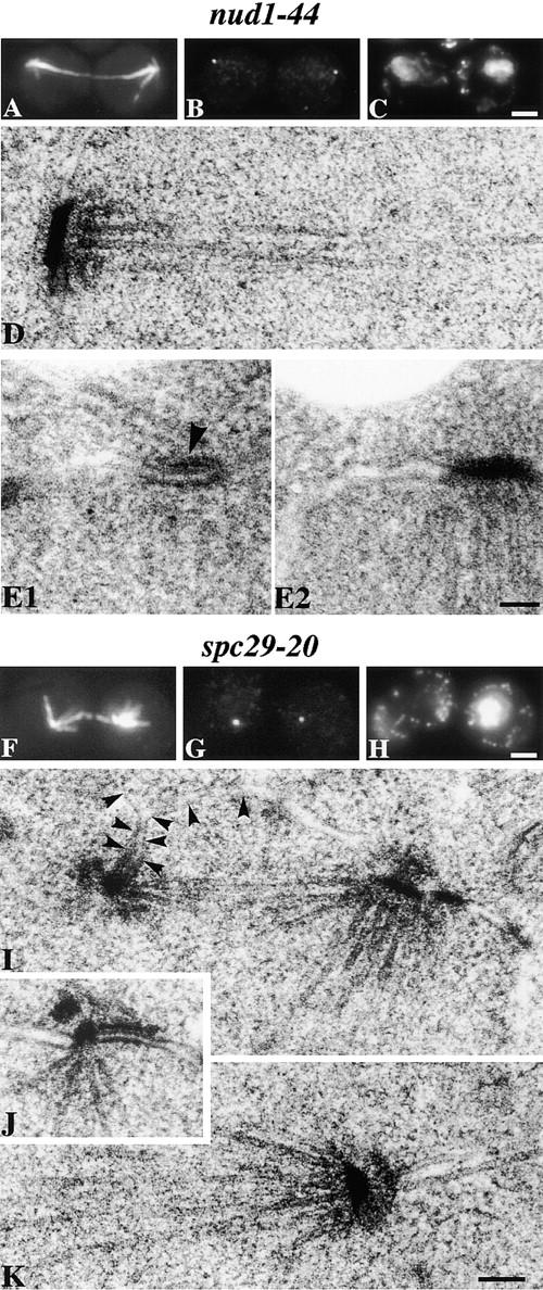
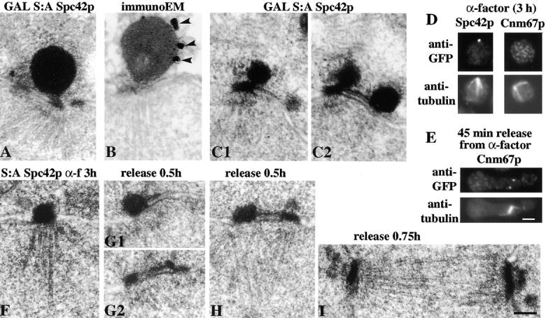
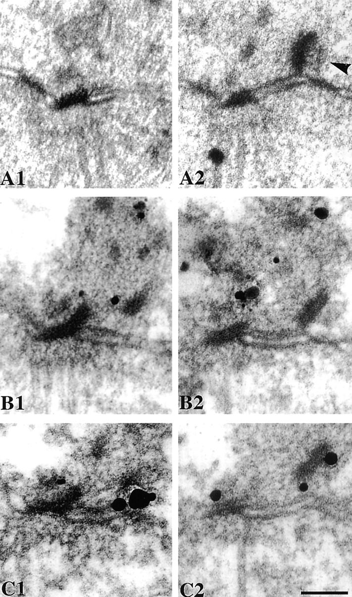
We have examined the process of spindle pole body (SPB) duplication in Saccharomyces cerevisiae by electron microscopy and found several stages. These include the assembly, probably from the satellite, of a large plaque-like structure, the duplication plaque, on the cytoplasmic face of the half-bridge and its insertion into the nuclear envelope. We analyzed the role of the main SPB components in the formation of these structures by identifying them from an SPB core fraction by mass spectrometry. Temperature-sensitive mutants for two of the components, Spc29p and Nud1p, were prepared to partly define their function. The composition of two of the intermediates in SPB duplication, the satellite and the duplication plaque, was examined by immunoelectron microscopy. Both contain cytoplasmic SPB components showing that duplication has already been partly achieved by the end of the preceding cell cycle when the satellite is formed. We show that by overexpression of SPB components the structure of the satellite can be changed and SPB duplication inhibited by disrupting the attachment of the plaque-like intermediate to the half-bridge. We present a model for SPB duplication where binding of SPB components to either end of the bridge structure ensures two separate SPBs.


Figures

Figure 1
Figure 1
Stages during SPB duplication as shown by EM of thin sections of the SPB region of cells arrested in α-factor (A), released for 30 min (B1–E), or 45 min (F). Arrows in A show the satellite and in C a pore structure attached to the fused half-bridge. Serial thin sections are shown in B1 and B2. In all of the EM figures in this paper except spindles, cytoplasm is in the top part of each panel and the existing SPB is on the left. Bar, 0.1 μm.
Figure 9
Figure 9
Diagram of the main core SPB components during the different stages of SPB duplication. Cytoplasmic microtubules which can grow from both the outer plaque and the half-bridge during SPB duplication are not shown.
Figure 2
Figure 2
Analysis of heparin-extracted SPB cores. (A) Thin section of the entire pellet. B is a higher magnification, and the inset in B shows the structural features of the lower right SPB core with arrowheads showing from the top down: the depleted outer plaque, IL1, IL2, and the central plaque. In SPBs from whole cells the IL2 and central plaque layers appear fused and together are called the central plaque. (C and D) Coomassie-stained SDS gradient gels of alkaline phosphatase (AP) or (E) λ phosphatase (λP)-digested SPB cores together with λ phosphatase alone. Identifications of the main bands by MALDI mass spectrometry are to the left of C. The SPB cores in C were isolated from ∼1011 cells. Lines beside the gel lanes in D and E show the positions of the same molecular weight standards used in C. Bars, 0.2 μm.
Figure 3
Figure 3
Localization of GFP-Spc29p by immunoEM in log phase cells. (A) Short spindle. (B and C) Single SPBs with half-bridges. About 200 SPBs were examined. Bars, 0.1 μm.
Figure 6
Figure 6
ImmunoEM localization of GFP-labeled Spc42p, Cnm67p, Nud1p, Spc72p, Spc29p, and Spc110p in cells arrested in G1 with α-factor (G1 α-f), G1 elutriated cells grown for 25 min (elut), and cells released from α-factor for 30 min to observe the duplication plaque (DP). Tub4p was detected with affinity-purified polyclonal antibodies. Adjacent serial sections have the same panel letter and are numbered. (P) Fluorescence of GFP-Spc29p and GFP-Spc110p in unfixed mps2 cells after 3 h at 37°C then treated with DAPI for 0.5 h at 37°C; (Q) immunofluorescence of the same cells with anti-GFP and anti-Tub4p; (R) EM of a thin section of a tetraploid MATa cell arrested with α-factor. The arrow indicates the satellite. Bars, 0.1 μm for the immunoEM and EM and 2 μm for the light microscopy.
Figure 4
Figure 4
Localization of Spc42p-GFP (42-GFP), Spc72p-GFP (72-GFP), and Nud1p-GFP (Nud-GFP) in cells containing a deletion of CNM67 (Δ67) with or without a rescuing plasmid carrying CNM67 (67), using either fluorescence of unfixed cells treated with DAPI (A–D, G, H, L, and M), immunofluorescence with anti-GFP (I) and anti-tubulin (J), or immunoEM (E, F, K, and N). About 200 SPBs were examined for E and K, and ∼30 SPBs for F and N. (O) Two-hybrid analysis of the interactions between Cnm67p, Spc42p, and Spc110p. The activation domain is on the left and the DNA-binding domain at the top. Bars, 0.1 μm for the immunoEM and 2 μm for the light microscopy.
Figure 5
Figure 5
ts phenotype of nud1-44 and spc29-20 alleles. nud1-44 cells were synchronized by elutriation and spc29-20 cells synchronized by α-factor (90 min at 23°C followed by 60 min at 37°C). Immunofluorescence of a synchronized nud1-44 cell (A–C) and an spc29-20 cell (F–H) after 4 h at 37°C with anti-tubulin (A and F), anti-Tub4p (B and G), and DAPI (C and H). (D) EM of a synchronized nud1-44 cell after 4 h at 37°C showing an SPB at the end of a postanaphase spindle. Adjacent serial sections of a synchronized nud1-44 cell containing a short spindle after 2 h at 37°C (E1 and E2) showing that a cytoplasmic microtubule is still interacting with the half-bridge (arrowhead). EM of synchronized spc29-20 cells after 2 h (I and J) and 4 h (K) at 37°C. Arrowheads in I indicate the inner nuclear membrane and show the small SPB is still inserted in the nuclear membrane. Bars, 0.1 μm for the EM and 2 μm for the immunofluorescence.
Figure 7
Figure 7
EM (A, C1, and C2) and immunoEM (B) of cells induced to overexpress S:A Spc42p under the control of the GAL promoter. C1 and C2 are adjacent serial sections. Arrowheads in B show the silver particles. (D) Immunofluorescence of Spc42p-GFP (left) and Cnm67p-GFP (right) in wild-type cells with anti-GFP (top) and anti-tubulin (bottom) after treatment with α-factor for 3 h at 30°C. (E) Immunofluorescence of Cnm67p-GFP 45 min after release from the α-factor treatment in D. (F) EM thin sections of S:A Spc42p cells treated with α-factor as in D and released for 0.5 h (G1, G2, and H) and 0.75 h (I). G1 and G2 are serial sections. These cells contained multiply integrated copies of S:A Spc42p because this increased the size of the balls and thus accentuated the morphological difference between the balls and plaques. In F–H, views were selected with the nuclear membrane bilayer in profile, thus excluding glancing sections of the central plaque. Bars, 0.1 μm for the EM and 2 μm for the immunofluorescence.
Figure 8
Figure 8
Thin section EM of cells overexpressing myc-ΔNLS-Spc110p for 2 h at 30°C under the control of the GAL promoter (A1 and A2), immunoEM with anti-myc to detect ΔNLS-Spc110p (B1 and B2), and with anti-GFP to detect Spc42p-GFP (C1 and C2). The numbers indicate serial sections. The arrowhead in A2 shows the outer plaque side of the displaced plaque. Bar, 0.1 μm.

References

    1. Anderson RGW, Brenner RM. The formation of basal bodies (centrioles) in the Rhesus monkey oviduct. J Cell Biol. 1971;50:10–34. - PMC - PubMed
    1. Baudin A, Ozier-Kalogeropoulos O, Denouel A, Lacroute F, Cullin C. A simple and efficient method for direct gene deletion in Saccharomyces cerevisiae. . Nucl Acids Res. 1993;21:3329–3330. - PMC - PubMed
    1. Beisson J, Sonneborn TM. Cytoplasmic inheritance of the organization of the cell cortex in . Paramecium aurelia Proc Natl Acad Sci USA. 1965;53:275–282. - PMC - PubMed
    1. Biggins S, Ivanovska I, Rose MD. Yeast ubiquitin-like genes are involved in the duplication of the microtubule organizing center. J Cell Biol. 1996;133:1331–1346. - PMC - PubMed
    1. Brachat A, Kilmartin JV, Wach A, Philippsen P. Saccharomyces cerevisiaecells with defective spindle pole body outer plaques accomplish nuclear migration via half-bridge-organized microtubules. Mol Biol Cell. 1998;9:977–991. - PMC - PubMed

Publication types

MeSH terms