. 1999 May 17;145(4):837-49.
doi: 10.1083/jcb.145.4.837.

p21-activated kinase 1 (Pak1) regulates cell motility in mammalian fibroblasts

Affiliations

p21-activated kinase 1 (Pak1) regulates cell motility in mammalian fibroblasts

M A Sells et al. J Cell Biol. .

Abstract

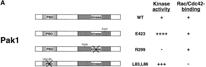
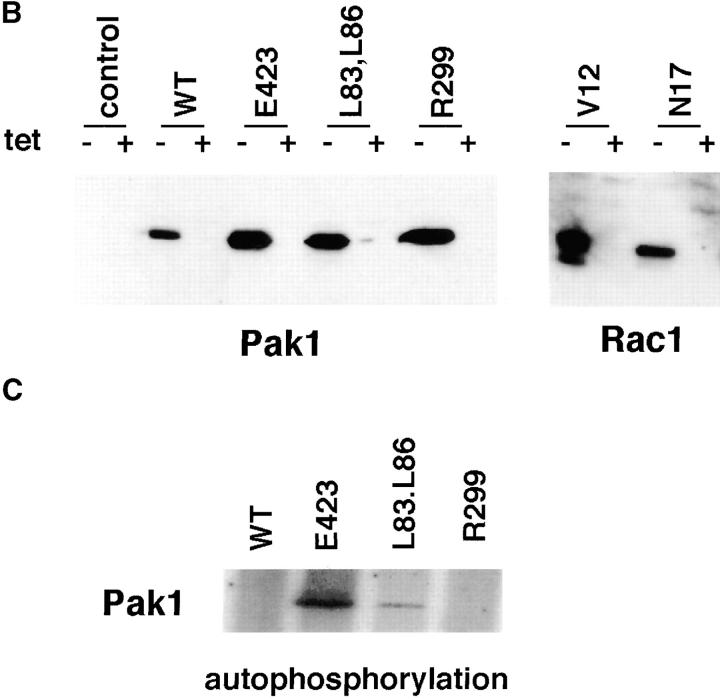
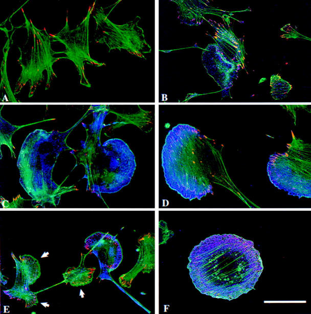
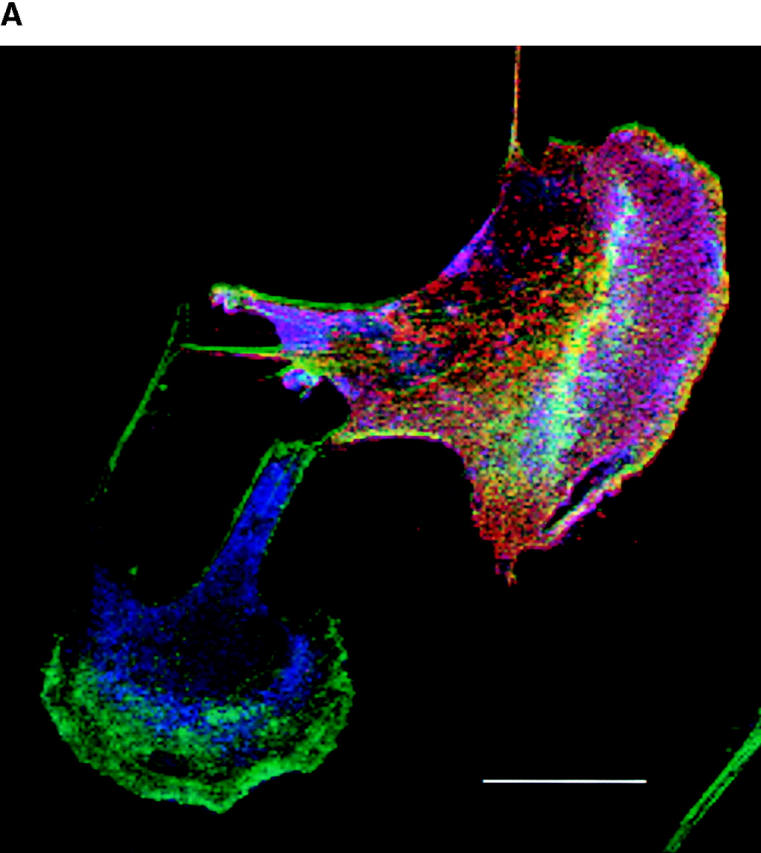
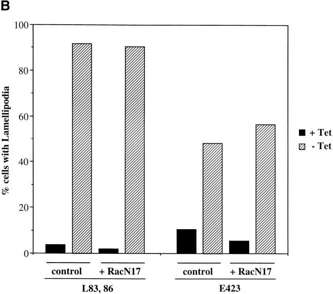
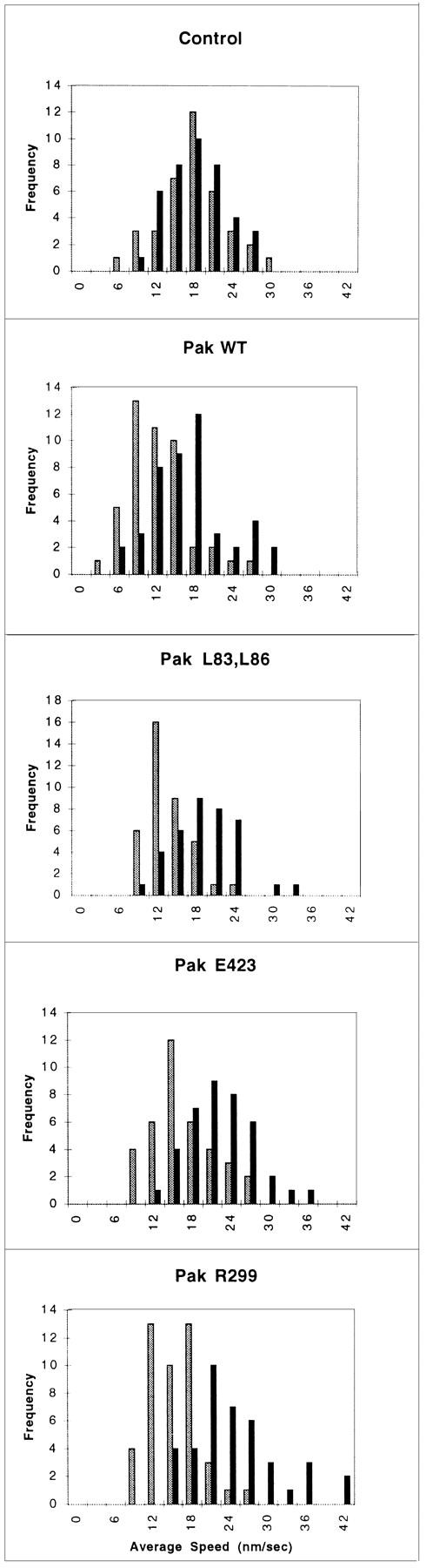
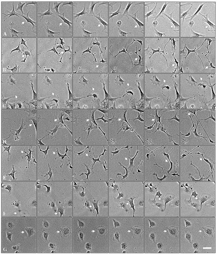
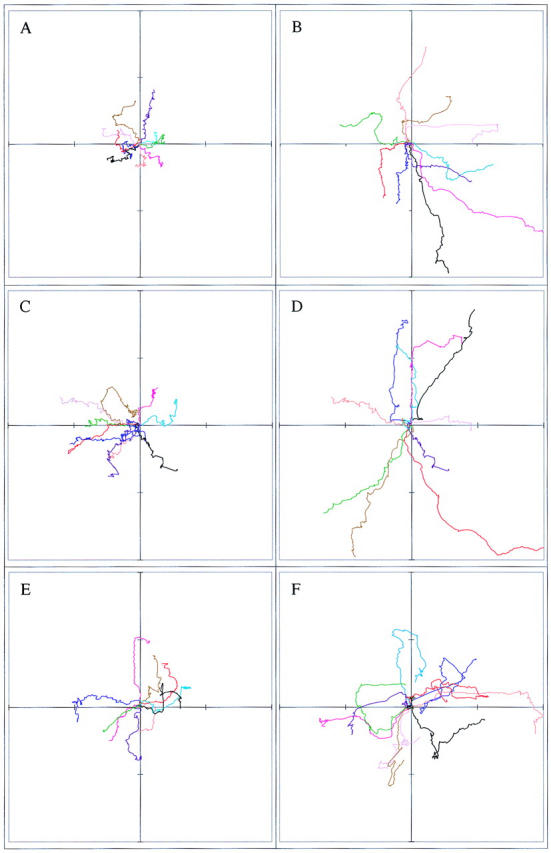
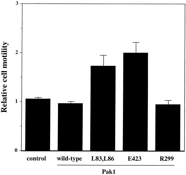
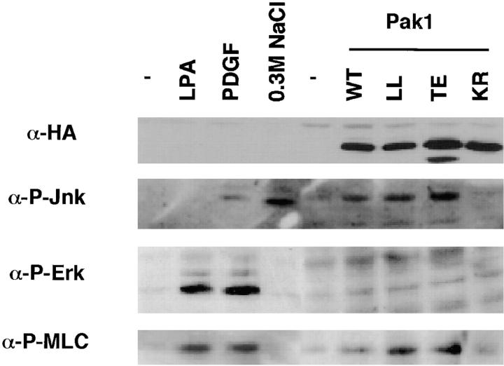
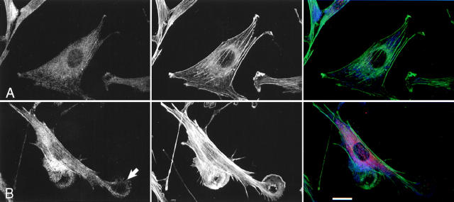
The p21 (Cdc42/Rac) activated kinase Pak1 regulates cell morphology and polarity in most, if not all, eukaryotic cells. We and others have established that Pak's effects on these parameters are mediated by changes in the organization of cortical actin. Because cell motility requires polarized rearrangements of the actin/myosin cytoskeleton, we examined the role of Pak1 in regulating cell movement. We established clonal tetracycline-regulated NIH-3T3 cell lines that inducibly express either wild-type Pak1, a kinase-dead, or constitutively-active forms of this enzyme, and examined the morphology, F-actin organization, and motility of these cells. Expression of any of these forms of Pak1 induced dramatic changes in actin organization which were not inhibited by coexpression of a dominant-negative form of Rac1. Cells inducibly expressing wild-type or constitutively-active Pak1 had large, polarized lamellipodia at the leading edge, were more motile than their normal counterparts when plated on a fibronectin-coated surface, and displayed enhanced directional movement in response to an immobilized collagen gradient. In contrast, cells expressing a kinase-dead form of Pak1 projected multiple lamellipodia emerging from different parts of the cell simultaneously. These cells, though highly motile, displayed reduced persistence of movement when plated on a fibronectin-coated surface and had defects in directed motility toward immobilized collagen. Expression of constitutively activated Pak1 was accompanied by increased myosin light chain (MLC) phosphorylation, whereas expression of kinase-dead Pak1 had no effect on MLC. These results suggest that Pak1 affects the phosphorylation state of MLC, thus linking this kinase to a molecule that directly affects cell movement.


Figures

Figure 1
Figure 1
Tetracycline regulated expression of various forms of Pak1 and Rac1 in NIH-3T3 clonal cell lines. (a) Structure and activities of Pak1 and Rac1 mutants used in this study. A diagrammatic representation of the wild-type and mutant forms of Pak1 and Rac1 are shown in the top panel with amino acid changes denoted by a single letter amino acid abbreviation of the mutant residue. For Pak1, domains shown include the p21 binding domain (PBD) and the catalytic domain (kinase). (b) Inducible expression of Pak1 and Rac1 proteins. S2-6 NIH-3T3 cells, containing the tTA transactivator, were transfected with tetracycline-regulatable expression plasmids bearing either no insert (control), or various forms of HA epitope–tagged Pak1 or Myc-tagged Rac1. Pak1 constructs: wild-type, WT; constitutively activated, E423; kinase-inactive, R299; constitutively activated and Cdc42/Rac-binding defective, L83,L86. Rac1 constructs: constitutively activated, V12; and dominant-negative, N17. Stable clones were isolated and characterized in both the presence and absence of tetracycline (tet) for Pak1 expression by immunoblot with anti-HA antisera, and for Rac1 expression by immunoblot with anti-Myc antisera. (c) Protein kinase activity of induced cell lines. HA-tagged Pak1 was immunoprecipitated from the indicated cell lines and tested for auto-kinase activity.
Figure 1
Figure 1
Tetracycline regulated expression of various forms of Pak1 and Rac1 in NIH-3T3 clonal cell lines. (a) Structure and activities of Pak1 and Rac1 mutants used in this study. A diagrammatic representation of the wild-type and mutant forms of Pak1 and Rac1 are shown in the top panel with amino acid changes denoted by a single letter amino acid abbreviation of the mutant residue. For Pak1, domains shown include the p21 binding domain (PBD) and the catalytic domain (kinase). (b) Inducible expression of Pak1 and Rac1 proteins. S2-6 NIH-3T3 cells, containing the tTA transactivator, were transfected with tetracycline-regulatable expression plasmids bearing either no insert (control), or various forms of HA epitope–tagged Pak1 or Myc-tagged Rac1. Pak1 constructs: wild-type, WT; constitutively activated, E423; kinase-inactive, R299; constitutively activated and Cdc42/Rac-binding defective, L83,L86. Rac1 constructs: constitutively activated, V12; and dominant-negative, N17. Stable clones were isolated and characterized in both the presence and absence of tetracycline (tet) for Pak1 expression by immunoblot with anti-HA antisera, and for Rac1 expression by immunoblot with anti-Myc antisera. (c) Protein kinase activity of induced cell lines. HA-tagged Pak1 was immunoprecipitated from the indicated cell lines and tested for auto-kinase activity.
Figure 2
Figure 2
Expression of mutant forms of Pak1 modulates F-actin and vinculin distribution in cells of NIH-3T3 clonal lines. Pak1 expression was induced by growth in tetracycline-free DME plus 10% CS for 8 h, followed by 16 h starvation in tetracycline-free DME plus 0.1% CS. Confocal images are shown. (a) control, (b) wild-type Pak1, (c) Pak1L83,L86, (d) Pak1E423, (e) Pak1R299 (arrows denote the three lamellipodia of a single cell), and (f) Rac1V12. Cells were stained for F-actin (FITC-phalloidin, green), vinculin (anti-vinculin, visualized with rhodamine X-labeled goat anti–mouse, red), and Pak1 or Rac1 (anti-HA or anti-Myc, visualized with Cy5-labeled goat anti–rabbit, blue). Bar, 25 μm.
Figure 3
Figure 3
Pak1-induced cytoskeletal changes are not mediated by activation of Rac1. (a) Cells conditionally expressing activated Pak1 were transfected with an constitutive expression vector for a dominant-negative (N17) form of Rac1. Pak1 expression was induced by growth in tetracycline-free DME plus 10% CS for 8 h, followed by 16 h starvation in tetracycline-free DME plus 0.5% CS. The cells were then fixed and stained for immunofluorescence. HA-tagged Pak1L83,L86 (blue), Myc-tagged Rac1N17 (red), F-actin (green). Bar, 25 μm. (b) At least 400 cells expressing Pak1 or Pak1 plus Rac1 were assessed for presence of lamellipodia. Values represent combined data from three separate experiments.
Figure 3
Figure 3
Pak1-induced cytoskeletal changes are not mediated by activation of Rac1. (a) Cells conditionally expressing activated Pak1 were transfected with an constitutive expression vector for a dominant-negative (N17) form of Rac1. Pak1 expression was induced by growth in tetracycline-free DME plus 10% CS for 8 h, followed by 16 h starvation in tetracycline-free DME plus 0.5% CS. The cells were then fixed and stained for immunofluorescence. HA-tagged Pak1L83,L86 (blue), Myc-tagged Rac1N17 (red), F-actin (green). Bar, 25 μm. (b) At least 400 cells expressing Pak1 or Pak1 plus Rac1 were assessed for presence of lamellipodia. Values represent combined data from three separate experiments.
Figure 4
Figure 4
Cell motility is stimulated by Pak1. Pak1 expression was induced by growth in tetracycline-free DME as described in Methods. After protein induction, the cells were treated with trypsin, washed in the presence of soybean-trypsin inhibitor, and then replated in tetracycline-free DME plus 0.1% BSA onto BSA-coated plates for 30 min. The cells were then transferred to a fibronectin-coated plate and allowed to attach for 30 min. A graphic representation of average speed of cells, expressed in nm/s is shown. Cell movement was tracked at 30-s intervals using Inovison Isee™ nano-tracking system. Results shown are representative of four independent experiments. (Gray bars) Plus tetracycline; (black bars) minus tetracycline.
Figure 5
Figure 5
Pak1 kinase activity affects character of movement. Pak1 expression was induced by growth in tetracycline-free DME as in Fig. 4. After attachment to fibronectin-coated plates, individual cells were tracked. Phase contrast micrographs shown were taken at 30-min intervals. (a) control cells; (b) wild-type Pak1, (c) Pak1L83,L86, (d) Pak1E423, (e) Pak1R299 (the cell denoted by an arrow displayed from two to four lamellipodia throughout the course of the experiment), (f) Rac1V12, and (g) Rac1N17. Arrows mark the track of a single motile cell for each sample. Bar, 50 μm.
Figure 6
Figure 6
Pak1 kinase activity affects persistence of movement. Pak1 expression was induced by growth in tetracycline-free DME and the cells plated and tracked as in Fig. 5. The paths of ten randomly-chosen cells were plotted for each experimental group. The cells tracked in a, c, and e were grown in the presence of tetracycline; those in b, d, and f were grown in the absence of tetracycline. (a and b) Pak1L83,L86, (c and d) Pak1E423, and (e and f) Pak1R299. Distance traversed from the origin is graphed in μm, with each hashmark representing 30 μm.
Figure 7
Figure 7
Pak1 kinase activity affects haptotaxis. Cells loaded into the top wells of a modified Boyden chamber and allowed to migrate in response to an immobilized collagen gradient. 18 h post-loading, the number of cells that traversed the membrane was quantified. Results shown are representative of three independent experiments, and are normalized to the results obtained for cells grown in the presence of tetracycline ± SE.
Figure 8
Figure 8
Activated Pak1 induces MLC phosphorylation in the absence of MAPK activation. Control NIH-3T3 cells or NIH-3T3 cells expressing various forms of Pak1 were starved overnight in media containing 0.5% CS, then treated with the indicated compounds (PDGF, 10 ng/ml or LPA 5 μg/ml) for 10 min. Immunoblots of cell lysates were probed with anti-HA (to detect transgene expression), and antibodies against the activated (phosphorylated) forms of Jnk, Erk, and MLC.
Figure 9
Figure 9
Pak1-induced phosphorylated MLC localizes to leading edge lamellipodia and to stress fibers. Immunofluorescent staining of a stable clonal line expressing a constitutively active form of Pak1 (Pak1L83,L86) in the presence (+tet; row a) or absence (−tet; row b) of tetracycline was performed after starvation overnight in media containing 0.5% CS. Arrow indicates punctate staining in edge ruffle. MLC phosphorylation (P-MLC) was detected by a polyclonal antibody specific for phospho-MLC (left and right panels, blue stain). Phalloidin stained F-actin is shown in the middle and right panels (green stain) whereas Pak1 expression is shown in the right panel (red stain). Bar, 25 μm.

Similar articles

Cited by

References

    1. Allen WE, Jones GE, Pollard JW, Ridley AJ. Rho, Rac, and Cdc42 regulate actin organization and cell adhesion in macrophages. J Cell Sci. 1997;110:707–720. - PubMed
    1. Allen WE, Zicha D, Ridley AJ, Jones GE. A role for Cdc42 in macrophage chemotaxis. J Cell Biol. 1998;141:1147–1157. - PMC - PubMed
    1. Amano M, Ito M, Kimura K, Fukata Y, Chihara K, Nakano T, Matsuura Y, Kaibuchi K. Phosphorylation and activation of myosin by Rho-associated kinase (Rho kinase) J Biol Chem. 1996;271:20246–20249. - PubMed
    1. Anand-Apte B, Zetter B, Viswanathan A, Qiu R-g, Chen J, Ruggieri R, Symons M. PDGF and fibronectin-stimulated migration are differentially regulated by the Rac and ERK pathways. J Biol Chem. 1997;272:30688–30692. - PubMed
    1. Bagrodia S, Taylor SJ, Jordon KA, Van Aelst L, Cerione RA. A novel regulator of p21-activated kinases. J Biol Chem. 1998;273:23633–23636. - PubMed

Publication types