. 1999 May 17;145(4):851-63.
doi: 10.1083/jcb.145.4.851.

Paxillin LD4 motif binds PAK and PIX through a novel 95-kD ankyrin repeat, ARF-GAP protein: A role in cytoskeletal remodeling

Affiliations

Paxillin LD4 motif binds PAK and PIX through a novel 95-kD ankyrin repeat, ARF-GAP protein: A role in cytoskeletal remodeling

C E Turner et al. J Cell Biol. .

Abstract

Paxillin is a focal adhesion adaptor protein involved in the integration of growth factor- and adhesion-mediated signal transduction pathways. Repeats of a leucine-rich sequence named paxillin LD motifs (Brown M.C., M.S. Curtis, and C.E. Turner. 1998. Nature Struct. Biol. 5:677-678) have been implicated in paxillin binding to focal adhesion kinase (FAK) and vinculin. Here we demonstrate that the individual paxillin LD motifs function as discrete and selective protein binding interfaces. A novel scaffolding function is described for paxillin LD4 in the binding of a complex of proteins containing active p21 GTPase-activated kinase (PAK), Nck, and the guanine nucleotide exchange factor, PIX. The association of this complex with paxillin is mediated by a new 95-kD protein, p95PKL (paxillin-kinase linker), which binds directly to paxillin LD4 and PIX. This protein complex also binds to Hic-5, suggesting a conservation of LD function across the paxillin superfamily. Cloning of p95PKL revealed a multidomain protein containing an NH2-terminal ARF-GAP domain, three ankyrin-like repeats, a potential calcium-binding EF hand, calmodulin-binding IQ motifs, a myosin homology domain, and two paxillin-binding subdomains (PBS). Green fluorescent protein- (GFP-) tagged p95PKL localized to focal adhesions/complexes in CHO.K1 cells. Overexpression in neuroblastoma cells of a paxillin LD4 deletion mutant inhibited lamellipodia formation in response to insulin-like growth fac- tor-1. Microinjection of GST-LD4 into NIH3T3 cells significantly decreased cell migration into a wound. These data implicate paxillin as a mediator of p21 GTPase-regulated actin cytoskeletal reorganization through the recruitment to nascent focal adhesion structures of an active PAK/PIX complex potentially via interactions with p95PKL.


Figures

Figure 8
Figure 8
Paxillin fusion proteins precipitate kinase activity that is stimulated by activated Cdc42. (A) GST–LD1 and LD4 fusion proteins were incubated with brain lysate and the washed precipitate subjected to in vitro kinase assays in the presence of GTPγS or GDP-loaded p21 GTPases. GST–LD4 precipitated kinase activity in the absence of added p21 as judged by phosphorylation of exogenous myelin basic protein and phosphorylation of coprecipitating proteins migrating between 70 and 120 kD. The kinase activity associated with LD4 was further stimulated by the addition of GTP-loaded Cdc42. (B) GST–54-313 previously has been shown to precipitate both tyrosine kinase (FAK) and serine kinase activity, resulting in phosphorylation of the paxillin fusion protein and proteins of 70, 95, and 120 kD. Repeating the assay with GST–54-313 dl LD4 failed to generate phosphorylated proteins of 70 and 95 kD consistent with the absence of p95PKL and PAK from these precipitates. Reduction in the intensity of the 120-kD band is consistent with the partial loss of FAK binding to the LD4 deletion mutant. Washing of the precipitates following the in vitro kinase reaction failed to release any of the coprecipitating phosphoproteins.
Figure 1
Figure 1
Differential binding of vinculin and FAK to paxillin LD motifs. (A) Schematic representation of the domain structure of paxillin showing the position of the LD motifs and the LIM domains. Previously reported sites of paxillin phosphorylation are also marked (Turner, 1998). (B) Individual GST–LD domains of paxillin were incubated with smooth muscle cell lysate and LD-binding proteins were affinity-isolated on glutathione agarose beads. A GST–paxillin fusion spanning aa 54–313, previously shown to contain vinculin and FAK binding sites, was included as a positive control. The coprecipitating proteins were resolved by SDS-PAGE and transferred to nitrocellulose. The membranes were probed with antibodies to FAK or vinculin. The FAK blot was then reprobed with an antibody to talin. FAK binding was restricted to GST–LD2, LD4, and the GST–paxillin-54–313 fusion protein. Vinculin bound preferentially to paxillin 54–313, LD1, and LD2. No binding of talin was detected to any of the paxillin fusion proteins. Ly, cell lysate; C, Coomassie blue stained gel of the GST–paxillin fusion proteins; MW, molecular weight standards.
Figure 1
Figure 1
Differential binding of vinculin and FAK to paxillin LD motifs. (A) Schematic representation of the domain structure of paxillin showing the position of the LD motifs and the LIM domains. Previously reported sites of paxillin phosphorylation are also marked (Turner, 1998). (B) Individual GST–LD domains of paxillin were incubated with smooth muscle cell lysate and LD-binding proteins were affinity-isolated on glutathione agarose beads. A GST–paxillin fusion spanning aa 54–313, previously shown to contain vinculin and FAK binding sites, was included as a positive control. The coprecipitating proteins were resolved by SDS-PAGE and transferred to nitrocellulose. The membranes were probed with antibodies to FAK or vinculin. The FAK blot was then reprobed with an antibody to talin. FAK binding was restricted to GST–LD2, LD4, and the GST–paxillin-54–313 fusion protein. Vinculin bound preferentially to paxillin 54–313, LD1, and LD2. No binding of talin was detected to any of the paxillin fusion proteins. Ly, cell lysate; C, Coomassie blue stained gel of the GST–paxillin fusion proteins; MW, molecular weight standards.
Figure 2
Figure 2
Differential binding of metabolically labeled proteins to paxillin LD motifs. CHO.K1 cells were metabolically labeled for 24 h with [35S]cysteine/methionine. Lysates were incubated with GST–paxillin LD motifs or GST–54-313 of paxillin, and binding proteins were precipitated using glutathione agarose beads. Coprecipitating proteins were resolved on SDS-PAGE gels and visualized by fluorography. Individual paxillin LD motifs precipitated a distinct set of labeled proteins. In a parallel experiment, proteins from unlabeled smooth muscle tissue specifically binding to LD4 that comigrated with the bands marked with asterisks (*) were processed for microsequence analysis. Ly, cell lysate.
Figure 3
Figure 3
PIX and clathrin bind selectively to paxillin LD4. (A) Individual GST–LD domains of paxillin were incubated with rat brain lysate and binding proteins were precipitated using glutathione agarose beads. The coprecipitating proteins were resolved on SDS-PAGE and transferred to nitrocellulose membrane. The membranes were probed with an antibody raised against PIX (Materials and Methods). PIX binding was restricted to the GST–LD4 fusion protein. Note that multiple isoforms of PIX have been reported, with brain expressing the most variants. The adaptor protein Crk failed to bind to any of the GST fusion proteins. A similar experiment was performed with gizzard smooth muscle lysate and probed with anticlathrin antibody. Clathrin bound exclusively to the paxillin GST–LD4 motif. (B) To confirm a role for LD4 in PIX-binding, the experiment was repeated using a fusion protein of the NH2 terminus of paxillin containing a deletion of LD4 (54–313 dl LD4). PIX failed to bind to the LD4 deletion mutant. Crk served as a negative control.
Figure 5
Figure 5
PAK and the SH3-SH3-SH3-SH2-adaptor protein Nck bind to paxillin LD4. (A) Individual GST–LD motifs and paxillin GST–54-313 were incubated with rat brain lysate and precipitated on glutathione agarose beads. Coprecipitating proteins were resolved by SDS-PAGE and transferred to nitrocellulose. The membrane was probed with antibodies to PAK and p130Cas. PAK was precipitated by GST fusions comprising paxillin 54–313 and LD4. No binding of p130Cas to any of the paxillin fusion proteins was detected. (B) A specific role for the LD4 motif in binding PAK and Nck was confirmed by comparison of binding to paxillin 54–313 and the paxillin 54–313 dl LD4 mutant in precipitation assays. Deletion of LD4 eliminated the binding of PAK and Nck. FAK binding was reduced, but not completely eliminated, consistent with the ability of FAK to bind to paxillin LD2 (Fig. 1), which remains intact in the 54–313 dl LD4 mutant.
Figure 5
Figure 5
PAK and the SH3-SH3-SH3-SH2-adaptor protein Nck bind to paxillin LD4. (A) Individual GST–LD motifs and paxillin GST–54-313 were incubated with rat brain lysate and precipitated on glutathione agarose beads. Coprecipitating proteins were resolved by SDS-PAGE and transferred to nitrocellulose. The membrane was probed with antibodies to PAK and p130Cas. PAK was precipitated by GST fusions comprising paxillin 54–313 and LD4. No binding of p130Cas to any of the paxillin fusion proteins was detected. (B) A specific role for the LD4 motif in binding PAK and Nck was confirmed by comparison of binding to paxillin 54–313 and the paxillin 54–313 dl LD4 mutant in precipitation assays. Deletion of LD4 eliminated the binding of PAK and Nck. FAK binding was reduced, but not completely eliminated, consistent with the ability of FAK to bind to paxillin LD2 (Fig. 1), which remains intact in the 54–313 dl LD4 mutant.
Figure 4
Figure 4
Sequence analysis of the p95 family of proteins. (A) Schematic representation of the domain structure of the p95 family. The NH2 terminus is comprised of a GCS/SAT zinc-finger-containing ARF–GAP domain, followed by three ankyrin-like repeats. A potential EF hand and partial IQ motifs are represented in red, with potential PBS highlighted in green. A region of low myosin homology, conserved in microtubule-vesicle linker proteins, is localized on the COOH terminus of p95PKL and CAT. (B) Multialign of the p95 family is presented. Peptides obtained from microsequence analysis of LD4-bound p95PKL are underlined. Amino acids constituting functional domains are color coded to match the schematic representation. The potential PBS regions are overlined in green.
Figure 4
Figure 4
Sequence analysis of the p95 family of proteins. (A) Schematic representation of the domain structure of the p95 family. The NH2 terminus is comprised of a GCS/SAT zinc-finger-containing ARF–GAP domain, followed by three ankyrin-like repeats. A potential EF hand and partial IQ motifs are represented in red, with potential PBS highlighted in green. A region of low myosin homology, conserved in microtubule-vesicle linker proteins, is localized on the COOH terminus of p95PKL and CAT. (B) Multialign of the p95 family is presented. Peptides obtained from microsequence analysis of LD4-bound p95PKL are underlined. Amino acids constituting functional domains are color coded to match the schematic representation. The potential PBS regions are overlined in green.
Figure 6
Figure 6
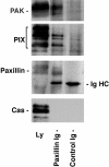
Endogenous PIX and PAK coprecipitate with paxillin. Paxillin was immunoprecipitated from a brain lysate. The paxillin precipitate and a mouse IgG control precipitate were subjected to SDS-PAGE followed by Western blotting. PIX and PAK were coprecipitated with paxillin, but not with the control IgG. The failure of the largest PIX isoform to precipitate efficiently with paxillin suggests that intact paxillin may discriminate between PIX family members for binding. No binding of p130Cas to either the paxillin or control IgG precipitate was observed. IgHC, immunoglobulin heavy chain.
Figure 7
Figure 7
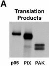
p95PKL directly binds to paxillin LD4 and PIX. To identify the components of the LD4-binding complex that bound directly to paxillin, p95PKL, p85PIX, and PAK were 35S metabolically labeled in vitro by coupled transcription/translation and tested for their ability to bind GST fusion proteins of paxillin (aa 54–313), PIX, and PAK. (A) Translation products of p95PKL, PIX, and PAK. (B) PAK bound to PIX as previously reported. However, no binding of either of these proteins to paxillin 54–313 or GST was detected. In contrast, p95PKL bound to both the paxillin and PIX fusion proteins. No binding of p95PKL to PAK or GST was detected. (C) Deletion of LD4 from the paxillin NH2 terminus fusion protein eliminated the binding to p95PKL. GST–LD4 was sufficient for p95PKL binding. These data provide evidence that p95PKL functions to link paxillin and PIX to PAK. (D) Two regions of p95PKL spanning aa 1–576 and 448–757, respectively, were generated by coupled in vitro transcription/translation and tested for paxillin and PIX binding. PIX bound selectively to the NH2 terminus of p95PKL, containing PBS1 and the ARF–GAP and ankyrin domains, while paxillin bound to the COOH-terminal region containing PBS2. (E) To demonstrate an association in vivo between paxillin and p95PKL, GFP– p95PKL was transfected into COS 7 cells. After lysis, a paxillin precipitate and a mouse IgG control precipitate were subjected to SDS-PAGE, followed by Western blotting with antibodies to GFP, paxillin, and p130Cas. GFP–p95PKL was coprecipitated with paxillin, but not with the control Ig. No binding of p130Cas to either the paxillin or control IgG precipitate was observed. IgHC, immunoglobulin heavy chain; Ly, cell lysate.
Figure 7
Figure 7
p95PKL directly binds to paxillin LD4 and PIX. To identify the components of the LD4-binding complex that bound directly to paxillin, p95PKL, p85PIX, and PAK were 35S metabolically labeled in vitro by coupled transcription/translation and tested for their ability to bind GST fusion proteins of paxillin (aa 54–313), PIX, and PAK. (A) Translation products of p95PKL, PIX, and PAK. (B) PAK bound to PIX as previously reported. However, no binding of either of these proteins to paxillin 54–313 or GST was detected. In contrast, p95PKL bound to both the paxillin and PIX fusion proteins. No binding of p95PKL to PAK or GST was detected. (C) Deletion of LD4 from the paxillin NH2 terminus fusion protein eliminated the binding to p95PKL. GST–LD4 was sufficient for p95PKL binding. These data provide evidence that p95PKL functions to link paxillin and PIX to PAK. (D) Two regions of p95PKL spanning aa 1–576 and 448–757, respectively, were generated by coupled in vitro transcription/translation and tested for paxillin and PIX binding. PIX bound selectively to the NH2 terminus of p95PKL, containing PBS1 and the ARF–GAP and ankyrin domains, while paxillin bound to the COOH-terminal region containing PBS2. (E) To demonstrate an association in vivo between paxillin and p95PKL, GFP– p95PKL was transfected into COS 7 cells. After lysis, a paxillin precipitate and a mouse IgG control precipitate were subjected to SDS-PAGE, followed by Western blotting with antibodies to GFP, paxillin, and p130Cas. GFP–p95PKL was coprecipitated with paxillin, but not with the control Ig. No binding of p130Cas to either the paxillin or control IgG precipitate was observed. IgHC, immunoglobulin heavy chain; Ly, cell lysate.
Figure 9
Figure 9
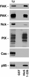
The paxillin family member, Hic-5, binds FAK, p95PKL, PIX, PAK, and Nck. The NH2 terminus of Hic-5 (1–226) was expressed as a GST fusion protein, incubated with rat brain lysate and precipitated on glutathione agarose beads. Coprecipitating proteins were resolved by SDS-PAGE and transferred to nitrocellulose. The membrane was probed with antibodies to FAK, PAK, Nck, PIX, and p130Cas. Alternatively, the GST–Hic-5 fusion protein was incubated with p95PKL that had been 35S metabolically labeled in vitro by coupled transcription/translation. As with paxillin, Hic-5 precipitated FAK, PAK, Nck, and PIX, but not p130Cas. Direct binding of Hic-5 to p95PKL was also detected.
Figure 10
Figure 10
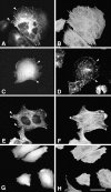
p95PKL localizes to focal adhesions. CHO.K1 cells, transfected with GFP–PKL (A and C), GFP–paxillin (E), or GFP alone (G) were double-labeled with rhodamine phalloidin (B, F, and H; to visualize actin filaments) or with antipaxillin antibody (D). GFP–PKL was enriched in small focal adhesions at the ends of actin stress fibers (A and B, arrowheads), similar to paxillin (E and F, arrowheads). In contrast, GFP alone demonstrated a diffuse cytoplasmic and nuclear distribution with no concentration at the end of actin bundles (G and H). GFP–PKL also colocalized with endogenous paxillin to focal adhesions (C and D, arrowheads). Bar, 5 μm.
Figure 11
Figure 11
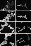
Overexpression of a paxillin LD4 deletion mutant in neuroblastoma SH-SY5Y cells inhibits cell spreading and lamellipodia formation in response to IGF-1. SH-SY5Y cells transfected with wild-type chicken paxillin (A, C, E, and G) or an LD4 deletion mutant of paxillin (B, D, F, and H) were either fixed before serum starvation (A–D) or serum-starved for 12 h (E and F), followed by stimulation for 5 min with 10 nM IGF-1 (G and H). Paxillin distribution (shown only for asynchronously growing cells) was visualized using a chicken paxillin-specific polyclonal antibody and a fluorescein-conjugated anti–rabbit secondary antibody (A and B). Actin distribution was visualized with rhodamine phalloidin (C–H). Both wild-type and mutant paxillin localized to focal adhesions in asynchronously growing cells (A and B, arrowheads). Cells containing the LD4 deletion mutant were generally less well-spread than those expressing wild-type paxillin. The LD4 deletion mutant exhibited significantly fewer lamellipodia (H, arrowheads) in response to treatment with IGF-1, as compared with the cells expressing wild-type paxillin (G, arrowheads). Bar, 5 μm.
Figure 12
Figure 12
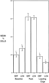
Microinjection of GST–LD4 into NIH3T3 cells retards migration. NIH3T3 cells were grown to confluence on glass coverslips and then half of the cells were removed with a teflon spatula. Cells at the edge of the wound were microinjected with either GST or GST– LD4, and then allowed to migrate into the wound for 24 h. Cells were fixed and stained with antibodies to GST. Microinjected cells (scored blind) were scored for position in one of three categories: baseline, cells remaining at the edge of the original wound; pack, cells which had migrated, but were not at the leading edge; and leading edge, cells at the front of the migrating pack. Four replicates were examined for each injected protein (>200 cells per treatment) and the data analyzed by t test.

Similar articles

Cited by

References

    1. Adam L, Vadlamuda R, Kondapaka B, Chernoff J, Mendelsohn J, Kumar R. Heregulin regulates cytoskeletal reorganization and cell migration through the p21-activated kinase-1 via phosphatidylinositol-3 kinase. J Biol Chem. 1998;273:28238–28246. - PubMed
    1. Ayi TC, Tsan JT, Hwang LY, Bowcock AM, Baer R. Conservation of function and primary structure in the BRCA1-associated RING domain (BARD1) protein. Oncogene. 1998;17:2143–2148. - PubMed
    1. Bagrodia S, Taylor SJ, Creasy CL, Chernoff J, Cerione RA. Identification of a mouse p21Cdc42/Racactivated kinase. J Biol Chem. 1995;270:22731–22737. - PubMed
    1. Bagrodia S, Taylor SJ, Jordan KA, Van Aelst L, Cerione RA. A novel regulator of p21-activated kinases. J Biol Chem. 1998;273:23633–23636. - PubMed
    1. Bagrodia, S., D. Bailey, Z. Leonard, M. Hart, J.-L. Guan, R.T. Premont, S.J. Taylor, and R.A. Cerione. 1999. A tyrosine phosphorylated protein that binds to an important regulatory region on the Cool family of Pak-binding proteins. J. Biol. Chem. In press. - PubMed

Publication types

MeSH terms

Substances

Associated data